


Product Monograph - Adruxe
RECENT MAJOR LABEL CHANGES
| NA | NA |
|---|
TABLE OF CONTENTS
Certain sections or subsections that are not applicable at the time of the preparation of the most recent authorized product monograph are not listed.
RECENT MAJOR LABEL CHANGES ............................................................................................ 2
TABLE OF CONTENTS .............................................................................................................. 2
Part I: Health Professional Information .................................................................................. 4
1 Indications ........................................................................................................................... 4
1.1 Pediatrics ................................................................................................................... 5
1.2 Geriatrics .................................................................................................................... 5
2 Contraindications ................................................................................................................ 5
3 Serious Warnings and Precautions Box ................................................................................ 5
4 Dosage and Administration ................................................................................................. 6
4.1 Dosing Considerations................................................................................................ 6
4.2 Recommended Dose and Dosage Adjustment ........................................................... 6
4.3 Reconstitution ............................................................................................................ 9
4.4 Administration ........................................................................................................... 9
4.5 Missed Dose ............................................................................................................. 11
5 Overdosage ....................................................................................................................... 11
6 Dosage Forms, Strengths, Composition and Packaging ...................................................... 11
7 Warnings and Precautions ................................................................................................. 12
7.1 Special Populations .................................................................................................. 21
8 Adverse Reactions ............................................................................................................. 22
8.1 Adverse Reaction Overview ..................................................................................... 22
8.2 Clinical Trial Adverse Reactions ................................................................................ 22
8.3 Less Common Clinical Trial Adverse Reactions ......................................................... 60
8.4 Abnormal Laboratory Findings: Hematologic, Clinical Chemistry and Other Quantitative Data ........................................................................................................... 61
8.5 Post-Market Adverse Reactions ............................................................................... 62
9 Drug Interactions ............................................................................................................... 64
9.1 Drug Interactions Overview ..................................................................................... 64
9.2 Drug-Drug Interactions ............................................................................................ 64
9.3 Drug-Food Interactions ............................................................................................ 65
9.4 Drug-Herb Interactions ............................................................................................ 65
9.5 Drug-Laboratory Test Interactions ........................................................................... 65
10 Clinical Pharmacology ...................................................................................................... 65
10.1 Mechanism of Action .......................................................................................... 65
10.2 Pharmacodynamics ............................................................................................ 66
10.3 Pharmacokinetics ............................................................................................... 66
11 Storage, Stability and Disposal ......................................................................................... 69
12 Special Handling Instructions ........................................................................................... 69
Part II: Scientific Information ................................................................................................ 69
13 Pharmaceutical Information ............................................................................................ 69
14 Clinical Trials .................................................................................................................... 70
14.1 Clinical Trials - Reference Biologic Drug ............................................................. 70
16 Non-Clinical Toxicology .................................................................................................... 97
17 Supporting Product Monographs ..................................................................................... 98
Patient Medication Information ........................................................................................... 99
Patient Medication Information ......................................................................................... 108
ADRUXE (rituximab for injection) is a biosimilar biologic drug (biosimilar) to Rituxan®. A biosimilar is a biologic drug that was granted authorization based on a demonstration of similarity to a version previously authorized in Canada, known as the reference biologic drug.
Part I: Health Professional Information
1 Indications
Non-Hodgkin’s Lymphoma (NHL)
ADRUXE (rituximab for injection) is indicated for:
- the treatment of patients with relapsed or refractory low-grade or follicular, CD20 positive, B-cell non-Hodgkin’s lymphoma.
- the treatment of patients with CD20 positive, diffuse large B-cell non-Hodgkin’s lymphoma (DLBCL) in combination with CHOP (cyclophosphamide, doxorubicin, vincristine, and prednisone) chemotherapy.
- the treatment of patients with previously untreated Stage III/IV follicular, CD20 positive, B-cell non-Hodgkin's lymphoma in combination with CVP (cyclophosphamide, vincristine and prednisolone) chemotherapy.
- the maintenance treatment of patients with follicular non-Hodgkin’s lymphoma who have responded to induction therapy with either CHOP or CHOP plus ADRUXE.
- single-agent maintenance treatment of previously untreated patients with advanced follicular non-Hodgkin’s lymphoma with high tumour burden and who have responded to induction therapy with either CHOP plus ADRUXE or CVP plus ADRUXE.
Chronic Lymphocytic Leukemia (CLL)
ADRUXE (rituximab for injection) is indicated for:
- the treatment of patients with previously untreated or previously treated B-cell chronic lymphocytic leukemia (B-CLL), Binet Stage B or C, in combination with fludarabine and cyclophosphamide.
The use of rituximab in CLL is based on an improvement in progression-free survival. Overall survival benefit has not been demonstrated in patients with previous treatment for CLL. The efficacy of treatment with R-FC (ADRUXE-fludarabine and cyclophosphamide) in CLL patients who were previously treated with ADRUXE in combination with fludarabine and cyclophosphamide has not been studied (see 14 Clinical Trials for details).
Rheumatoid Arthritis (RA)
ADRUXE (rituximab for injection) in combination with methotrexate is indicated in adult patients:
- to reduce signs and symptoms in adult patients with moderately to severely active rheumatoid arthritis who have had an inadequate response or intolerance to one or more tumour necrosis factor (TNF) inhibitor therapies.
Rituximab in combination with methotrexate has been shown to reduce the rate of progression of joint damage as measured by x-ray.
Granulomatosis with Polyangiitis (GPA, also known as Wegener's Granulomatosis) and Microscopic Polyangiitis (MPA)
ADRUXE (rituximab for injection) in combination with glucocorticoids is indicated for
- the induction of remission in adult patients with severely active Granulomatosis with Polyangiitis (GPA, also known as Wegener's Granulomatosis) and Microscopic Polyangiitis (MPA).
Consideration should be given to current treatment guidelines for vasculitis.
1.1 Pediatrics
Pediatrics (< 18 years of age): The safety and effectiveness of ADRUXE in pediatric patients have not been established; therefore, Health Canada has not authorized an indication for pediatric use.
1.2 Geriatrics
Geriatrics (≥ 65 years of age): In the CLL setting, exploratory subgroup analysis indicates that use in the geriatric population is associated with differences in efficacy and safety. See 14.1 Clinical Trials - Reference Biologic Drug and for details.
2 Contraindications
- ADRUXE (rituximab) is contraindicated in patients with known Type I hypersensitivity or anaphylactic reactions to murine proteins, Chinese Hamster Ovary (CHO) cell proteins, (See), or to any component of this product. For a complete listing, see 6 Dosage Forms, Strengths, Composition and Packaging.
- ADRUXE is also contraindicated in patients who have or have had progressive multifocal leukoencephalopathy (PML).
- ADRUXE is not recommended for use in patients with severe, active infections.
3 Serious Warnings and Precautions Box
Serious Warnings and Precautions
- General: Rituximab is a potent drug. Several adverse reactions are associated with rituximab, some of which are severe and life-threatening (see 7 Warnings and Precautions). This drug should only be used by health professionals experienced in treating Non-Hodgkin’s Lymphoma (NHL), Chronic Lymphocytic Leukemia (CLL), Rheumatoid Arthritis (RA), Granulomatosis with Polyangiitis (GPA, also known as Wegener's Granulomatosis) and Microscopic Polyangiitis (MPA). Patients should be treated in a setting where full resuscitation facilities are immediately available, and where medications and supportive care measures for the treatment of hypersensitivity reactions (e.g., epinephrine, antihistamines, glucocorticoids) are immediately available in the event of an allergic reaction during administration (see 4 Dosage and Administration).
- Infusion Reactions: Deaths within 24 hours of rituximab infusion have occurred. Approximately 80% of fatal infusion reactions occurred in association with the first infusion. Carefully monitor patients during infusions. Discontinue rituximab infusion and provide medical treatment for Grade 3 or 4 infusion reactions (see , Infusion-Related Events).
Progressive Multifocal Leukoencephalopathy (PML): Patients with RA, NHL and CLL who received treatment with rituximab may have an increased risk of PML. PML can cause disability or death. Healthcare professionals should monitor patients on rituximab for any new sign or
- symptom that may be suggestive of PML. Further treatment with rituximab should be withheld immediately at the first sign or symptom suggestive of PML (see , Progressive Multifocal Leukoencephalopathy).
- Tumor Lysis Syndrome (TLS): Acute renal failure requiring dialysis with instances of fatal outcome has been reported in the setting of TLS following treatment of NHL patients with rituximab (see , Infusion-Related Events).
- Hepatitis B Virus (HBV) Reactivation: HBV reactivation has occurred in patients treated with rituximab, in some cases resulting in fulminant hepatitis, hepatic failure, and death. All patients should be screened for HBV infection before treatment initiation, and should be monitored during and after treatment with rituximab. In the event of HBV reactivation, rituximab and concomitant medications should be discontinued.
- Mucocutaneous Reactions: Severe, including fatal, mucocutaneous reactions including Toxic Epidermal Necrolysis (TEN) and Stevens Johnson Syndrome (SJS) have occurred in patients treated with rituximab. Patients experiencing a severe mucocutaneous reaction should discontinue treatment with rituximab and seek prompt medical evaluation (see 7 Warnings and Precautions, Skin).
- Infections: Serious, including fatal, bacterial, fungal, and new or reactivated viral infections can occur during or following the completion of rituximab-based therapy. rituximab treatment should not be initiated in patients with severe active infections. Patients should be screened for infectious disease history (see 7 Warnings and Precautions, Infections).
- Cardiovascular: Serious and potentially fatal cardiovascular events have been reported rarely following administration of rituximab (see 7 Warnings and Precautions).
4 Dosage and Administration
4.1 Dosing Considerations
- Hypersensitivity reactions and severe infusion-related reaction may occur with administration of ADRUXE (see 7 Warnings and Precautions). Since transient hypotension may occur during infusion with ADRUXE, consideration should be given to withholding anti-hypertensive medications 12 hours prior to and throughout infusion with ADRUXE.
- Patients who develop clinically significant arrhythmias should undergo cardiac monitoring during and after subsequent infusions of rituximab. Patients with pre-existing cardiac conditions such as angina and arrhythmias should be monitored during and after the infusion of ADRUXE.
4.2 Recommended Dose and Dosage Adjustment
Non-Hodgkin’s Lymphoma
Usual Dose
Low Grade or Follicular Non-Hodgkin’s Lymphoma
Premedication
Premedication consisting of an analgesic or anti-pyretic (e.g. acetaminophen) and an antihistaminic drug (e.g. diphenhydramine) should always be administered before each infusion of ADRUXE.
Premedication with glucocorticoids should also be considered, particularly if ADRUXE is not given in combination with steroid-containing chemotherapy (See 7 Warnings and Precautions: Non-Hodgkin’s Lymphoma and Chronic Lymphocytic Leukemia-Infusion- Related Events).
Initial treatment
The recommended dosage of ADRUXE as a single agent is 375 mg/m2 given as an IV infusion once weekly for four doses (on days 1, 8, 15, and 22).
The recommended dosage of ADRUXE in combination with CVP chemotherapy is 375 mg/m2 for 8 cycles (21 days/cycle), administered as an IV infusion on day 1 of each chemotherapy cycle after IV administration of the corticosteroid component of CVP.
Maintenance treatment
In previously untreated patients with advanced high-tumour burden follicular lymphoma, after complete or partial response to induction treatment the recommended dose of ADRUXE maintenance therapy is 375 mg/m2 body surface area. ADRUXE maintenance therapy should be initiated eight weeks following completion of ADRUXE in combination with chemotherapy.
ADRUXE as a single agent should be administered every 8 weeks for a maximum of 12 doses (two years).
The recommended dose of ADRUXE for relapsed or refractory patients after response to induction treatment is 375 mg/m2 every 3 months until disease progression or for a maximum period of two years.
Diffuse Large B-cell Non-Hodgkin’s Lymphoma
Premedication
Premedication consisting of an analgesic/anti-pyretic (e.g. acetaminophen) and an antihistaminic drug (e.g. diphenhydramine) should always be administered before each infusion of ADRUXE.
Premedication with glucocorticoids should also be considered, particularly if ADRUXE is not given in combination with steroid-containing chemotherapy (See 7 Warnings and Precautions/ Non-Hodgkin’s Lymphoma and Chronic Lymphocytic Leukemia/Infusion- Related Events).
ADRUXE should be used in combination with CHOP chemotherapy. The recommended dosage of ADRUXE is 375 mg/m2 administered on day 1 of each chemotherapy cycle after IV administration of the glucocorticoid component of CHOP. The other components of CHOP (cyclophosphamide, doxorubicin, vincristine) should be given after the administration of ADRUXE.
Chronic Lymphocytic Leukemia
Premedication
Premedication consisting of an analgesic or anti-pyretic (e.g. acetaminophen) and an antihistaminic drug (e.g. diphenhydramine) should always be administered before each infusion of ADRUXE.
Premedication with glucocorticoids should also be considered, particularly if ADRUXE is not given in combination with steroid-containing chemotherapy (See 7 Warnings and Precautions: Non-Hodgkin’s Lymphoma and Chronic Lymphocytic Leukemia-Infusion- Related Events).
The recommended dosage of ADRUXE in combination with chemotherapy for previously untreated and previously treated patients is 375 mg/m2 body surface area administered on day 1 of the first treatment cycle followed by 500mg/m2 body surface area administered on day 1 of each subsequent cycle for 6 cycles in total. The chemotherapy should be given after ADRUXE infusion.
Prophylaxis with adequate hydration and administration of uricostatics (such as allopurinol) starting 48 hours prior to start of therapy is recommended for CLL patients to reduce the risk of tumour lysis syndrome. For CLL patients whose lymphocyte counts are > 25 x109/L it is recommended to administer methylprednisolone IV shortly before infusion with ADRUXE to decrease the rate and severity of acute infusion reactions and/or cytokine release syndrome. In study ML17102 an equivalent of 80mg of methylprednisolone (100 mg prednisone IV) was given prior to infusions with rituximab for injection. Seventy-four percent (74%) of patients in the R-FC arms of study ML17102 received at least one dose of corticosteroids, with 27% receiving two or more doses.
Dosage Adjustments During Treatment
No dose reductions of ADRUXE are recommended but 47% of patients in the clinical trial ML17102 for CLL required a delayed and/or slowed infusion, and 17% required their first dose split over two days. When ADRUXE is given in combination with CHOP chemotherapy, standard dose reductions for the chemotherapeutic drugs should be applied. When ADRUXE is given as maintenance treatment, treatment should be delayed in case of significant clinical toxicity according to standard practice.
ADRUXE as a Component of ZEVALIN® (Ibritumomab Tiuxetan) Therapeutic Regimen
As a required component of the ZEVALIN therapeutic regimen, ADRUXE is administered twice. The first administration of ADRUXE is a single infusion of 250 mg/m2 and should precede the second administration by 7-9 days. At the second administration, ADRUXE 250 mg/m2 should be infused within 4 hours prior to the administration of 90Y-ibritumomab tiuxetan. Refer to the ZEVALIN product monograph for full prescribing information.
Rheumatoid Arthritis
Premedication
Premedication consisting of an analgesic or anti-pyretic (e.g. acetaminophen) and an antihistaminic drug (e.g. diphenhydramine) should always be administered before each infusion of rituximab. Premedication with glucocorticoids should also be administered in order to reduce the frequency and severity of infusion-related reactions. Patients should receive 100 mg IV methylprednisolone to be completed 30 minutes prior to each ADRUXE infusion (See 7 Warnings and Precautions: Rheumatoid Arthritis-Infusion-Related Events).
Usual Dose
A course of ADRUXE consists of two 1000 mg IV infusions. The recommended dosage of ADRUXE is 1000 mg by IV infusion followed two weeks later by the second 1000 mg IV infusion.
Retreatment in Patients with RA
The need for further courses should be evaluated 24 weeks following the previous course with retreatment given based on residual disease or disease activity returning to a level above a DAS28-ESR of 2.6 (treatment to remission). Patients may receive further courses no sooner than 16 weeks following the previous course.
Granulomatosis with Polyangiitis (GPA, also known as Wegener’s Granulomatosis) and Microscopic Polyangiitis (MPA)
The recommended dosage of ADRUXE for treatment of GPA/MPA is 375 mg/m2 body surface area, administered as an IV infusion once weekly for 4 weeks.
Glucocorticoids administered as methylprednisolone 1000 mg intravenously per day for 1 to 3 days followed by oral prednisone 1 mg/kg/day (not to exceed 80 mg/day and tapered per clinical need) are recommended to treat severe vasculitis symptoms. This regimen should begin within 14 days prior to or with the initiation of ADRUXE and may continue during and after the 4 week course of ADRUXE treatment.
The efficacy and safety of subsequent courses of ADRUXE have not been established (see 7 Warnings and Precautions and 14 Clinical Trials).
4.3 Reconstitution
Parenteral Products:
Preparation for Administration:
Use appropriate aseptic technique. Use a sterile needle and syringe to prepare ADRUXE. ADRUXE does not contain any preservative or bacteriostatic agent. Withdraw the necessary amount of ADRUXE and dilute to a final concentration of 1 to 4 mg/mL into an infusion bag containing either 0.9% Sodium Chloride Injection USP or 5% Dextrose Injection USP. To avoid foaming, gently invert the bag to mix the solution. Discard any unused portion left in the vial. Parenteral drug products should be inspected visually for particulate matter and discoloration prior to administration. For in-use stability information refer to the 11 Storage, Stability and Disposal.
Incompatibilities
No incompatibilities between ADRUXE and polyvinylchloride or polyethylene bags have been observed.
4.4 Administration
ADRUXE infusions should be administered in a setting where full resuscitation facilities (see 3 Serious Warnings and Precautions Box) are immediately available, and under the close supervision of someone experienced and capable of dealing with severe infusion-related reactions. ADRUXE should be administered as an IV infusion through a dedicated line. Do not administer as an intravenous push or bolus.
Chronic Lymphocytic Leukemia
Do not administer as an intravenous push or bolus. Premedication with glucocorticoids should be considered, particularly if ADRUXE is not given in combination with steroid-containing chemotherapy. Premedication may attenuate infusion-related events. In the clinical trial ML17102 for CLL, the equivalent of 80 mg methylprednisolone (100 mg prednisone IV) was given to most patients prior to each infusion.
First Infusion
The ADRUXE solution for infusion should be administered intravenously at an initial rate of 50 mg/hr. ADRUXE should not be mixed or diluted with other drugs. If hypersensitivity or infusion-related events do not occur, escalate the infusion rate in 50 mg/hr increments every 30 minutes, to a maximum of 400 mg/hr. This rate corresponds to an administration time of 4.25 hours. If hypersensitivity or an infusion related event develops, the infusion should be temporarily slowed or interrupted (see 7 Warnings and
Precautions). The infusion can continue at one-half the previous rate upon improvement of patient symptoms.
Subsequent Infusions
Subsequent infusions of ADRUXE can be administered at an initial rate of 100 mg/hr, and increased by 100 mg/hr increments at 30-minute intervals, to a maximum of 400 mg/hr as tolerated. This rate corresponds to an administration time of 3.25 hours.
Rheumatoid Arthritis
First infusion of each course
The recommended initial rate for infusion is 50 mg/hr; after the first 30 minutes, it can be escalated in 50 mg/hr increments every 30 minutes, to a maximum of 400 mg/hr. This rate corresponds to an administration time of 4.25 hours.
Second infusion of each course
Subsequent doses of ADRUXE can be infused at an initial rate of 100 mg/hr, and increased by 100 mg/hr increments at 30 minutes intervals, to a maximum of 400 mg/hr. This rate corresponds to an administration time of 3.25 hours.
Alternative 120-minute subsequent infusions with the concentration of 4 mg/mL in a 250 mL volume (Rheumatoid Arthritis Only)
If patients did not experience a serious infusion-related adverse event during the previous infusion administered using the standard administration schedule, an alternative 120-minute infusion of a concentration at 4 mg/mL in a 250 mL volume can be administered for the second infusion. Initiate at a rate of 62.5 mL/hour (125 mg) given in the first 30 minutes and 150 mL/hour (875 mg) given over the next 90 minutes. If the 120-minute infusion is tolerated, the same alternative 120-minute infusion rate can be used when administering subsequent infusions and courses.
Patients who have clinically significant cardiovascular disease including arrhythmias or previous serious infusion reactions to any prior biologic therapy or to rituximab, should not be administered the alternative 120-minute infusion.
The Reddy2Assist program has been established to facilitate the administration of ADRUXE in RA and GPA/MPA. Information about the Reddy2Assist program can be obtained by calling 1-877-938-0670.
Granulomatosis with Polyangiitis (GPA, also known as Wegener’s Granulomatosis) and Microscopic Polyangiitis (MPA)
First infusion
The recommended initial infusion rate for ADRUXE is 50 mg/h; subsequently, the rate can be escalated in 50 mg/h increments every 30 minutes to a maximum of 400 mg/h. This rate corresponds to an administration time of 4.25 hours.
Subsequent infusions
Subsequent infusions of ADRUXE can be started at a rate of 100 mg/h and increased by 100 mg/h increments every 30 minutes to a maximum of 400 mg/h. This rate corresponds to an administration time of 3.25 hours.
Pneumocystis Jiroveci Pneumonia (PCP) prophylaxis is recommended for patients with GPA/MPA during and following treatment.
The Reddy2Assist program has been established to facilitate the administration of ADRUXE in RA and GPA/MPA. Information about the Reddy2Assist program can be obtained by calling 1-877-938-0670.
4.5 Missed Dose
Missed or delayed doses should not be omitted but administered at a later time point, based on professional judgment observing the total number of planned cycles and the planned interval between doses.
5 Overdosage
There has been no experience with overdosage in human clinical trials. Single doses higher than 1000 mg have not been tested in controlled clinical studies. The highest dose tested to date is 5g in patients with chronic lymphocytic leukemia. No additional safety signals were identified. Patients who experience overdose should have immediate interruption of their infusion and be closely monitored. Consideration should be given to the need for regular monitoring of blood cell count and for increased risk of infections while patients are B cell-depleted.
For the most recent information in the management of a suspected drug overdose, contact your regional poison control centre or Health Canada’s toll-free number, 1-844-POISON-X (1-844-764-7669).
6 Dosage Forms, Strengths, Composition and Packaging
To help ensure the traceability of biologic products, including biosimilars, health professionals should recognise the importance of recording both the brand name and the non-proprietary (active ingredient) name as well as other product-specific identifiers such as the Drug Identification Number (DIN) and the batch/lot number of the product supplied.
Table 1 Dosage Forms, Strengths, Composition and Packaging
ADRUXE is a clear to opalescent, colorless to yellowish liquid for intravenous (IV) administration. ADRUXE is supplied at a concentration of 10 mg/mL in either 100 mg (10 mL) or 500 mg (50 mL) single-use vials.
100 mg: Each Carton contains one 100 mg/10 mL vial (10 mg/mL), with an orange flip-off cap.
500 mg: Each Carton contains one 500 mg/50 mL vial (10 mg/mL), with a purple flip-off cap.
| Route of Administration | Dosage Form / Strength/Composition | Non-medicinal Ingredients |
| Intravenous infusion | Concentrate for solution, for infusion/10 mg/mL (100 mg/10 mL or 500 mg/50 mL, single-use vials) | Citric acid monohydrate, Polysorbate 80, Sodium chloride, Trisodium citrate dihydrate, Water for injection |
The drug product is formulated as a sterile, preservative-free, concentrated aqueous solution for infusion composed of 10 mg/mL of the active ingredient in citrate buffer (7.17 mg/mL sodium citrate dihydrate, 0.164 mg/mL citric acid monohydrate), 9.0 mg/mL sodium chloride and 0.7 mg/mL polysorbate 80 and water for injection, pH 6.5.
7 Warnings and Precautions
Please see 3 Serious Warnings and Precautions Box
Carcinogenesis and Mutagenesis
No long-term animal studies have been performed to establish the carcinogenic or mutagenic potential of rituximab, or to determine its effects on fertility in males or females. Individuals of childbearing potential should use effective contraceptive methods during treatment and for up to 12 months following therapy with ADRUXE.
Cardiovascular
Non-Hodgkin’s Lymphoma and Chronic Lymphocytic Leukemia
Since transient hypotension may occur during infusion with rituximab, consideration should be given to withholding anti-hypertensive medications 12 hours prior to and throughout infusion with ADRUXE. Serious and potentially fatal cardiovascular events have been reported rarely following administration of rituximab. These events included: angina pectoris, cardiac arrhythmias such as atrial flutter and fibrillation, heart failure, myocardial infarction and cardiogenic shock. Infusions with ADRUXE should be discontinued in the event of serious or life threatening cardio-pulmonary events. Patients who develop clinically significant cardiovascular events should undergo cardiac monitoring during and after subsequent infusions of ADRUXE. Patients with pre-existing cardiac conditions including arrhythmias and angina have had recurrences of these events during therapy with rituximab and should be monitored throughout the infusion and immediate post-infusion period.
Rheumatoid Arthritis (RA) and Granulomatosis with Polyangiitis (GPA, also known as Wegener's Granulomatosis) and Microscopic Polyangiitis (MPA)
Since hypotension may occur during infusion with rituximab, consideration should be given to withholding anti-hypertensive medications 12 hours prior to the infusion of ADRUXE.
Angina pectoris, cardiac arrhythmias such as atrial flutter and fibrillation, heart failure or myocardial infarction have occurred in patients treated with rituximab. Patients who develop clinically significant arrhythmias should undergo cardiac monitoring during and after subsequent infusions of ADRUXE. Patients with a history of cardiac disease such as angina and arrhythmias should be monitored closely (see 4 Dosage and Administration).
Driving and Operating Machinery
It is not known whether ADRUXE has an effect on the ability to drive and operate machines, though the pharmacologic activity and adverse events reported to date do not indicate that such an effect is to be expected.
Endocrine and Metabolism
Non-Hodgkin’s Lymphoma and Chronic Lymphocytic Leukemia
Tumour Lysis Syndrome
Rituximab mediates the rapid lysis of benign and malignant CD20 positive cells. Signs and symptoms (e.g., hyperuricemia, hyperkalemia, hypocalcemia, hyperphosphatemia, acute renal failure, elevated LDH, high fevers) consistent with Tumour Lysis Syndrome (TLS) have been reported to occur within 1 to 2 hours though initial reports of TLS were not diagnosed until 12-24 hours after the first infusion in NHL patients with high numbers of circulating malignant lymphocytes. Acute renal failure requiring dialysis with instances of fatal outcome has been reported in the setting of TLS in NHL patients. Prophylaxis for TLS should be considered for patients at risk of developing rapid tumour lysis (e.g. patients with a high tumour burden or with a high number (>25 x 109/L) of circulating malignant cells such as patients with CLL and mantle cell lymphoma). These patients should be followed closely and appropriate laboratory monitoring performed. Appropriate medical therapy should be provided for patients who develop signs and symptoms consistent with rapid tumour lysis. Following treatment for and complete resolution of signs and symptoms, subsequent rituximab therapy has been administered in conjunction with prophylactic therapy for TLS in a limited number of cases.
Gastrointestinal
Non-Hodgkin’s Lymphoma and Chronic Lymphocytic Leukemia
Abdominal pain, bowel obstruction and perforation, in some cases leading to death, were observed in patients receiving rituximab in combination with chemotherapy for DLBCL. A causal association with rituximab has not been established.
In post-marketing reports, which include both patients with low-grade or follicular NHL and DLBCL, the mean time to onset of symptoms was 6 days (range 1-77) in patients with documented gastro-intestinal perforation. Complaints of abdominal pain, especially early in the course of treatment, should prompt a thorough diagnostic evaluation and appropriate treatment.
Hematologic
Non-Hodgkin’s Lymphoma and Chronic Lymphocytic Leukemia
Myelosuppression
Although rituximab is not myelosuppressive in monotherapy, caution should be exercised when considering treatment of patients with neutrophil counts < 1.5 x 10⁹/L and/or platelet counts of <75 x 109/L, as clinical experience with such patients is limited. Rituximab has been used in patients who underwent autologous bone marrow transplantation and in other risk groups with a presumable reduced bone marrow function without inducing myelotoxicity.
Grade 3-4 neutropenia and decreased white blood cell counts were very common in ML17102 with combination therapy of rituximab with fludarabine and cyclophosphamide. Grade 4 lymphopenia was not captured. Neutropenia and febrile neutropenia occurred in higher frequencies in the R-FC arm. This increase did not result in a statistically significant increase in hospitalization rates.
Immune
Non-Hodgkin’s Lymphoma and Chronic Lymphocytic Leukemia
HAMA/HACA Formation
Human anti-murine antibody (HAMA) was not detected in 67 patients evaluated. Of 356 patients evaluated for human anti-chimeric antibody (HACA), 1.1% (4 patients) were positive. Patients who develop HAMA/HACA titers may have allergic or hypersensitivity reactions when treated with rituximab or other murine or chimeric monoclonal antibodies.
Immunization
The safety of immunization with live viral vaccines, following therapy with rituximab has not been studied. Therefore, vaccination with live virus vaccines is not recommended while receiving ADRUXE or during peripheral B-cell depletion.
Patients treated with ADRUXE may receive non-live vaccinations. However, with non-live vaccines response rates to the vaccination could be reduced. In a non-randomized study, patients with relapsed or refractory low-grade NHL who received rituximab monotherapy when compared to healthy untreated controls had a lower rate of response to vaccination with tetanus recall antigen (16% vs 81%) and Keyhole Limpet Haemocyanin (KLH) neoantigen (4% vs 76%) when assessed for >2-fold increase in antibody titer.
Mean pre-therapeutic antibody titers against a panel of antigens (Streptococcus pneumoniae, influenza A, mumps, rubella, varicella) were maintained for at least 6 months after treatment with rituximab.
Infections
Infections have been reported in some patients with prolonged hypogammaglobulinemia (defined as hypogammaglobulinemia >11 months after rituximab exposure).
Hepatitis B Reactivation with Related Fulminant Hepatitis
Cases of Hepatitis B virus (HBV) reactivation, occasionally with fulminant hepatitis, hepatic failure, and death has been reported in some patients with hematologic malignancies treated with rituximab. The majority of patients received rituximab in combination with chemotherapy. Isolated cases have been reported in patients who either had evidence of antibodies against Hepatitis B surface antigen before treatment or did not have any such antibodies. The median time to diagnosis of hepatitis was approximately 4 months after the initiation of rituximab and approximately one month after the last dose (see 8 Adverse Reactions).
Hepatitis B reactivation can occur in oncology patients even if Hepatitis B surface antigen status is normal. HBV screening should be performed in all patients before initiation of treatment with ADRUXE. At minimum, this should include HBsAg-status and HBcAb-status. These can be complemented with other appropriate markers as per local guidelines. Reactivation of HBV infection is a well-known complication in patients with chronic hepatitis B, especially in those receiving cytotoxic or immunosuppressive therapy. In addition, non-Hodgkin’s lymphoma of itself may be an independent risk factor for HBV reactivation. Patients with active hepatitis B disease should not be treated with ADRUXE. Patients with positive hepatitis B serology should consult liver disease experts before start of treatment and should be monitored and managed following local medical standards to prevent hepatitis B reactivation.
In patients who develop reactivation of viral hepatitis B, ADRUXE and any concomitant chemotherapy should be discontinued and appropriate treatment including antiviral therapy initiated. There are insufficient data regarding the safety of resuming therapy with rituximab in patients who develop hepatitis subsequent to HBV reactivation.
Additional Serious Viral Infections
The following additional serious viral infections, either new, reactivated or exacerbated, have been identified in clinical studies or post-marketing reports. The majority of patients were profoundly immune-suppressed. These viral infections included JC virus [progressive multifocal leukoencephalopathy (PML) (see 7 Warnings and Precautions: Non-Hodgkin’s Lymphoma and Chronic Lymphocytic Leukemia, Progressive Multifocal Leukoencephalopathy)], cytomegalovirus, herpes simplex virus, parvovirus B19, varicella zoster virus, West Nile virus, and hepatitis C. In some cases, the viral infections occurred up to one year following discontinuation of rituximab and have resulted in death. ADRUXE treatment should not be initiated in patients with an active and/or severe infection or severely immunocompromised patients.
Tuberculosis Reactivation
In the CLL clinical trial ML17102, one patient treated with rituximab plus fludarabine and cyclophosphamide experienced reactivation of tuberculosis. Patients who develop reactivation of tuberculosis should be treated as per current medical practice and ADRUXE should be discontinued. There are no data regarding the safety of resuming therapy with ADRUXE in patients who develop tuberculosis reactivation.
Pneumocystis Jiroveci Pneumonia
Cases of Pneumocystis Jiroveci Pneumonia (PJP) have been reported in patients receiving rituximab in combination with chemotherapy. These cases included patients with multiple risk factors for PJP, including the underlying disease state and other immunosuppressive therapies. The use of PJP prophylaxis should be considered according to local guidelines.
Infusion-Related Events
Rituximab is associated with infusion-related reactions, which may be related to release of cytokines and/or other chemical mediators. Severe infusion-related reactions might be clinically indistinguishable from hypersensitivity reactions or cytokine release syndrome. Severe infusion-related reactions with fatal outcome have been reported during post-marketing use. Severe infusion-related reactions usually manifested within 30 minutes to 2 hours after starting the first infusion with rituximab. These reactions were characterized by pulmonary events, and included, in some cases, rapid tumour lysis and features of tumour lysis syndrome in addition to fever, chills, rigors, hypotension, urticaria, bronchospasm, acute respiratory distress syndrome, angioedema and other symptoms (see 8 Adverse Reactions: Experience From Clinical Trials in Hemato-Oncology).
Infusion related deaths (death within 24 hours of infusion) have been reported at a rate of approximately 0.04-0.07% (4-7 per 10,000 patients treated). Nearly all fatal events occurred in association with the first infusion.
Patients with a high number (> 25 X 109/L) of circulating malignant cells or high tumour burden such as patients with CLL and mantle cell lymphoma, who may be at higher risk of especially severe cytokine release syndrome, should only be treated with extreme caution and when other therapeutic alternatives have been exhausted. These patients should be very closely monitored throughout the first infusion. Consideration should be given to the use of a reduced infusion rate for the first infusion in these patients or a split dosing over two days during the first cycle and any subsequent cycles if the lymphocyte count is still >25 x 109/L; in the CLL ML17102 trial, 47% of patients required a delayed and/or slowed infusion, and 17% of patients required split dosing.
Premedication consisting of an anti-pyretic and an antihistaminic (e.g. acetominophen and diphenhydramine) should always be administered before each infusion of ADRUXE. Premedication with glucocorticoids should also be considered, particularly if ADRUXE is not given in combination with steroid-containing chemotherapy (see 4 Dosage and Administration).
Medications for the treatment of hypersensitivity reactions, e.g., epinephrine, antihistamines and glucocorticoids should be available for immediate use in the event of a reaction during administration. In the CLL clinical trials, most patients received high-dose boluses intravenous corticosteroids [100 mg Prednisone IV] before each rituximab infusion.
Patients should be monitored closely throughout the infusion. Patients with a high tumour burden or with a high number (>25 x 109/L) of circulating malignant cells such as patients with CLL and mantle cell lymphoma may be at higher risk of developing severe infusion-related reactions. If mild, the symptoms are usually reversible with interruption of ADRUXE infusion. Treatment of infusion-related symptoms with diphenhydramine and acetaminophen is recommended. Additional treatment with bronchodilators or IV saline or IV corticosteroids may be indicated and should be immediately available. In patients with severe reaction, the infusion should be interrupted immediately (see 4 Dosage and Administration) and they should receive aggressive symptomatic treatment. Since initial improvement may be followed by deterioration, these patients should be closely monitored until Tumour Lysis Syndrome (TLS) and pulmonary infiltration have been ruled out. In most cases, the infusion can be resumed at a 50% reduction in rate (e.g., from 100 mg/hr to 50 mg/hr) when symptoms have completely resolved. Most patients who have experienced non-life-threatening reactions have been able to complete the full course of therapy (see 4 Dosage and Administration). Further treatment of patients after complete resolution of signs and symptoms has rarely resulted in repeated severe infusion-related reactions. In the patients with a severe reaction, the decision to administer further infusions should be made by the treating physician on a case-by-case basis after assessing the risk versus benefit to the patient.
Anaphylaxis
Anaphylactic reactions, including fatalities, have been reported in patients treated with rituximab. These reactions may be clinically indistinguishable from severe infusion-related reactions, other hypersensitivity reactions or cytokine release syndrome. True hypersensitivity reactions typically occur after starting the second or subsequent infusion of rituximab. Epinephrine, antihistamines and glucocorticoids should be available for immediate use in the event of a hypersensitivity reaction to ADRUXE.
Rheumatoid Arthritis (RA) and Granulomatosis with Polyangiitis (GPA, also known as Wegener's Granulomatosis) and Microscopic Polyangiitis (MPA)
A total of 96/1039 (9.2%) patients with rheumatoid arthritis tested positive for HACA in clinical studies. The emergence of HACA was not associated with clinical deterioration or with an increased risk of reactions to subsequent infusions in the majority of patients. The presence of HACA could be associated with worsening of infusion or allergic reactions after the second infusion of subsequent courses. Such events could include hypersensitivity or anaphylactic reactions or anaphylactic shock. Failure to deplete B cells after receipt of further treatment courses has also been observed rarely.
A total of 23/99 (23%) rituximab-treated patients with GPA/MPA tested positive for HACA by 18 months. The clinical relevance of HACA formation in rituximab-treated patients is unclear.
Immunization
Physicians should review the patient’s vaccination status and patients should, if possible, be brought up-to-date with all immunizations in agreement with current immunization guidelines prior to initiating ADRUXE therapy. Vaccinations should be completed at least 4 weeks prior to first administration of ADRUXE.
The safety of immunization with live viral vaccines following ADRUXE therapy has not been studied. Therefore, vaccination with live virus vaccines is not recommended while on ADRUXE or while peripherally B cell depleted.
Patients treated with ADRUXE may receive non-live vaccinations. However, response rates to non-live vaccines may be reduced. In a randomized study, patients with RA treated with rituximab and methotrexate had comparable response rates to tetanus recall antigen (39% vs 42%), reduced rates to pneumococcal polysaccharide vaccine (43% vs 82% to at least 2 pneumococcal antibody serotypes), and KLH neoantigen (47% vs 93%), when given at least 6 months after rituximab as compared to patients only receiving methotrexate. Should non-live vaccinations be required while receiving ADRUXE therapy, these should be completed at least 4 weeks prior to commencing the next course of ADRUXE.
In the overall experience of rituximab repeat treatment over one year, the proportions of patients with positive antibody titers against S. pneumoniae, influenza, mumps, rubella, varicella and tetanus toxoid were generally similar to the proportions at baseline.
Infections
Infections have been reported in some patients with prolonged hypogammaglobulinemia (defined as hypogammaglobulinemia >11 months after rituximab exposure).
Serious infections can occur during therapy with rituximab. Based on the mechanism of action of rituximab and the knowledge that B cells play an important role in maintaining normal immune response, patients may have increased risk of infection following ADRUXE therapy (see 10 Clinical Pharmacology). ADRUXE should not be administered to patients with an active and/or severe infection or severely immuno-compromised patients (e.g. AIDS where levels of CD4 or CD8 are very low). Physicians should exercise caution when considering the use of ADRUXE in patients with a history of recurring or chronic infections or with underlying conditions which may further predispose patients to serious infection (see 8 Adverse Reactions: Rheumatoid Arthritis). Patients who develop infection following therapy with ADRUXE should be promptly evaluated and treated appropriately.
Hepatitis B Reactivation
Cases of hepatitis B reactivation including those with a fatal outcome, have been reported in RA and GPA/MPA patients receiving rituximab.
Hepatitis B virus (HBV) screening should be performed in all patients before initiation of treatment with ADRUXE. At minimum, this should include HBsAg-status and HBcAb-status. These can be complemented with other appropriate markers as per local guidelines. Patients with active hepatitis B disease should not be treated with ADRUXE. Patients with positive hepatitis B serology should consult liver disease experts before start of treatment and should be monitored and managed following local medical standards to prevent hepatitis B reactivation.
Infusion-Related Events
Rituximab is associated with infusion-related reactions, which may be related to release of cytokines and/or other chemical mediators. For RA patients premedication consisting of an anti-pyretic and an antihistaminic (e.g. acetaminophen and diphenhydramine) should always be administered before each infusion of ADRUXE. For RA patients, premedication with glucocorticoids should also be administered before each infusion of ADRUXE, in order to reduce the frequency and severity of infusion-related reactions (see 8 Adverse Reactions: Rheumatoid Arthritis and DOSAGE AND ADMINISTRATION: Rheumatoid Arthritis).
For GPA/MPA patients, rituximab was given in combination with higher doses of IV glucocorticoids (see 4 Dosage and Administration: GPA/MPA), which may reduce the incidence and severity of these events. Infusion related reactions (IRRs) in the GPA/MPA clinical study were defined as any adverse event occurring within 24 hours of an infusion and considered to be infusion-related by investigators in the safety population. Ninety nine patients were treated with rituximab and 12% experienced at least one
IRR. All IRRs were CTC Grade 1 or 2. The most common IRRs included cytokine release syndrome, flushing, throat irritation, and tremor.
Rituximab has caused severe infusion reactions. In spontaneous reports, fatal infusion reactions were reported very rarely in patients with autoimmune diseases and other co-morbidities (e.g. pulmonary fibrosis and Systemic Lupus Erythematosus (SLE)). The co-morbidities may have contributed to the fatal outcome (see 7 Warnings and Precautions: Non-Hodgkin’s Lymphoma and Chronic Lymphocytic Leukemia).
Severe infusion-related reactions with fatal outcome have been reported in the post-marketing setting (see 8 Adverse Reactions: Rheumatoid Arthritis, Post-Market Adverse Drug Reactions) and co- morbidities may have contributed to the fatal outcome. Closely monitor patients with pre-existing cardiac conditions and those who experienced prior cardiopulmonary adverse reactions.
In clinical studies, 10/990 (1%) patients with rheumatoid arthritis who received a first infusion of rituximab at any dose experienced a serious reaction during the infusion. Four out of ten patients that experienced serious infusion reactions did not receive premedication with IV steroids. No infusion reactions in the RA population were fatal. Most infusion events reported were mild to moderate in severity. The most common symptoms were headache, pruritus, throat irritation, flushing, rash, urticaria, hypertension, and pyrexia. In general, the proportion of patients experiencing any infusion reaction was higher following the first infusion of any treatment course than following the second infusion. Subsequent rituximab infusions were better tolerated by patients than the initial infusion. Less than 1% of patients experienced serious IRRs, with most of these reported during the first infusion of the first course. (See 8 Adverse Reactions: Rheumatoid Arthritis). The infusion-related reactions reported with rituximab were usually reversible with a reduction in rate, or interruption, of the infusion and administration of appropriate symptomatic treatment, if required. In most cases, the infusion can be resumed at a 50% reduction in rate (e.g. from 100 mg/h to 50 mg/h) when symptoms have completely resolved.
Depending on the severity of the infusion-related reaction and the required interventions, temporarily or permanently discontinue ADRUXE.
Anaphylaxis
Anaphylactic and other hypersensitivity reactions have been reported following the IV administration of proteins to patients. Medicinal products for the treatment of hypersensitivity reactions, e.g., epinephrine, antihistamines and glucocorticoids, should be available for immediate use in the event of an allergic reaction during administration of rituximab.
Concomitant use with Biologic Agents and Disease-Modifying Antirheumatic Drugs (DMARDs) other than Methotrexate in RA
Limited data are available on the safety of the use of biologic agents or DMARDs other than methotrexate in patients exhibiting peripheral B cell depletion following treatment with rituximab. Patients should be closely observed for signs of infection if biologic agents and/or DMARDs are used concomitantly.
Use in Patients with RA who had no Prior Inadequate Response to TNF Antagonists
A favourable benefit-risk relationship has not been established in patients with RA with prior inadequate responses to non-biologic DMARDs, and in MTX-naïve patients. The use of ADRUXE in patients with RA who have no prior inadequate response to one or more TNF antagonists is not recommended.
Retreatment in Patients with GPA and MPA
The safety and efficacy of retreatment with ADRUXE have not been established.
The efficacy and safety of ADRUXE for the treatment of autoimmune diseases other than rheumatoid arthritis and GPA/MPA has not been established.
Monitoring and Laboratory Tests
Non-Hodgkin’s Lymphoma and Chronic Lymphocytic Leukemia
Complete blood counts (CBC) and platelet counts should be obtained at regular intervals in patients with hematologic malignancies during therapy with ADRUXE and more frequently in patients who develop cytopenias (see 8 Adverse Reactions).
Neurologic
Non-Hodgkin’s Lymphoma and Chronic Lymphocytic Leukemia
Four cases of stroke or cerebral ischemia originated from a clinical study (GELA, LNH98-5) and concerned patients from 72 to 79 years of age, who had received rituximab in combination with CHOP chemotherapy, all with a history of cardiovascular disease or cardiovascular risk factors. In particular, lacunar lesions were seen in two patients, both of whom had a medical history of hypertension, the major risk factor of such small vessel disease. In 2 of these reports, the events were fatal and in the other two, the events were reported to have resolved. Furthermore, if the accepted definition of transient ischemic attack (TIA) (duration of signs/symptoms <24 hours) is applied, then one of the four patients with reported stroke experienced a TIA.
Progressive Multifocal Leukoencephalopathy
Cases of Progressive Multifocal Leukoencephalopathy have been reported during the use of rituximab in hematologic malignancies (NHL, CLL) (see 8 Adverse Reactions). The majority of patients had received rituximab in combination with chemotherapy or as part of a hematopoietic stem cell transplant.
Patients being treated with ADRUXE should be instructed to report any new neurological signs or symptoms to their physician. Physicians treating patients with non-Hodgkin’s lymphoma and chronic lymphocytic leukemia should be alert to any new signs or symptoms that may be suggestive of PML and consider PML in the differential diagnosis of patients reporting new-onset neurological symptoms.
Consultation with a neurologist should be considered as clinically indicated. Symptoms of PML are diverse, progress over days to weeks, and can include progressive weakness on one side of the body or clumsiness of limbs, disturbance of vision, and changes in thinking, memory and orientation leading to confusion and personality changes. Further treatment with ADRUXE should be withheld immediately at the first sign or symptom suggestive of PML and an evaluation that includes a magnetic resonance imaging (MRI) scan without and, where clinically indicated, with gadolinium-enhancement of the brain should be performed. Cerebrospinal fluid analysis for JC viral DNA is recommended to confirm a diagnosis of PML. Discontinue ADRUXE and consider discontinuation or reduction of any concomitant chemotherapy or immunosuppressive therapy in patients with confirmed PML.
The absolute risk for PML in patients treated with ADRUXE cannot be precisely estimated and factors that might increase an individual patient's risk for PML have not been identified. There are no known interventions that can reliably prevent or adequately treat PML if it occurs. It is not known whether early detection of PML and discontinuation of ADRUXE will mitigate the disease. The relationship between the risk of PML and the duration of treatment is unknown.
Rheumatoid Arthritis (RA) and Granulomatosis with Polyangiitis (GPA, also known as Wegener's Granulomatosis) and Microscopic Polyangiitis (MPA)
Progressive Multifocal Leukoencephalopathy
Cases of fatal progressive multifocal leukoencephalopathy have been reported following use of rituximab for the treatment of autoimmune diseases (including RA). Several, but not all of the reported cases had potential risk factors for PML, including the underlying disease, long-term immunosuppressive therapy or chemotherapy. PML has also been reported in patients with autoimmune disease not treated with rituximab.
Patients being treated with ADRUXE should be instructed to report any new neurological signs or symptoms to their physician. Physicians treating patients with autoimmune diseases should be alert to any new signs or symptoms that may be suggestive of PML and consider PML in the differential diagnosis of patients reporting new-onset neurological symptoms. Consultation with a neurologist should be considered as clinically indicated. Symptoms of PML are diverse, progress over days to weeks, and can include progressive weakness on one side of the body or clumsiness of limbs, disturbance of vision, and changes in thinking, memory and orientation leading to confusion and personality changes. Further treatment with ADRUXE should be withheld immediately at the first sign or symptom suggestive of PML and an evaluation that includes a magnetic resonance imaging (MRI) scan without and, where clinically indicated, with gadolinium-enhancement of the brain should be performed. Cerebrospinal fluid analysis for JC viral DNA is recommended to confirm a diagnosis of PML. Discontinue ADRUXE and consider discontinuation or reduction of any concomitant chemotherapy or immunosuppressive therapy in patients with confirmed PML.
The absolute risk for PML in patients treated with ADRUXE cannot be precisely estimated and factors that might increase an individual patient's risk for PML have not been identified. There are no known interventions that can reliably prevent or adequately treat PML if it occurs. It is not known whether early detection of PML and discontinuation of ADRUXE will mitigate the disease. The relationship between the risk of PML and the duration of treatment is unknown.
Respiratory
Non-Hodgkin’s Lymphoma and Chronic Lymphocytic Leukemia
Pulmonary Events
Pulmonary events have included hypoxia, lung infiltration and acute respiratory failure. Some of these events have been preceded by severe bronchospasm and dyspnea. Patients with a history of pulmonary insufficiency or those with pulmonary tumour infiltration may be at greater risk of poor outcome and should be treated with increased caution.
Acute respiratory failure may be accompanied by events such as pulmonary interstitial infiltration or edema, visible on a chest x-ray. The syndrome usually manifests itself within one or two hours of initiating the first infusion. Patients who experience severe pulmonary events should have their infusion interrupted immediately (see 4 Dosage and Administration) and should receive aggressive symptomatic treatment. In some cases, symptoms worsened over time, while in others initial improvement was followed by clinical deterioration. Therefore, patients experiencing pulmonary events or other severe infusion-related symptoms should be closely monitored until complete resolution of their symptoms.
Skin
Non-Hodgkin’s Lymphoma and Chronic Lymphocytic Leukemia
Severe mucocutaneous reactions including Stevens Johnson Syndrome (SJS), lichenoid dermatitis, vesiculobullous dermatitis, Toxic Epidermal Necrolysis (TEN) and paraneoplastic pemphigus have been reported rarely. Some of these cases were fatal. The onset varied from days to several months following exposure to rituximab. Patients experiencing a severe mucocutaneous reaction should discontinue treatment with ADRUXE and seek prompt medical evaluation. In case of such an event, with a suspected relationship to ADRUXE, treatment should be permanently discontinued. Skin biopsy may help to establish a diagnosis and guide subsequent treatment.
Rheumatoid Arthritis (RA) and Granulomatosis with Polyangiitis (GPA, also known as Wegener's Granulomatosis) and Microscopic Polyangiitis (MPA)
Severe skin reactions such as Toxic Epidermal Necrolysis (TEN) and Stevens-Johnson Syndrome (SJS), some with fatal outcome, have been reported. In case of such an event, with a suspected relationship to ADRUXE, treatment should be permanently discontinued.
7.1 Special Populations
7.1.1 Pregnancy
IgG immunoglobulins are known to pass the placental barrier. Developmental toxicity studies performed in cynomolgus monkeys revealed no evidence of embryotoxicity in utero. Newborn offspring of maternal animals exposed to rituximab were noted to have depleted B-cell populations during the postnatal phase. B cell levels in human neonates following maternal exposure to rituximab have not been studied in clinical trials. There are no adequate and well-controlled data from studies in pregnant women, however, transient B-cell depletion and lymphocytopenia have been reported in some infants born to mothers exposed to rituximab during pregnancy. For these reasons, ADRUXE should not be administered to pregnant women unless the possible benefit outweighs the potential risk.
Women of childbearing age should employ effective contraceptive methods during and for up to 12 months after treatment with ADRUXE.
The potential risk of transmissible maternal infections either recently acquired or reactivated through the use of ADRUXE should also be considered when prescribing ADRUXE to pregnant women.
7.1.2 Breastfeeding
There are limited data on the presence of rituximab in human milk and the effect on the breastfed child, and there are no data on the effect on milk production. Rituximab is detected in the milk of lactating cynomolgus monkeys, and maternal IgG is present in human breast milk. Rituximab has also been reported to be excreted at low concentrations in human breast milk. Given that the clinical significance of this finding for children is not known, advise women not to breastfeed during treatment with ADRUXE and for 6 months after the last dose due to the potential of serious adverse reactions in breastfed children.
7.1.3 Pediatrics
Pediatrics (< 18 years of age): The safety and effectiveness of ADRUXE in pediatric patients have not been established; therefore, Health Canada has not authorized an indication for pediatric use.
Hypogammaglobulinemia has been observed in pediatric patients treated with rituximab, in some cases severe and requiring long-term immunoglobulin substitution therapy.
7.1.4 Geriatrics
No dose adjustment is required in geriatric patients (aged > 65 years). In diffuse large B-cell lymphoma clinical studies, no overall differences in effectiveness were observed between elderly and younger subjects. However, geriatric patients were more likely to experience cardiac adverse events, mostly supraventricular arrhythmias. Serious pulmonary adverse events were also more common among the elderly, including pneumonia and pneumonitis.
In low-grade or follicular lymphoma clinical studies, no overall differences in safety or effectiveness were observed between geriatric and younger subjects.
In the trial of previously untreated CLL patients, patients over the age of 65 had, in general, more Grade 3/4 AEs with increasing age, and more AEs were recorded in the R-FC arm compared with FC alone.
Similar patterns were observed for SAEs (See 8 Adverse Reactions). The effect of rituximab when added to FC seems to be most pronounced with younger age. Due to the small size of the subgroup of patients over the age of 70 (FC n=25, R-FC n=33), no meaningful conclusion can be drawn for the effect rituximab might have in this age category (see 14 Clinical Trials).
Safety findings were similar in the BO17072 trial in previously treated CLL patients. Grade 3/4 AEs and SAEs generally increased with age in both arms of the study and were more frequently reported in the R-FC arm than the FC arm. However, the incidence of Grade 3/4 AEs was the same in R-FC and FC- treated patients over the age of 70 years (see 14 Clinical Trials).
In RA clinical studies, adverse reactions, including incidence, severity and type of adverse reaction were similar between older and younger patients.
8 Adverse Reactions
8.1 Adverse Reaction Overview
HEMATO-ONCOLOGY
Clinical trials have been conducted in patients with various malignancies and benign disorders in hematology treated with rituximab, predominantly in combination with chemotherapy. Across all hematologic indications, the most frequently observed serious adverse drug reactions were:
- bacterial infections, viral infections, bronchitis
- neutropenia, leucopenia, febrile neutropenia, thrombocytopenia
- infusion related reactions, angioedema
The majority of serious infusion-related reactions occurred during the first infusion of rituximab.
8.2 Clinical Trial Adverse Reactions
Clinical trials are conducted under very specific conditions. Therefore, the frequencies of adverse reactions observed in the clinical trials may not reflect frequencies observed in clinical practice and should not be compared to frequencies reported in clinical trials of another drug.
Experience from Clinical Trials in Hemato-Oncology
The frequencies of adverse drug reactions (ADRs) reported with rituximab alone or in combination with chemotherapy are summarised in the tables below and are based on data from clinical trials. These
ADRs had either occurred in single-arm studies or had occurred with at least a 2% difference compared to the control-arm in at least one of the major randomized clinical trials. ADRs are added to the appropriate category in the tables below according to the highest incidence seen in any of the major clinical trials. Within each frequency grouping ADRs are listed in descending order of severity. Frequencies are defined as very common ≥1/10, common ≥1/100 to <1/10 and uncommon ≥1/1,000 to <1/100.
Rituximab Monotherapy/Maintenance Therapy
The ADRs in Table 2 are based on data from single-arm studies including 356 patients with low-grade or follicular lymphoma treated with rituximab weekly as single-agent for the treatment or re-treatment of non-Hodgkin’s lymphoma up to 4 weeks in most patients and from 25 patients who received doses other than 375 mg/m2 four doses and up to 500 mg/m2 single dose in the Phase I setting. The table also contains ADRs based on data from 671 patients with follicular lymphoma who received rituximab as maintenance therapy for up to 2 years following response to initial induction with CHOP or R-CHOP, R-CVP or R-FCM (see 14 Clinical Trials section for further details). The ADRs were reported up to 12 months after treatment with monotherapy and up to 1 month after treatment with rituximab maintenance.
Table 2 Summary of ADRs Reported in Patients with Low-Grade or Follicular Lymphoma Receiving Rituximab Monotherapy (N = 356) or Rituximab maintenance Treatment (N = 166) in Clinical Trials
| System Organ Class | Very Common (≥ 10%) | Common (≥ 1% to <10%) | Uncommon (≥ 0.1% to <1%) |
|---|---|---|---|
| System Organ Class | Very Common (≥ 10%) | Common (≥ 1% to <10%) | Uncommon (≥ 0.1% to <1%) |
| Infections and infestations | Bacterial infections, viral infections | Sepsis, +pneumonia, +febrile infection, +herpes zoster, +respiratory tract infection, fungal infections, infections of unknown etiology | |
| Blood and the lymphatic system disorders | neutropenia, leucopenia | anemia, thrombocytopenia | coagulation disorders, transient aplastic anemia, hemolytic anemia, lymphadenopathy |
| Immune system disorders | angioedema | hypersensitivity | |
| Metabolism and nutrition disorders | hyperglycemia, weight decrease, peripheral edema, face edema, increased LDH, hypocalcemia | ||
| Psychiatric disorders | depression, nervousness | ||
| Nervous system disorders | paresthesia, hypoesthesia, agitation, insomnia, vasodilatation, dizziness, anxiety | dysgeusia | |
| Eye disorders | lacrimation disorder, conjunctivitis | ||
| Ear and labyrinth disorders | tinnitus, ear pain | ||
| Cardiac disorders | +myocardial infarction, arrhythmia, +atrial fibrillation, tachycardia, +cardiac disorder | +left ventricular failure, +supraventricular tachycardia, +ventricular tachycardia, +angina, +myocardial ischemia, bradycardia | |
| Vascular disorders | hypertension, orthostatic hypotension, hypotension | ||
| Respiratory, thoracic and mediastinal disorders | bronchospasm, respiratory disease, chest pain, dyspnea, cough, rhinitis | asthma, bronchiolitis obliterans, lung disorder, hypoxia | |
| Gastrointestinal disorders | nausea | vomiting, diarrhea, abdominal pain, dysphagia, stomatitis, constipation dyspepsia, anorexia, throat irritation | abdominal enlargement |
Skin and subcutaneous tissue disorders | pruritus, rash | urticaria, +alopecia, sweating, night sweats | |
| System Organ Class | Very Common (≥ 10%) | Common (≥ 1% to <10%) | Uncommon (≥ 0.1% to <1%) |
Musculoskeletal, connective tissue and bone disorders | hypertonia, myalgia, arthralgia, back pain, neck pain, pain | ||
| General disorders and administration site conditions | fever, chills, asthenia, headache | tumour pain, flushing, malaise, cold syndrome | Infusion site pain |
| Investigations | decreased IgG levels | ||
| For each term, the frequency count was based on reactions of all grades (from mild to severe), except for terms marked with "+" where the frequency count was based only on severe (≥ Grade 3 NCI common toxicity criteria) reactions. Only the highest frequency observed in either trial is reported. | |||
Rituximab as Monotherapy
The adverse events listed below were considered by the investigator to be related or of unknown relationship to rituximab and were reported during or up to 12 months after treatment. Adverse events were graded according to the four scale National Cancer Institute (NCI) Common Toxicity Criteria.
Table 3 Summary of Adverse Events Reported in ≥ 1% of 356 NHL Patients Receiving Rituximab Monotherapy in Clinical Trials
| All grades | Grade 3 and 4 | |||||||||
|---|---|---|---|---|---|---|---|---|---|---|
Body system Adverse event | N | % | N | % | ||||||
| Any adverse event | 324 | 91.0 | 63 | 17.7 | ||||||
| Blood and lymphatic system | ||||||||||
| Leukopenia | 44 | 12.4 | 10 | 2.8 | ||||||
| Neutropenia | 40 | 11.2 | 15 | 4.2 | ||||||
| Thrombocytopenia | 34 | 9.6 | 6 | 1.7 | ||||||
| Anemia | 13 | 3.7 | 4 | 1.1 | ||||||
| Body as a whole | ||||||||||
| Fever | 172 | 48.3 | 2 | 0.6 | ||||||
| Chills | 113 | 31.7 | 8 | 2.2 | ||||||
| Asthenia | 64 | 18.0 | 1 | 0.3 | ||||||
| Headache | 45 | 12.6 | 2 | 0.6 | ||||||
| Throat irritation | 27 | 7.6 | - | - | ||||||
| Abdominal pain | 25 | 7.0 | 2 | 0.6 | ||||||
| Back pain | 16 | 4.5 | 1 | 0.3 | ||||||
| Flushing | 15 | 4.2 | - | - | ||||||
| Pain | 15 | 4.2 | - | - | ||||||
| Chest pain | 8 | 2.2 | - | - | ||||||
| Infection | 7 | 2.0 | 2 | 0.6 | ||||||
| Malaise | 7 | 2.0 | - | - | ||||||
| Tumour pain | 6 | 1.7 | - | - | ||||||
| Cold syndrome | 5 | 1.4 | - | - | ||||||
| Neck pain | 4 | 1.1 | - | - | ||||||
| Cardiovascular system | ||||||||||
| Hypotension | 35 | 9.8 | 3 | 0.8 | ||||||
| Hypertension | 16 | 4.5 | 1 | 0.3 | ||||||
| Arrhythmia | 5 | 1.4 | 2 | 0.6 | ||||||
| Tachycardia | 5 | 1.4 | - | - | ||||||
| Hypotension orthostatic | 4 | 1.1 | - | - | ||||||
| Digestive system | ||||||||||
| Nausea | 61 | 17.1 | 1 | 0.3 | ||||||
| Vomiting | 24 | 6.7 | 1 | 0.3 | ||||||
| Diarrhea | 15 | 4.2 | - | - | ||||||
| Anorexia | 10 | 2.8 | - | - | ||||||
| Dyspepsia | 10 | 2.8 | - | - | ||||||
| Dysphagia | 5 | 1.4 | 1 | 0.3 | ||||||
| Stomatitis | 5 | 1.4 | - | - | ||||||
| Constipation | 4 | 1.1 | - | - | ||||||
Metabolic and nutritional disorders | ||||||||||
| Angioedema | 38 | 10.7 | 1 | 0.3 | ||||||
| Hyperglycemia | 19 | 5.3 | 1 | 0.3 | ||||||
| Peripheral edema | 17 | 4.8 | - | - | ||||||
| Hypocalcemia | 8 | 2.2 | - | - | ||||||
Increased lactate- dehydrogenase | 8 | 2.2 | - | - | ||||||
| Face edema | 4 | 1.1 | - | - | ||||||
| Decreased weight | 4 | 1.1 | - | - | ||||||
| Musculoskeletal system | ||||||||||
| Myalgia | 29 | 8.1 | 1 | 0.3 | ||||||
| Arthralgia | 21 | 5.9 | 2 | 0.6 | ||||||
| Hypertonia | 5 | 1.4 | - | - | ||||||
| Pain | 4 | 1.1 | 1 | 0.3 | ||||||
| Nervous system | ||||||||||
| Dizziness | 26 | 7.3 | - | - | ||||||
| Paresthesia | 9 | 2.5 | - | - | ||||||
| Anxiety | 8 | 2.2 | - | - | ||||||
| Insomnia | 8 | 2.2 | - | - | ||||||
| Vasodilatation | 6 | 1.7 | - | - | ||||||
| Agitation | 5 | 1.4 | - | - | ||||||
| Hypesthesia | 5 | 1.4 | - | - | ||||||
| Respiratory system | ||||||||||
| Bronchospasm | 28 | 7.9 | 5 | 1.4 | ||||||
| Rhinitis | 26 | 7.3 | 1 | 0.3 | ||||||
| Increased cough | 18 | 5.1 | 1 | 0.3 | ||||||
| Dyspnea | 8 | 2.2 | 3 | 0.8 | ||||||
| Pneumonia | 7 | 2.0 | 1 | 0.3 | ||||||
| Infection | 6 | 1.7 | 1 | 0.3 | ||||||
| Sinusitis | 6 | 1.7 | - | - | ||||||
| Pharyngitis | 5 | 1.4 | - | - | ||||||
| Bronchitis | 4 | 1.1 | - | - | ||||||
| Chest pain | 4 | 1.1 | - | - | ||||||
| Respiratory disease | 4 | 1.1 | - | - | ||||||
| Skin and appendages | ||||||||||
| Pruritus | 44 | 12.4 | 1 | 0.3 | ||||||
| Rash | 40 | 11.2 | 1 | 0.3 | ||||||
| Urticaria | 26 | 7.3 | 3 | 0.8 | ||||||
| Sweat | 10 | 2.8 | - | - | ||||||
| Night sweat | 10 | 2.8 | - | - | ||||||
| Herpes zoster | 8 | 2.2 | 1 | 0.3 | ||||||
| Herpes simplex | 5 | 1.4 | 1 | 0.3 | ||||||
| Special senses | ||||||||||
| Lacrimation disorder | 11 | 3.1 | - | - | ||||||
| Conjunctivitis | 5 | 1.4 | - | - | ||||||
| Ear pain | 4 | 1.1 | - | - | ||||||
| Tinnitus | 4 | 1.1 | - | - | ||||||
Subpopulations
Elderly patients (≥ 65 years): The incidence of any adverse event and of Grade 3 and 4 adverse events was similar in elderly (N=94) and younger (N=237) patients (88.3% versus 92.0% for any adverse event and 16.0% versus 18.1% for Grade 3 and 4 adverse events).
Bulky disease: Patients with bulky disease (N=39) had a higher incidence of Grade 3 and 4 adverse events than patients without bulky disease (N=195; 25.6% versus 15.4%). The incidence of any adverse event was similar in these two groups (92.3% in bulky disease versus 89.2% in non-bulky disease).
Retreatment: The percentage of patients reporting any adverse event and Grade 3 and 4 adverse events upon re-treatment (N=60) with further courses of rituximab was similar to the percentage of patients reporting any adverse event and Grade 3 and 4 adverse events upon initial exposure (N=203; 95.0% versus 89.7% for any adverse event and 13.3% versus 14.8% for Grade 3 and 4 adverse events).
Rituximab Maintenance Treatment
Previously Untreated Follicular Non-Hodgkin's Lymphoma
In a study (MO18264) of patients with previously untreated Follicular non-Hodgkin's Lymphoma (see 14 Clinical Trials), detailed safety data collection was limited to Grade ≥ 2 infections, Grade ≥ 3 adverse events, and serious adverse events (see Table 4).
Table 4 Summary of Adverse Events Reported in ≥ 1% of Patients Receiving Rituximab Maintenance Therapy in MO18264
Body System Adverse Event | Observation N = 508 n (%) | Rituximab N = 501 n (%) |
|---|---|---|
| All Body Systems | 179 (35.2) | 263 (52.5) |
| Infections and Infestations | 114 (22.4) | 184 (36.7) |
| Bronchitis | 24 (4.7) | 47 (9.4) |
| Upper respiratory tract infection | 11 (2.2) | 26 (5.2) |
| Sinusitis | 8 (1.6) | 19 (3.8) |
| Infection | 10 (2.0) | 12 (2.4) |
| Nasopharyngitis | 14 (2.8) | 8 (1.6) |
| Urinary tract infection | 8 (1.6) | 13 (2.6) |
| Oral herpes | 2 (0.4) | 10 (2.0) |
| Rhinitis | 2 (0.4) | 10 (2.0) |
| Lung infection | 4 (0.8) | 7 (1.4) |
| Pharyngitis | 4 (0.8) | 7 (1.4) |
| Pneumonia | 4 (0.8) | 7 (1.4) |
| Respiratory tract infection | 3 (0.8) | 8(1.6) |
| Viral infection | 3 (0.6) | 5 (1.0) |
| Ear infection | 1 (0.2) | 5 (1.0) |
| Gastroenteritis | 1 (0.2) | 5 (1.0) |
Blood and Lymphatic System Disorders | 7 (1.4) | 26 (5.2) |
| Neutropenia | 5 (1.0) | 19 (3.8) |
| Leukopenia | 1 (0.2) | 8 (1.6) |
Neoplasms Benign, Malignant and Unspecified (incl. cysts and polyps) | 19 (3.7) | 22 (4.4) |
| Basal cell carcinoma | 4 (0.8) | 5 (1.0) |
Table 5 Summary of Grade 3–5 AEs by Age Group (MSAP) in MO18264
| Age Group (years) | Observation N = 508 n (%) | Rituximab N = 501 n (%) |
|---|---|---|
| < 65 | n = 387 | n = 379 |
| Total patients with at least one Grade 3/4 AE | 54 (13.9) | 84 (22.2) |
| Total patients with at least one Grade 3/4 Infection & Infestations AE | 2 (0.5) | 16 (4.2) |
| Total patients with a Grade 5 AE | 1 (0.2) | 2 (0.5) |
| Total patients with a Grade 5 Infection & Infestations AE | ─ | ─* |
| 65–74 inclusive | n = 97 | n = 99 |
| Total patients with at least one Grade 3/4 AE | 18 (18.6) | 24 (24.2) |
| Total patients with at least one Grade 3/4 Infection & Infestations AE | 2 (2.1) | 4 (4.0) |
| Total patients with a Grade 5 AE | 1 (1.0) | ─ |
| Total patients with a Grade 5 Infection & Infestations AE | ─ | ─ |
| ≥ 75 | n = 24 | n = 23 |
| Total patients with at least one Grade 3/4 AE | 9 (37.5) | 6 (26.1) |
| Total patients with at least one Grade 3/4 Infection & Infestations AE | 1 (4.2) | 2 (8.7) |
| Total patients with a Grade 5 AE | ─ | 1 (4.3) |
| Total patients with a Grade 5 Infection & Infestations AE | ─ | ─ |
MSAP: Maintenance Safety Analysis Population Percentages are based on the corresponding number (n).
* One patient died of fulminant hepatitis B (categorized as a hepatobiliary AE rather than an Infection & Infestation AE).
The results of rituximab maintenance treatment in patients older than 75 years of age should be interpreted with caution due to the small number of patients in this subgroup.
Relapsed/Refractory Follicular Non-Hodgkin's Lymphoma
The following data are from a Phase III clinical trial where patients with relapsed or refractory follicular non-Hodgkin’s lymphoma were randomized in a first phase to induction treatment with CHOP (cyclophosphamide, doxorubicin, vincristine, prednisone) or rituximab plus CHOP (R- CHOP). Patients who responded to induction treatment with CHOP or R-CHOP were randomized in a second phase to receive no further treatment (observation) or maintenance treatment with rituximab.
In the induction phase of the trial, a total of 462 patients (228 on CHOP, 234 on R-CHOP) contributed to the safety evaluation of the two induction regimens.
Table 6 Induction Phase: Summary of NCIC-CTC Grade 3 and 4 Adverse Events Reported in ≥ 1% of 462 Patients in Either Treatment Group (CHOP or R- CHOP)
| System Organ Class | Incidence N (%) | |||||
|---|---|---|---|---|---|---|
| CHOP | R-CHOP | |||||
| Adverse Event | 152 (67) | 185 (79) | ||||
| Blood and Lymphatic System Disorders | ||||||
| Neutropenia* | 108 (47) | 129 (55) | ||||
| Leucopenia | 106 (46) | 111 (47) | ||||
| Thrombocytopenia | 18 (8) | 17 (7) | ||||
| Febrile neutropenia* | 8 (4) | 14 (6) | ||||
| Hematotoxicity | 12 (5) | 9 (4) | ||||
| Anemia | 5 (2) | 6 (3) | ||||
| Lymphopenia | 3 (1) | 2 (<1) | ||||
| Cardiac Disorders | ||||||
| Cardiac disorder | 6 (3) | 2 (<1) | ||||
| Gastrointestinal Disorders | ||||||
| Nausea* | 9 (4) | 13 (6) | ||||
| Vomiting | 8 (4) | 7 (3) | ||||
| Diarrhea | 5 (2) | 6 (3) | ||||
| Abdominal pain | 6 (3) | 4 (2) | ||||
| Constipation* | 1 (<1) | 7 (3) | ||||
| Stomatitis* | 1 (<1) | 4 (2) | ||||
| General Disorders and Administration Site Conditions | ||||||
| Asthenia | 10 (4) | 5 (2) | ||||
| Pyrexia | 6 (3) | 7 (3) | ||||
| Pain | 1 (<1) | 3 (1) | ||||
| Immune System Disorders | - | 10 (4) | ||||
| Hypersensitivity* | ||||||
| Infections and Infestations | ||||||
| Neutropenic infection | 18 (8) | 15 (6) | ||||
| Sepsis | 5 (2) | 3 (1) | ||||
| Urinary tract infection | 4 (2) | 3 (1) | ||||
| Pneumonia | - | 3 (1) | ||||
| Metabolism and Nutrition Disorders | 5 (2) | 4 (2) | ||||
| Hyperglycemia | ||||||
| Musculoskeletal and Connective Tissue Disorders | ||||||
| Back pain* | 1 (<1) | 4 (2) | ||||
| Pain in extremity | 3 (1) | - | ||||
| Nervous System Disorders | 4 (2) | 7 (3) | ||||
| Sensory disturbance | ||||||
| Respiratory, Thoracic and Mediastinal Disorders | 6 (3) | 3 (1) | ||||
| Dyspnea | ||||||
| Skin and Subcutaneous Tissue Disorders | ||||||
| Alopecia* | 15 (7) | 30 (13) | ||||
| Skin disorder* | 2 (<1) | 4 (2) | ||||
| Vascular Disorders | 3 (1) | 2 (<1) | ||||
| Deep vein thrombosis | ||||||
*Adverse events that were reported at a higher incidence (≥ 2% difference) in the R-CHOP group compared to the CHOP group and, therefore, may be attributable to rituximab.
A total of 333 patients (167 observations, 166 rituximab) were included in the safety evaluation of the maintenance phase of the study. Maintenance treatment with rituximab consisted of a single infusion of rituximab at 375 mg/m2 body surface area administered every 3 months for a maximum period of 2 years or until disease progression.
Table 7 Maintenance Phase: Summary of NCIC-CTC Adverse Events (Grades 1–4 and Grades 3-4) Reported in ≥ 1% of 333 Patients in Either Treatment Group (Observation or Rituximab Maintenance)
| System Organ Class | Incidence | ||||||||||||||||||
|---|---|---|---|---|---|---|---|---|---|---|---|---|---|---|---|---|---|---|---|
| Observation N = 167 | Rituximab N = 166 | ||||||||||||||||||
Grades 1-4 N (%) | Grades 3-4 N (%) | Grades 1-4 N (%) | Grades 3-4 N (%) | ||||||||||||||||
Adverse Event Total patients with at least one adverse event | 138 (83) | 41 (25) | 151 (91) | 64 (39) | |||||||||||||||
Blood and Lymphatic System Disorders | |||||||||||||||||||
| Leukopenia* # | 37 (22) | 4 (2) | 50 (30) | 9 (5) | |||||||||||||||
| Neutropenia* # | 22 (13) | 8 (5) | 40 (24) | 18 (11) | |||||||||||||||
| Thrombocytopenia | 23 (14) | 2 (1) | 20 (12) | 1 (<1) | |||||||||||||||
| Hematotoxicity | 4 (2) | 4 (2) | 2 (1) | 2 (1) | |||||||||||||||
| Lymphopenia | 2 (1) | - | 2 (1) | - | |||||||||||||||
| Cardiac Disorders | |||||||||||||||||||
| Cardiac disorder# | 9 (5) | 4 (2) | 10 (6) | 6 (4) | |||||||||||||||
| Palpitations* | - | - | 3 (2) | - | |||||||||||||||
| Angina pectoris | 2 (1) | 2 (1) | - | - | |||||||||||||||
| Arrhythmia | - | - | 2 (1) | - | |||||||||||||||
| Ear and Labyrinth Disorders | 1 (<1) | - | 2 (1) | - | |||||||||||||||
| Hearing impaired | |||||||||||||||||||
| Eye Disorders | - | - | 3 (2) | - | |||||||||||||||
| Conjunctivitis* | |||||||||||||||||||
| Gastrointestinal Disorders | |||||||||||||||||||
| Diarrhea* | 14 (8) | 2 (1) | 17 (10) | 2 (1) | |||||||||||||||
| Abdominal pain* | 11 (7) | - | 17 (10) | - | |||||||||||||||
| Nausea | 14 (8) | - | 14 (8) | - | |||||||||||||||
| Stomatitis* | 2 (1) | - | 14 (8) | - | |||||||||||||||
| Dyspepsia | 6 (4) | - | 8 (5) | - | |||||||||||||||
| Vomiting* | 4 (2) | - | 9 (5) | - | |||||||||||||||
| Constipation* | 2 (1) | - | 8 (5) | - | |||||||||||||||
| Abdominal pain upper | 3 (2) | - | 4 (2) | - | |||||||||||||||
| Abdominal distension | 3 (2) | - | 2 (1) | - | |||||||||||||||
| Dry mouth | 3 (2) | - | 2 (1) | - | |||||||||||||||
| Reflux esophagitis | 3 (2) | - | - | - | |||||||||||||||
| Gastric ulcer | 2 (1) | - | - | - | |||||||||||||||
| Gastrointestinal ulcer | - | - | 2 (1) | - | |||||||||||||||
| Intestinal obstruction | - | - | 2 (1) | 2 (1) | |||||||||||||||
General Disorders and Administration Site Conditions | 43 (26) | 4 (2) | 50 (30) | 1 (<1) | |||||||||||||||
| Asthenia* | |||||||||||||||||||
Pyrexia* Influenza like illness* Pain* Chest Pain Edema due to cardiac disease Edema peripheral Chills* Chest discomfort | 6 (4) 6 (4) 2 (1) 5 (3) 3 (2) 3 (2) - 1 (<1) | 1 (<1) - - - - - - - | 12 (7) 10 (6) 7 (4) 3 (2) 4 (2) 3 (2) 5 (3) 2 (1) | 2 (1) - - - - - - - | |||||||||||||||
Immune System Disorders Hypersensitivity* | 1 (<1) | - | 12 (7) | - | |||||||||||||||
Infections and Infestations Nasopharyngitis* Upper respiratory tract infection* Sinusitis* Herpes zoster* Bronchitis Lower Respiratory tract infection* Urinary tract infection Herpes simplex* Influenza Pharyngitis* Pneumonia* Respiratory tract infection* Candidiasis Gastroenteritis Lung infection Rhinitis Cystitis Diverticulitis Ear infection Eye infection* Localized infection Onychomycosis Oral infection Vaginal candidiasis Viral infection* Cellulitis Febrile infection Infection Otitis externa | 5 (3) 4 (2) 2 (1) 4 (2) 6 (4) 2 (1) 4 (2) 2 (1) 3 (2) 1 (<1) 2 (1) - 1 (<1) 2 (1) 1 (<1) 1 (<1) 1 (<1) 1 (<1) 1 (<1) 1 (<1) 1 (<1) 1 (<1) 1 (<1) 1 (<1)
- | - - - - - - - - - - 1 (<1) - - - - - - - - - - - - - - - - - - | 14 (8) 13 (8) 10 (6) 7 (4) 4 (2) 7 (4) 5 (3) 6 (4) 5 (3) 6 (4) 5 (3) 7 (4) 3 (2) 2 (1) 3 (2) 3 (2) 2 (1) 2 (1) 2 (1) 3 (2) 2 (1) 2 (1) 2 (1) 2 (1) 3 (2)
| - -
- - - - -
3 (2) - - - - - - - - - - - - -
- - | |||||||||||||||
Investigations Weight decreased Weight increased* Blood lactate dehydrogenase increased | 6 (4) 3 (2) 1 (<1) | - - - | 8 (5) 7 (4) 3 (2) | - - - | |||||||||||||||
| Blood alkaline phosphatase increased | - | - | 2 (1) | - | |||||||||||||||
Metabolism and Nutrition Disorders Anorexia Hyperglycemia Hypokalemia Diabetes mellitus Gout | 8 (5) 3 (2) 2 (1) 2 (1) - | - - - - - | 5 (3) 2 (1) 1 (<1)
| - - - - - | |||||||||||||||
Musculoskeletal and Connective Tissue Disorders | |||||||||||||||||||
Arthralgia* Myalgia* Back pain Pain in extremity* Bone pain Shoulder Pain Groin pain Musculoskeletal pain Neck pain Flank pain Muscle spasms Muscular weakness | 13 (8) 12 (7) 8 (5) 2 (1) 5 (3) 2 (1) 2 (1) 3 (2) 1 (<1) - - - | - - - - - - - - - - - - | 20 (12) 17 (10) 12 (7) 11 (7) 7 (4) 5 (3) 4 (2) 1 (<1) 2 (1) 2 (1) 2 (1) 2 (1) | - - - - - - - - - - - - | |||||||||||||||
Neoplasms Benign, Malignant and Unspecified (including Cysts and Polyps) Cancer pain | 1 (<1) | - | 2 (1) | - - | |||||||||||||||
Nervous System Disorders Sensory Disturbance Headache Dizziness Insomnia Dysgeusia Vertigo Syncope | 40 (24) 8 (5) 6 (4) 5 (3) 2 (1) 1 (<1) 2 (1) | 2 (1) - - - - - | 38 (23) 9 (5) 3 (2) 4 (2) 1 (<1) 2 (1) - | 3 (2) - - - - - - | |||||||||||||||
Psychiatric Disorders Anxiety Depression Mood altered | 6 (4) 4 (2) 1 (<1) | - - - | 6 (4) 4 (2) 2 (1) | - - - | |||||||||||||||
Renal and Urinary Disorders Dysuria Pollakisuria Nephrolithiasis | 3 (2) 1 (<1) 2 (1) | - - - | 4 (2) 4 (2) 1 (<1) | - - - | |||||||||||||||
Nocturia Hematuria Renal colic Urinary incontinence | 1 (<1) - - 2 (1) | - - - - | 2 (1) 2 (1) 2 (1) - | - - - - | |||||||||||||||
Reproductive System and Breast Disorders Amenorrhea Testicular pain |
| - - | 2 (1) - | - - | |||||||||||||||
Respiratory, Thoracic and Mediastinal Disorders Cough* Dyspnea Dyspnea exertional Rhinitis allergic Nasal congestion Pharyngolaryngeal pain Lung disorder Pleural effusion Pleuritic pain | 15 (9) 7 (4) 2 (1) 2 (1) - -
- | - - - - - - - - - | 22 (13) 5 (3) 4 (2) 2 (1) 3 (2) 3 (2) 2 (1)
| 2 (1) - - - - - - - - | |||||||||||||||
Skin and Subcutaneous Tissue Disorders Alopecia Rash Hyperhidrosis Night sweats Pruritus Skin disorder Rash pruritic Nail disorder Dermatitis Psoriasis Rash erythematous Periorbital edema | 12 (7) 11 (7) 10 (6) 10 (6) 6 (4) 4 (2) 3 (2) 2 (1) 1 (<1) 3 (2) 1 (<1) 2 (1) | -
- - - - - - - - | 12 (7) 10 (6) 7 (4) 6 (4) 6 (4) 3 (2) 3 (2) 2 (1) 2 (1)
- | 3 (2) - - - - - - - - - - | |||||||||||||||
Vascular Disorders Hot Flush* Hemorrhage Hypertension Lymphedema | 3 (2) 3 (2) 3 (2) - | -
- | 7 (4) 3 (2) 3 (2) 2 (1) | -
- | |||||||||||||||
*Adverse events (Grades 1-4) that were reported at a higher incidence (≥ 2% difference) in the rituximab maintenance group compared to observation and, therefore, may be attributable to rituximab.
#Adverse events (Grades 3-4) that were reported at a higher incidence (≥ 2% difference) in the rituximab maintenance group compared to observation and, therefore, may be attributable to rituximab.
Rituximab in Combination with Chemotherapy in NHL and CLL
The ADRs listed in the table below are based on rituximab-arm data from controlled clinical trials that occurred in addition to those seen with monotherapy/maintenance therapy and/or at a higher frequency grouping: 202 patients with diffuse large B-cell lymphoma (DLBCL) treated with R-CHOP, and from 234 and 162 patients with follicular lymphoma treated with R-CHOP or R-CVP, respectively, and from 397 previously untreated CLL patients and 274 previously treated CLL patients, treated with rituximab in combination with fludarabine and cyclophosphamide (R-FC) (see 14.1 Clinical Trials - Reference Biologic Drug for further details).
Table 8 Summary of Severe ADRs Reported in Patients Receiving R-CHOP in DLBCL (N = 202), R-CHOP in Follicular Lymphoma (N = 234) and R-CVP in Follicular Lymphoma (N = 162) and R-FC in Previously Untreated CLL (N = 397) or Previously Treated CLL (N = 274)
| System Organ Class | Very Common (≥ 10%) | Common (≥ 1% to <10%) |
|---|---|---|
| Infections and infestations | bronchitis | acute bronchitis, sinusitis, hepatitis B* |
| Blood and the lymphatic system disorders | neutropenia# febrile neutropenia thrombocytopenia | pancytopenia granulocytopenia |
Skin and subcutaneous tissue disorders | alopecia | skin disorder |
| General disorders and administration site conditions | fatigue, shivering |
* includes reactivation and primary infections; frequency based on R-FC regimen in previously treated CLL Frequency count was based on only severe reactions defined in clinical trials as ≥ Grade 3 NCI common toxicity criteria
Only the highest frequency observed in any trial is reported
# prolonged and/or delayed onset neutropenia after completion of an R-FC course in previously untreated or relapsed/refractory CLL
Rituximab in Combination with CVP Chemotherapy
The following data are based on 321 patients from a randomized phase III clinical trial comparing rituximab plus CVP (R-CVP) to CVP alone (162 R-CVP, 159 CVP). Differences between the treatment groups with respect to the type and incidence of adverse event were mainly accounted for by typical adverse events associated with rituximab monotherapy.
Table 9 Summary of Adverse Events (all Intensities) Reported in ≥ 1% of 321 Patients in Either Treatment Group (CVP or R-CVP)
| Incidence | |||||||
|---|---|---|---|---|---|---|---|
| Incidence | |||||||
| Incidence | |||||||
| Incidence | |||||||
| Incidence | |||||||
CVP N=159 | R-CVP N=162 | ||||||
CVP N=159 | R-CVP N=162 | ||||||
CVP N=159 | R-CVP N=162 | ||||||
CVP N=159 | R-CVP N=162 | ||||||
CVP N=159 | R-CVP N=162 | ||||||
| Body System | N (%) | N (%) | |||||
| Body System | N (%) | N (%) | |||||
| Body System | N (%) | N (%) | |||||
| Body System | N (%) | N (%) | |||||
| Body System | N (%) | N (%) | |||||
| Blood and Lymphatic System Disorders | |||||||
| Neutropenia | 3 (1.9) | 13 (8.0) | |||||
| Anemia NOS | 4 (2.5) | 4 (2.5) | |||||
| Leukopenia NOS | - | 2 (1.2) | |||||
| Lymphadenopathy | 2 (1.3) | - | |||||
| Cardiac Disorders | |||||||
| Palpitations | 2 (1.3) | 2 (1.2) | |||||
| Tachycardia NOS | 1 (0.6) | 2 (1.2) | |||||
| Ear and Labyrinth Disorders | |||||||
| Ear Pain | 3 (1.9) | 4 (2.5) | |||||
| Tinnitus | 1 (0.6) | 2 (1.2) | |||||
| Vertigo | 2 (1.3) | - | |||||
| Eye Disorders | |||||||
| Vision Blurred | 4 (2.5) | 5 (3.1) | |||||
| Eye Pain | 1 (0.6) | 4 (2.5) | |||||
| Dry Eye NOS | 1 (0.6) | 2 (1.2) | |||||
| Eye Irritation | 2 (1.3) | 1 (0.6) | |||||
| Gastrointestinal Disorders | |||||||
| Nausea | 56 (35.2) | 55 (24.0) | |||||
| Constipation | 43 (27.0) | 42 (25.9) | |||||
| Abdominal Pain NOS | 21 (13.2) | 23 (14.2) | |||||
| Vomiting NOS | 25 (15.7) | 19 (11.7) | |||||
| Dyspepsia | 16 (10.1) | 23 (14.2) | |||||
| Diarrhea NOS | 19 (11.9) | 19 (11.7) | |||||
| Abdominal Pain Upper | 10 (6.3) | 11 (6.8) | |||||
| Stomatitis | 11 (6.9) | 7 (4.3) | |||||
| Oral Pain | 3 (1.9) | 9 (5.6) | |||||
| Abdominal Distension | 3 (1.9) | 4 (2.5) | |||||
| Abdominal Discomfort | 2 (1.3) | 4 (2.5) | |||||
| Flatulence | 2 (1.3) | 4 (2.5) | |||||
| Mouth Ulceration | 3 (1.9) | 3 (1.9) | |||||
| Ascites | 3 (1.9) | 1 (0.6) | |||||
| Gastritis NOS | 1 (0.6) | 3 (1.9) | |||||
| Abdominal Pain Lower | 2 (1.3) | 1 (0.6) | |||||
| Aphthous Stomatitis | 1 (0.6) | 2 (1.2) | |||||
| Gastroesophageal Reflux Disease | 1 (0.6) | 2 (1.2) | |||||
| Rectal Hemorrhage | 2 (1.3) | 1 (0.6) | |||||
| Toothache | 2 (1.3) | 1 (0.6) | |||||
| Dysphagia | - | 2 (1.2) | |||||
| Hypoesthesia Oral | - | 2 (1.2) | |||||
| Loose Stools | 2 (1.3) | - | |||||
| Tongue Ulceration | 2 (1.3) | - | |||||
| General Disorders and Administration Site Conditions | |||||||
| Fatigue | 39 (24.5) | 38 (23.5) | |||||
| Pyrexia | 14 (8.8) | 21 (13.0) | |||||
| Asthenia | 14 (8.8) | 8 (4.9) | |||||
| Lethargy | 9 (5.7) | 12 (7.4) | |||||
| Influenza like illness | 7 (4.4) | 13 (8.0) | |||||
| Rigors | 3 (1.9) | 16 (9.9) | |||||
| Pain NOS | 5 (3.1) | 12 (7.4) | |||||
| Chest Pain | 5 (3.1) | 11 (6.8) | |||||
| Chest Tightness | 2 (1.3) | 11 (6.8) | |||||
| Edema Peripheral | 8 (5.0) | 5 (3.1) | |||||
| Mucosal Inflammation NOS | 4 (2.5) | 5 (3.1) | |||||
| Axillary Pain | 4 (2.5) | - | |||||
| Feeling Hot | 1 (0.6) | 2 (1.2) | |||||
| Malaise | 1 (0.6) | 2 (1.2) | |||||
| Chest Discomfort | - | 2 (1.2) | |||||
| Hyperpyrexia | - | 2 (1.2) | |||||
| Immune System Disorders | |||||||
| Hypersensitivity NOS | 1 (0.6) | 5 (3.1) | |||||
| Seasonal Allergy | 1 (0.6) | 2 (1.2) | |||||
| Infections and Infestations | |||||||
| Nasopharyngitis | 11 (6.9) | 15 (9.3) | |||||
| Upper Respiratory Tract Infection NOS | 9 (5.7) | 4 (2.5) | |||||
| Urinary Tract Infection NOS | 6 (3.8) | 6 (3.7) | |||||
| Herpes Simplex | 4 (2.5) | 4 (2.5) | |||||
| Pneumonia NOS | 2 (1.3) | 6 (3.7) | |||||
| Lower Respiratory Tract Infection NOS | 1 (0.6) | 6 (3.7) | |||||
| Influenza | 4 (2.5) | 2 (1.2) | |||||
| Pharyngitis | 3 (1.9) | 1 (0.6) | |||||
| Viral Infection NOS | - | 4 (2.5) | |||||
| Gastroenteritis Viral NOS | 1 (0.6) | 2 (1.2) | |||||
| Herpes Zoster | 2 (1.3) | 1 (0.6) | |||||
| Oral Candidiasis | 1 (0.6) | 2 (1.2) | |||||
| Tooth Abscess | 2 (1.3) | 1 (0.6) | |||||
| Infection NOS | - | 2 (1.2) | |||||
| Neutropenic Sepsis | 2 (1.3) | - | |||||
| Respiratory Tract Infection NOS | - | 2 (1.2) | |||||
| Sinusitis NOS | 2 (1.3) | - | |||||
| Injury, Poisoning and Procedural Complications | |||||||
| Excoriation | 3 (1.9) | 1 (0.6) | |||||
| Joint Sprain | 2 (1.3) | 1 (0.6) | |||||
| Investigations | |||||||
| Weight Increased | 2 (1.3) | 6 (3.7) | |||||
| Weight Decreased | 4 (2.5) | 3 (1.9) | |||||
| Blood Glucose Increased | 2 (1.3) | - | |||||
| Blood Lactate Dehydrogenase Increased | 2 (1.3) | - | |||||
| Metabolism and Nutrition Disorders | |||||||
| Anorexia | 5 (3.1) | 2 (1.2) | |||||
| Appetite Increased NOS | 2 (1.3) | 2 (1.2) | |||||
| Hyperglycemia NOS | - | 2 (1.2) | |||||
| Musculoskeletal and Connective Tissue Disorders | |||||||
| Back Pain | 16 (10.1) | 13 (8.0) | |||||
| Arthralgia | 11 (6.9) | 14 (8.6) | |||||
| Pain in Extremity | 9 (5.7) | 10 (6.2) | |||||
| Myalgia | 7 (4.4) | 9 (5.6) | |||||
| Muscle Cramp | 3 (1.9) | 10 (6.2) | |||||
| Bone Pain | 5 (3.1) | 5 (3.1) | |||||
| Groin Pain | 5 (3.1) | 2 (1.2) | |||||
| Pain in Jaw | 3 (1.9) | 4 (2.5) | |||||
| Neck Pain | 6 (3.8) | - | |||||
| Chest Wall Pain | 2 (1.3) | 3 (1.9) | |||||
| Joint Swelling | 3 (1.9) | 2 (1.2) | |||||
| Buttock Pain | 2 (1.3) | - | |||||
| Facial Pain | - | 2 (1.2) | |||||
| Nervous System Disorders | |||||||
| Headache | 30 (18.9) | 29 (17.9) | |||||
| Peripheral Neuropathy NOS | 25 (15.7) | 30 (18.5) | |||||
| Paresthesia | 25 (15.7) | 28 (17.3) | |||||
| Hypoesthesia | 11 (6.9) | 14 (8.6) | |||||
| Dizziness | 13 (8.2) | 9 (5.6) | |||||
| Dysgeusia | 8 (5.0) | 11 (6.8) | |||||
| Peripheral Sensory Neuropathy | 5 (3.1) | 1 (0.6) | |||||
| Polyneuropathy NOS | 3 (1.9) | 2 (1.2) | |||||
| Neuropathy NOS | 2 (1.3) | 2 (1.2) | |||||
| Parosmia | 4 (2.5) | - | |||||
| Dysphonia | 2 (1.3) | 1 (0.6) | |||||
| Hyperesthesia | 1 (0.6) | 2 (1.2) | |||||
| Paresthesia oral | - | 3 (1.9) | |||||
| Tremor | 1 (0.6) | 2 (1.2) | |||||
| Burning Sensation NOS | - | 2 (1.2) | |||||
| Sinus Headache | 2 (1.3) | - | |||||
| Psychiatric Disorders | |||||||
| Insomnia | 16 (10.1) | 20 (12.3) | |||||
| Depression | 7 (4.4) | 4 (2.5) | |||||
| Anxiety | 4 (2.5) | 3 (1.9) | |||||
| Mood Alteration NOS | 1 (0.6) | 3 (1.9) | |||||
| Sleep Disorder NOS | 1 (0.6) | 2 (1.2) | |||||
| Irritability | - | 2 (1.2) | |||||
| Renal and Urinary Disorders | |||||||
| Dysuria | 4 (2.5) | 2 (1.2) | |||||
| Pollakiuria | 2 (1.3) | 4 (2.5) | |||||
| Micturition Urgency | 2 (1.3) | 3 (1.9) | |||||
| Cystitis NOS | 2 (1.3) | 2 (1.2) | |||||
| Hematuria | - | 2 (1.2) | |||||
| Renal Failure Acute | - | 2 (1.2) | |||||
| Urinary Retention | - | 2 (1.2) | |||||
| Reproductive System and Breast Disorders | |||||||
| Breast Pain | 1 (0.6) | 2 (1.2) | |||||
| Vaginal Hemorrhage | 2 (1.3) | 1 (0.6) | |||||
| Amenorrhea NOS | - | 2 (1.2) | |||||
| Respiratory, Thoracic and Mediastinal Disorders | |||||||
| Cough | 8 (5.0) | 25 (15.4) | |||||
| Pharyngolaryngeal Pain | 15 (9.4) | 17 (10.5) | |||||
| Dyspnea | 9 (5.7) | 14 (8.6) | |||||
| Bronchitis NOS | 3 (1.9) | 6 (3.7) | |||||
| Nasal Congestion | 3 (1.9) | 4 (2.5) | |||||
| Throat Irritation | - | 6 (3.7) | |||||
| Asthma NOS | 3 (1.9) | 1 (0.6) | |||||
| Dyspnea Exertional | 3 (1.9) | 1 (0.6) | |||||
| Pleural Effusion | 2 (1.3) | 2 (1.2) | |||||
| Rhinitis NOS | 3 (1.9) | 1 (0.6) | |||||
| Throat Tightness | - | 4 (2.5) | |||||
| Bronchospasm NOS | - | 3 (1.9) | |||||
| Hiccups | 2 (1.3) | 1 (0.6) | |||||
| Hoarseness | 2 (1.3) | 1 (0.6) | |||||
| Productive Cough | 1 (0.6) | 2 (1.2) | |||||
| Respiratory Tract Congestion | 1 (0.6) | 2 (1.2) | |||||
| Wheezing | 1 (0.6) | 2 (1.2) | |||||
| Sinus Pain | 2 (1.3) | - | |||||
| Skin and Subcutaneous Tissue Disorders | |||||||
| Alopecia | 21 (13.2) | 22 (13.6) | |||||
| Rash NOS | 7 (4.4) | 22 (13.6) | |||||
| Pruritus | 1 (0.6) | 15 (9.3) | |||||
| Night Sweats | 8 (5.0) | 5 (3.1) | |||||
| Sweating Increased | 5 (3.1) | 6 (3.7) | |||||
| Urticaria NOS | - | 9 (5.6) | |||||
| Erythema | - | 5 (3.1) | |||||
| Acne NOS | - | 4 (2.5) | |||||
| Dry Skin | 1 (0.6) | 3 (1.9) | |||||
| Hypotrichosis | 1 (0.6) | 3 (1.9) | |||||
| Rash Generalized | 2 (1.3) | 2 (1.2) | |||||
| Contusion | 2 (1.3) | 1 (0.6) | |||||
| Psoriasis | 2 (1.3) | 1 (0.6) | |||||
| Rash Pruritic | 1 (0.6) | 2 (1.2) | |||||
| Skin Lesion NOS | - | 3 (1.9) | |||||
| Pain of Skin | 2 (1.3) | - | |||||
| Vascular Disorders | |||||||
| Flushing | 4 (2.5) | 21 (13.0) | |||||
| Hypertension NOS | 3 (1.9) | 8 (4.9) | |||||
| Hypotension NOS | 1 (0.6) | 6 (3.7) | |||||
| Lymphedema NOS | 2 (1.3) | - | |||||
| Phlebitis NOS | - | 2 (1.2) | |||||
Rituximab in Combination with CHOP Chemotherapy
The following table shows all Grade 3 to 4 clinical adverse events, including Grade 2 infections, reported in ≥ 1% of patients in either treatment group (CHOP and rituximab plus CHOP [R- CHOP]) in a randomized phase III clinical trial in the total safety population (n=398). Adverse events were graded according to the four-scale National Cancer Institute of Canada (NCIC) Common Toxicity Criteria.
Table 10 Summary of Grade 3 and 4 Adverse Events (Including Grade 2 Infections) Reported in ≥ 1% of 398 Patients in Either Treatment Group (CHOP or R- CHOP)
| Any Grade 3 and 4 Adverse Event (including Grade 2 Infections) | Incidence | ||||||||
|---|---|---|---|---|---|---|---|---|---|
CHOP N=196 | R-CHOP N=202 | ||||||||
| N (%) | N (%) | ||||||||
| Body System | 148 (75.5) | 164 (81.2) | |||||||
| Blood and Lymphatic System Disorders | |||||||||
| Febrile neutropenia# | 47 (24.0) | 46 (22.8) | |||||||
| Neutropenia | 10 (5.1) | 11 (5.4) | |||||||
| Anemia | 10 (5.1) | 9 (4.5) | |||||||
| Pancytopenia | 2 (1.0) | 2 (1.0) | |||||||
| Thrombocytopenia | 2 (1.0) | 2 (1.0) | |||||||
| Cardiac Disorders | |||||||||
| Cardiac failure | 11 (5.6) | 9 (4.5) | |||||||
| Atrial fibrillation* | 1 (0.5) | 5 (2.5) | |||||||
| Pulmonary edema | 2 (1.0) | 4 (2.0) | |||||||
| Tachycardia | 1 (0.5) | 3 (1.5) | |||||||
| Cardiomyopathy | 3 (1.5) | - | |||||||
| Left ventricular dysfunction | 2 (1.0) | - | |||||||
| Endocrine Disorders | 4 (2.0) | 2 (1.0) | |||||||
| Diabetes mellitus inadequate control | |||||||||
| Gastrointestinal Disorders | |||||||||
| Vomiting | 13 (6.6) | 8 (4.0) | |||||||
| Abdominal Pain* | 9 (4.6) | 13 (6.4) | |||||||
| Constipation | 8 (4.1) | 6 (3.0) | |||||||
| Nausea | 9 (4.6) | 4 (2.0) | |||||||
| Diarrhea | 5 (2.6) | 5 (2.5) | |||||||
| Gastrointestinal disorder | 3 (1.5) | 2 (1.0) | |||||||
| Abdominal Pain Upper | 2 (1.0) | - | |||||||
| Dysphagia | 2 (1.0) | - | |||||||
| Gastritis | 2 (1.0) | - | |||||||
| Ileus paralytic | 2 (1.0) | - | |||||||
| Malaena | 2 (1.0) | - | |||||||
| General Disorders and Administration Site Conditions | |||||||||
| Pyrexia | 34 (17.3) | 26 (12.9) | |||||||
| Fatigue | 14 (7.1) | 9 (4.5) | |||||||
| General physical health deterioration | 10 (5.1) | 10 (5.0) | |||||||
| Mucosal Inflammation | 5 (2.6) | 8 (4.0) | |||||||
| Shivering | 2 (1.0) | 7 (3.5) | |||||||
| Chest Pain | 4 (2.0) | 4 (2.0) | |||||||
| Influenza like illness | 3 (1.5) | 4 (2.0) | |||||||
| Fall | 4 (2.0) | 3 (1.5) | |||||||
| Malaise | 4 (2.0) | 2 (1.0) | |||||||
| Multi-organ failure | 4 (2.0) | 2 (1.0) | |||||||
| Asthenia | 1 (0.5) | 4 (2.0) | |||||||
| Edema lower limb | 1 (0.5) | 4 (2.0) | |||||||
| Edema | - | 3 (1.5) | |||||||
| Ulcer | 2 (1.0) | 1 (0.5) | |||||||
| Hepato-Billiary Disorders | 1 (0.5) | 3 (1.5) | |||||||
| Cholestasis | |||||||||
| Infections and Infestations | |||||||||
| Bronchitis* | 16 (8.2) | 24 (11.9) | |||||||
| Urinary Tract Infection | 18 (9.2) | 20 (9.9) | |||||||
| Pneumonia | 15 (7.7) | 11 (5.4) | |||||||
| Sepsis | 7 (3.6) | 4 (2.0) | |||||||
| Septic shock | 7 (3.6) | 4 (2.0) | |||||||
| Herpes zoster | 3 (1.5) | 8 (4.0) | |||||||
| Implant infection | 5 (2.6) | 4 (2.0) | |||||||
| Staphylococcal septicemia | 3 (1.5) | 5 (2.5) | |||||||
| Superinfection lung | 4 (2.0) | 5 (2.5) | |||||||
| Acute bronchitis* | 1 (0.5) | 5 (2.5) | |||||||
| Lung infection | 4 (2.0) | 2 (1.0) | |||||||
| Sinusitis* | - | 5 (2.5) | |||||||
| Herpes simplex | 3 (1.5) | 3 (1.5) | |||||||
| Tonsillitis | 3 (1.5) | 3 (1.5) | |||||||
| Infection | 3 (1.5) | 2 (1.0) | |||||||
| Nasopharyngitis | 3 (1.5) | 2 (1.0) | |||||||
| Cystitis | 2 (1.0) | 1 (0.5) | |||||||
| Erysipelas | 2 (1.0) | 1 (0.5) | |||||||
| Gastroenteritis helicobacter | 2 (1.0) | - | |||||||
| Septicemia escherichial | 2 (1.0) | - | |||||||
| Tooth Infection | 2 (1.0) | - | |||||||
| Injury and Poisoning | 2 (1.0) | 2 (1.0) | |||||||
| Femoral neck fracture | |||||||||
| Investigations | |||||||||
| Abnormal ejection fraction | 4 (2.0) | 4 (2.0) | |||||||
| Positive blood cultures | 4 (2.0) | 1 (0.5) | |||||||
| Metabolism and Nutrition Disorders | |||||||||
| Anorexia | 5 (2.6) | 4 (2.0) | |||||||
| Dehydration | 2 (1.0) | - | |||||||
| Hyperglycemia | 2 (1.0) | - | |||||||
Musculoskeletal, Connective Tissue and Bone Disorders | |||||||||
| Back Pain* | 2 (1.0) | 5 (2.5) | |||||||
| Sciatica | 2 (1.0) | 2 (1.0) | |||||||
| Nervous System Disorders | |||||||||
| Paresthesia | 2 (1.0) | 5 (2.5) | |||||||
| Dizziness (excluding vertigo) | 3 (1.5) | 2 (1.0) | |||||||
| Cerebrovascular accident | 1 (0.5) | 3 (1.5) | |||||||
| Polyneuropathy | 2 (1.0) | 2 (1.0) | |||||||
| Depressed level of consciousness | 2 (1.0) | - | |||||||
| Psychiatric Disorders | |||||||||
| Confusion | 5 (2.6) | - | |||||||
| Depression | 2 (1.0) | 2 (1.0) | |||||||
| Renal and Urinary Disorders | |||||||||
| Renal colic | 2 (1.0) | 2 (1.0) | |||||||
| Urinary Retention | 2 (1.0) | 1 (0.5) | |||||||
| Renal Failure | 2 (1.0) | - | |||||||
| Respiratory, Thoracic and Mediastinal Disorders | 7 (3.6) | 18 (8.9) | |||||||
| Dyspnea* | |||||||||
| Cough | 7 (3.6) | 8 (4.0) | |||||||
| Rhinitis | 5 (2.6) | 2 (1.0) | |||||||
| Rhinorrhea | 4 (2.0) | 1 (0.5) | |||||||
| Skin and Subcutaneous Tissue Disorders | 3 (1.5) | 3 (1.5) | |||||||
| Pruritus | |||||||||
| Vascular Disorders | |||||||||
| Venous thrombosis deep limb | 6 (3.1) | 6 (3.0) | |||||||
| Hypotension | 3 (1.5) | 5 (2.5) | |||||||
| Hypertension* | 1 (0.5) | 5 (2.5) | |||||||
| Pulmonary embolism | 3 (1.5) | 2 (1.0) | |||||||
| Venous thrombosis | 1 (0.5) | 4 (2.0) | |||||||
| Peripheral ischemia | 2 (1.0) | - | |||||||
| Phlebitis | 2 (1.0) | - | |||||||
* Adverse events that were reported at a higher incidence (≥ 2% difference) in the R-CHOP group as compared to the CHOP group and, therefore, may be attributable to R-CHOP.
# Febrile neutropenia as reported by investigators: Fever and neutropenia with or without documented infection (see below, subsection Infections).
The following terms have been reported as adverse events, however, were reported at a similar (< 2% difference between the groups) or lower incidence in the rituximab-arms compared to control arms: Hematotoxicity, neutropenic infection, urinary tract infection, septic shock, superinfection lung, implant infection, septicemia staphylococcal, lung infection, rhinorrhea, pulmonary edema, cardiac failure, sensory disturbance, venous thrombosis, mucosal inflammation NOS, influenza-like illness, edema lower limb, abnormal ejection fraction, pyrexia, general physical health deterioration, fall, multi-organ failure, venous thrombosis deep limb, abnormal ejection fraction, positive blood culture, anorexia, diabetes mellitus inadequate control.
The safety profile for rituximab in combination with other chemotherapies (e.g. MCP, CHVP- IFN) is comparable to the safety profile as described for the combination of rituximab and CVP, CHOP or FC in equivalent populations.
Rituximab in Combination with FC Chemotherapy
The following table shows all Grade 3 to 4 clinical adverse events and serious adverse events reported with a ≥ 2% difference in frequency between either treatment group (R-FC and FC) in ML17102 and BO17072. Grade 1 and 2 adverse events and Grade 4 lymphocytopenia were not captured in study ML17102. A total of 550 SAEs in 344 patients were reported across the two arms in the primary analysis of ML17102. Infections and infestations (15% in FC vs 18% in R- FC) and blood and lymphatic system disorders (11% in FC vs 17% in R-FC) were reported at higher frequencies, as expected, for the rituximab-containing arm. One case of tuberculosis was recorded as an adverse event in the R-FC arm. In the updated overall survival results (final analysis) of study ML17102 after a median of 66.4 months of observation (additional four years of follow-up data beyond that for the primary analysis), the safety profile of rituximab in combination with FC remained unchanged compared with that reported at the time of the primary analysis.
Table 11 Summary of Grade 3 & 4 Adverse Events and Serious Adverse Events that Occurred with a Difference in Incidence of ≥ 2% Between Either the R-FC Arm or the FC Arm
| Incidence | |||||||||||||||||
|---|---|---|---|---|---|---|---|---|---|---|---|---|---|---|---|---|---|
ML17102 (previously untreated CLL**) | BO17072 (previously treated CLL) | ||||||||||||||||
FC N = 396 | R-FC N = 397 | FC N = 272 | R-FC N = 274 | ||||||||||||||
| N (%) | N (%) | N (%) | N (%) | ||||||||||||||
Any Grade 3 and 4 Adverse Event* | |||||||||||||||||
| Blood and Lymphatic System Disorders | |||||||||||||||||
| Neutropenia | 75 (18.9) | 119 (30.0) | 108 (39.7) | 116 (42.3) | |||||||||||||
| Leukopenia | 46 (11.6) | 93 (23.4) | - | - | |||||||||||||
| Thrombocytopenia | 39 (9.8) | 26 (6.5) | - | - | |||||||||||||
| Febrile neutropenia | 22 (5.6) | 37 (9.3) | 32 (11.8) | 40 (14.6) | |||||||||||||
| Anemia | 26 (6.6) | 16 (4.0) | - | - | |||||||||||||
| Pancytopenia | 5 (1.3) | 13 (3.3) | - | - | |||||||||||||
| Granulocytopenia | 12 (4.4) | 18 (6.6) | |||||||||||||||
General Disorders and Administration Site Conditions | |||||||||||||||||
| Pyrexia | 21 (5.3) | 12 (3.0) | - | - | |||||||||||||
| Infections and Infestations | |||||||||||||||||
| Hepatitis B | - | - | - | 6 (2.2) | |||||||||||||
| Any Serious Adverse Event* | |||||||||||||||||
Blood and Lymphatic System Disorders | |||||||||||||||||
| Febrile neutropenia | 22 (5.6) | 30 (7.6) | 21 (7.7) | 29 (10.6) | |||||||||||||
| Anemia | - | - | 11 (4.0) | 3 (1.1) | |||||||||||||
*Grade 1 and 2 adverse events and Grade 4 lymphocytopenia were not captured in ML17102.
**Primary analysis: 20.7 months median observation time.
Table 12 Summary of Grade 3 or 4 Adverse Events and Deaths by Binet Stage in ML17102 (Primary Analysis: 20.7 Months Median Observation Time)
| Binet Stage | FC | R-FC |
|---|---|---|
| Overall Incidence | 246 (62%) | 304 (77%) |
| Binet Stage A | ||
| N | 20 | 18 |
| Total patients with at least one AE (%) | 14 (70%) | 13 (72%) |
| Deaths (%) | 3 (15%) | 1 (6%) |
| Binet Stage B | ||
| N | 253 | 256 |
| Total patients with at least one AE (%) | 144 (57%) | 189 (74%) |
| Deaths (%) | 32 (13%) | 13 (5%) |
| Binet Stage C | ||
| N | 122 | 123 |
| Total patients with at least one AE (%) | 87 (71%) | 102 (83%) |
| Deaths (%) | 12 (10%) | 19 (15%) |
In the subgroup analysis of Binet stage, in both arms of ML17102, the rate of Grade 3 or 4 AEs slightly increased from Binet stage B to Binet stage C. In the Binet stage A subgroup, there was no difference in the incidence of Grade 3 or 4 AEs between the FC and R-FC arms. In Binet stage B and C patients, the rates of Grade 3 or 4 AEs were higher in the R-FC arm compared to the FC arm. Similar patterns were observed for SAEs.
Table 13 Summary of Grade 3 or 4 Adverse Events and Fatal AEs by Binet Stage in BO17072
| Binet Stage | FC | R-FC |
|---|---|---|
| Binet Stage A | ||
| N | 31 | 24 |
| Total patients with at least one Grade 3/4 AE (%) | 20 (65%) | 18 (75%) |
| Fatal AEs (%) | 4 (13%) | 4 (17%) |
| Binet Stage B | ||
| N | 157 | 164 |
| Total patients with at least one Grade 3/4 AE (%) | 109 (69%) | 127 (77%) |
| Fatal AEs (%) | 12 (8%) | 16 (10%) |
| Binet Stage C | ||
| N | 84 | 86 |
| Total patients with at least one Grade 3/4 AE (%) | 71 (85%) | 74 (86%) |
| Fatal AEs (%) | 10 (12%) | 16 (19%) |
Table 14 Summary of Grade 3 or 4 Adverse Events and Deaths by Age in ML17102 (Primary Analysis: 20.7 Months Median Observation Time)
| Age (years old) | FC | R-FC |
|---|---|---|
| < 65 | ||
| N | 280 | 275 |
| Total patients with at least one AE (%) | 168 (60%) | 203 (74%) |
| Deaths (%) | 31 (11%) | 26 (9%) |
| ≥ 65 - ≤ 70 | ||
| N | 91 | 90 |
| Total patients with at least one AE (%) | 59 (65%) | 72 (80%) |
| Deaths (%) | 15 (16%) | 6 (7%) |
| >70 | ||
| N | 25 | 32 |
| Total patients with at least one AE (%) | 19 (76%) | 29 (91%) |
| Deaths (%) | 1 (4%) | 1 (3%) |
In the subgroup analysis of age in ML17102, Grade 3 or 4 AEs tended to increase with increasing age > 65 years, especially for > 70 years and more AEs were recorded in the R-FC arm compared with FC alone. Similar patterns were observed for SAEs.
Table 15 Summary of Grade 3 or 4 Adverse Events and Fatal AEs by Age in BO17072
| Age (years old) | FC | R-FC |
|---|---|---|
| < 65 | ||
| N | 159 | 154 |
| Total patients with at least one Grade 3/4 AE (%) | 105 (66%) | 109 (71%) |
| Fatal AEs (%) | 12 (8%) | 5 (3%) |
| ≥ 65 - ≤ 70 | ||
| N | 68 | 74 |
| Total patients with at least one Grade 3/4 AE (%) | 53 (78%) | 67 (91%) |
| Fatal AEs (%) | 6 (9%) | 19 (26%) |
| >70 | ||
| N | 45 | 46 |
| Total patients with at least one Grade 3/4 AE (%) | 42 (93%) | 43 (93%) |
| Fatal AEs (%) | 8 (18%) | 12 (26%) |
Further Information on Selected, Serious Adverse Drug Reactions-- Non-Hodgkin’s Lymphoma and Chronic Lymphocytic Leukemia Patients
Infusion-Related Reactions
Monotherapy – 4 weeks treatment
Hypotension, fever, chills, rigors, urticaria, bronchospasm, sensation of tongue or throat swelling (angioedema), nausea, fatigue, headache, pruritus, dyspnea, rhinitis, vomiting, flushing, and pain at disease sites have occurred in association with rituximab infusion as part of an infusion- related symptom complex. Such infusion-related symptoms occurred in the majority of patients during the first infusion with rituximab. The incidence of infusion-related symptoms decreased from 77% (7% Grade 3/4) with the first infusion to approximately 30% (2% Grade 3/4) with the fourth infusion and to 14% (no Grade 3/4 events) with the eighth infusion. Some features of tumor lysis syndrome have also been observed (see 7 Warnings and Precautions: Tumor Lysis Syndrome).
Maintenance Treatment (NHL) up to 2 years
Non-serious signs and symptoms suggestive of an infusion-related reaction were reported in 41% of patients under general disorders (mainly asthenia, pyrexia, influenza-like illness, pain) and in 7% of patients for immune system disorders (hypersensitivity). Serious infusion-related reactions occurred in <1% of patients (see 7 Warnings and Precautions: Infusion -Related Events).
Combination Therapy (R-CVP in NHL; R-CHOP in DLBCL, R-FC in CLL)
Severe infusion-related reactions occurred in up to 12% of all patients at the time of the first treatment cycle with rituximab in combination with chemotherapy. The incidence of severe infusion-related reactions decreased to less than 1% by the eighth cycle of therapy. The signs and symptoms were consistent with those observed during monotherapy (see 7 Warnings and Precautions), but also included dyspepsia, rash, hypertension, tachycardia, features of tumour lysis syndrome. Additional reactions reported in isolated cases at the time of R-CHOP therapy were myocardial infarction, atrial fibrillation, pulmonary edema and acute reversible thrombocytopenia.
Infections
Monotherapy 4 weeks treatment
These were usually common, non-opportunistic and mild. Rituximab induced B-cell depletion in 70 to 80% of patients but was associated with decreased serum immunoglobulins in only a minority of patients. Infectious events, irrespective of causal assessment, occurred in 30.3% of 356 patients: 18.8% of patients had bacterial infections, 10.4% had viral infections, 1.4% had fungal infections, and 5.9% had infections of unknown etiology. Severe infectious events (grade 3 or 4), including sepsis occurred in 3.9% of patients; in 1.4% during the treatment period and in 2.5% during the follow up period.
Maintenance Treatment (NHL) up to 2 years
The proportion of patients with Grade 1 to 4 infections was 26% in the observation group and 47% in the rituximab group with severe (Grade 3/4) infections in 2% of patients on observation and 11% receiving rituximab maintenance treatment. Severe infections reported in ≥ 1% of patients in the rituximab arm were pneumonia (2%), respiratory tract infection (2%), febrile infection (1%), and herpes zoster (1%). In a large proportion of infections (all grades), the infectious agent was not specified or isolated, however, where an infectious agent was specified, the most frequently reported underlying agents were bacterial (observation 2%, rituximab 11%), viruses (observation 8%, rituximab 11 %) and fungi (observation 3%, rituximab 4%). There was no cumulative toxicity in terms of infections reported over the 2 -year maintenance period.
Data from a phase III clinical trial included two cases of fatal PML in NHL patients that occurred after disease progression and retreatment (see 7 Warnings and Precautions).
Combination Therapy (R-CVP in NHL; R-CHOP in DLBCL, R-FC in CLL)
In the R-CVP study the overall proportion of patients with infections or infestations during treatment and for 28 days after trial treatment end was comparable between the treatment groups (33% R-CVP, 32% CVP). The most common infections were upper respiratory tract infections which were reported for 12.3% patients on R-CVP and 16.4% patients receiving CVP; most of these infections were nasopharyngitis. Serious infections were reported in 4.3% of the patients receiving R-CVP and 4.4% of the patients receiving CVP. No life-threatening infections were reported during this study.
In the R-CHOP study the overall incidence of Grade 2 to 4 infections was 45.5% in the R-CHOP group and 42.3% in the CHOP group. Grade 2 to 4 fungal infections were more frequent in the R-CHOP group (4.5% vs 2.6% in the CHOP group); this difference was due to a higher incidence of localized Candida infections during the treatment period. The incidence of Grade 2 to 4 herpes zoster, including ophthalmic herpes zoster, was higher in the R-CHOP group (4.5%) than in the CHOP group (1.5%), with 7 of a total of 9 cases in the R-CHOP group occurring during the treatment phase [20, 61]. The proportion of patients with Grade 2 to 4 infections and/or febrile neutropenia was 55.4% in the R-CHOP group and 51.5% in the CHOP group.
Febrile neutropenia (i.e. no report of concomitant documented infection) was reported only during the treatment period, in 20.8% in the R-CHOP group and 15.3% in the CHOP group.
In patients with CLL, the overall incidence of Grade 3 or 4 infections during treatment and for 28 days after the end of trial treatment was comparable between the treatment groups both in the previously untreated (18% R-FC, 17% FC) and in the previously treated setting (19% R-FC, 18% FC). The incidence of Grade 3 or 4 hepatitis B infection (reactivation and primary infection) was 2% R-FC vs. 0% FC.
Hematologic Events
Monotherapy 4 weeks
Hematologic adverse events occur in a minority of patients and are usually mild and reversible. Severe neutropenia was reported in 4.2% of patients, severe anemia was reported in 1.1% of patients and severe thrombocytopenia was reported in 1.7% of patients. A single occurrence of transient aplastic anemia (pure red cell aplasia) and two occurrences of hemolytic anemia following therapy with rituximab were reported.
Maintenance Treatment (NHL) up to 2 years
Leucopenia (all grades) occurred in 26% of patients on observation vs 32% of patients in the rituximab arm, and neutropenia was reported in 14% of patients on observation and in 25% of patients on rituximab. There was a higher incidence of Grade 3-4 leucopenia (observation 2%, rituximab 5%) and neutropenia (observation 5%, rituximab 11%) in the rituximab arm compared to the observation arm. The incidence of Grade 3 to 4 thrombocytopenia (observation 1%, rituximab <1%) was low. In approximately half of the patients with available data on B-cell recovery after end of rituximab induction treatment, it took 12 months or more for their B-cell levels to return to normal values.
Combination Therapy (R-CVP in NHL; R-CHOP in DLBCL, R-FC in CLL)
Severe (Grade 3/4) Adverse Events Neutropenia: There was a higher incidence of Grade 3-4 neutropenia in the rituximab containing study arms compared to the chemotherapy arms. In the R-CVP study, the incidence of neutropenia was 24% in the R-CVP arm versus 14% in the CVP arm. These laboratory findings were reported as adverse events and resulted in medical intervention in 3.1% of patients on R-CVP and 0.6% of patients on CVP. The higher incidence of neutropenia in the R-CVP group was not associated with a higher incidence of infections and infestations. In the R-CHOP study, the incidence of severe neutropenia was 97% in the R- CHOP arm versus 88% in the CHOP arm. In previously untreated patients with CLL, Grade 3/ 4 neutropenia was reported as an adverse event in 30% of patients in the R-FC arm and in 19% of patients in the FC arm. In patients with previously treated CLL, the incidence of Grade 3/4 neutropenia adverse events was slightly higher in the R-FC arm (42% R-FC) compared to FC arm (40%).
Severe (Grade 3/4) Adverse Events Leucopenia: In the R-CHOP study, the incidence of severe leucopenia was 88% in the R-CHOP arm versus 79% in the CHOP arm. In previously untreated CLL, more patients receiving R-FC experienced Grade 3/ 4 adverse events of leucopenia (23%) compared with patients receiving FC (12%). In patients with previously treated CLL, the overall incidence of Grade 3/4 leucopenia adverse events was comparable between the treatment arms (4% R-FC, 3% FC).
Studies in previously untreated and relapsed refractory CLL have established that in some cases neutropenia was prolonged or with late onset following treatment in the rituximab plus FC group.
Severe (Grade 3/4) Adverse Events Anemia and Thrombocytopenia: No relevant difference between the treatment arms was observed with respect to Grade 3 and 4 anemia or thrombocytopenia. In the R-CVP study, the incidence of anemia was 0.6% in the R-CVP arm versus 1.9% in the CVP arm. The incidence of thrombocytopenia was 1.2% in the R-CVP arm versus 0% in the CVP arm. In the R-CHOP study, the incidence of anemia was 14% in the R- CHOP arm versus 19% in the CHOP arm. The incidence of thrombocytopenia was 15% in the R-CHOP arm versus 16% in the CHOP arm. The time to recovery from all hematological abnormalities was comparable in the two treatment groups. In the CLL first -line study, grade 3/4 anemia was reported by 4% of patients treated with R-FC compared to 7% of patients receiving FC, and Grade 3/ 4 thrombocytopenia was reported by 7% of patients in the R-FC group compared to 10% of patients in the FC group. In the previously treated CLL study, adverse events of Grade 3/4 anemia were reported in 12% of patients treated with R-FC compared to 13% of patients receiving FC and Grade 3/4 thrombocytopenia was reported by 11% of patients in the R-FC group compared to 9% of patients in the FC group.
Cardiovascular Events (see 7 Warnings and Precautions)
Monotherapy 4 weeks treatment
Cardiovascular events were reported in 18.8% of patients during the treatment period. The most frequently reported events were hypotension and hypertension. Two patients (0.6%) experienced grade 3 or 4 arrhythmia (including ventricular and supraventricular tachycardia) during an infusion with rituximab and one patient with a history of myocardial infarction experienced angina pectoris, evolving into myocardial infarction 4 days later.
Maintenance Treatment (NHL) up to 2 years
The incidence of Grade 3 to 4 cardiac disorders was comparable between the two treatment groups (5% in observation, 7% in rituximab). Cardiac events were reported as serious adverse event in <1% of patients on observation and in 3% of patients on rituximab: atrial fibrillation (1%), myocardial infarction (1%), left ventricular failure (<1%), myocardial ischemia (<1%).
Combination Therapy (R-CVP in NHL; R-CHOP in DLBCL, R-FC in CLL)
In the R-CVP study the overall incidence of cardiac disorders in the safety population was low (4% R-CVP, 5% CVP), with no relevant differences between the treatment groups.
In the R-CHOP study the incidence of Grade 3 and 4 cardiac arrhythmias, predominantly supraventricular arrhythmias such as tachycardia and atrial flutter/fibrillation, was higher in the R-CHOP group (14 patients, 6.9%) as compared to the CHOP group (3 patients, 1.5%). All of these arrhythmias either occurred in the context of a rituximab infusion or were associated with predisposing conditions such as fever, infection, acute myocardial infarction or pre-existing respiratory and cardiovascular disease (see 7 Warnings and Precautions). No difference between the R-CHOP and CHOP group was observed in the incidence of other Grade 3 and 4 cardiac events including heart failure, myocardial disease and manifestations of coronary artery disease.
In CLL, the overall incidence of Grade 3 or 4 cardiac disorders was low both in previously untreated patients (4% R-FC, 3% FC) and in previously treated patients (4% R-FC, 4% FC).
IgG Levels
Maintenance Treatment (NHL) up to 2 years
After induction treatment, median IgG levels were below the lower limit of normal (LLN) (<7 g/L) in both the observation and the rituximab groups. In the observation group, the median IgG level subsequently increased to above the LLN, but remained constant during rituximab treatment.
The proportion of patients with IgG levels below the LLN was about 60% in the rituximab group throughout the 2-year treatment period, while it decreased in the observation group (36% after 2 years).
Neurologic Events
Combination Therapy (R-CVP in NHL; R-CHOP in DLBCL, R-FC in CLL)
During the treatment period, (2% of patients) in the R-CHOP group, all with cardiovascular risk factors, experienced thromboembolic cerebrovascular accidents during the first treatment cycle. There was no difference between the treatment groups in the incidence of other thromboembolic events. In contrast, (1.5% of patients) had cerebrovascular events in the CHOP group, all of which occurred during the follow-up period.
In CLL, the overall incidence of Grade 3 or 4 nervous system disorders was low both in previously untreated patients (4% R-FC, 4% FC) and in previously treated patients (3% R-FC, 3% FC).
Pulmonary Events (see 7 Warnings and Precautions)
Three pulmonary events have been reported in temporal association with rituximab infusion as a single agent: acute, infusion-related bronchospasm, an acute pneumonitis presenting 1-4 weeks post infusion with rituximab, and bronchiolitis obliterans. The bronchiolitis obliterans was associated with progressive pulmonary symptoms and culminated in death several months following the last infusion with rituximab. The safety of resumption or continued administration of rituximab in patients with pneumonitis or bronchiolitis obliterans is unknown.
Malignancy
Combination Therapy (R-CVP in NHL; R-CHOP in DLBCL, R-FC in CLL)
In the CLL previously untreated study, the incidence of malignancy following exposure to rituximab was 4.5% compared to 3.8% in patients not exposed to rituximab.
Rituximab in Combination with FC Chemotherapy
The following table shows all serious clinical adverse events reported in ≥ 1% of patients in either treatment group (R-FC and FC) in ML17102 and BO17072. In ML17102 Grade 1 and 2 adverse events and Grade 4 lymphocytopenia were not captured in the study.
Table 16 Summary of Serious Adverse Events that Occurred with an Incidence of ≥ 1%
| Incidence | |||||||||||||
|---|---|---|---|---|---|---|---|---|---|---|---|---|---|
ML17102# (previously untreated CLL***) | BO17072 (previously treated CLL) | ||||||||||||
FC N = 396 | R-FC N = 397 | FC N = 272 | R-FC N = 274 | ||||||||||
| N (%) | N (%) | N (%) | N (%) | ||||||||||
Blood and Lymphatic System Disorders* | |||||||||||||
| Febrile neutropenia | 22 (6) | 30 (8) | 21 (8) | 29 (11) | |||||||||
| Anemia | 9 (2) | 6 (2) | 11 (4) | 3 (1) | |||||||||
| Anemia hemolytic autoimmune | 5 (2) | 2 (<1) | |||||||||||
| Hemolytic anemia | 3 (1) | 2 (<1) | |||||||||||
| Leukopenia | 3 (<1) | 9 (2) | 1 (<1) | 3 (1) | |||||||||
| Neutropenia | 3 (<1) | 8 (2) | 7 (3) | 8 (3) | |||||||||
| Thrombocytopenia | 5 (1) | 6 (2) | |||||||||||
| Autoimmune thrombocytopenia | 4 (1) | 2 (<1) | |||||||||||
| Pancytopenia | 3 (<1) | 6 (2) | 5** (2) | 5 (2) | |||||||||
| Febrile bone marrow aplasia | 2 (<1) | 3 (1) | |||||||||||
| Infections and Infestations | |||||||||||||
| Pneumonia | 20 (5) | 18 (5) | 18 (7) | 15 (5) | |||||||||
| Herpes Zoster | 6 (2) | 8 (2) | 3 (1) | 1 (<1) | |||||||||
| Sepsis | 8 (2) | 5 (1) | 3 (1) | 4 (1) | |||||||||
| Bronchitis | 5 (1) | 5 (1) | 2 (<1) | 6 (2) | |||||||||
| Infection | 2 (<1) | 5 (1) | |||||||||||
| Sinusitis | 1 (<1) | 4 (1) | |||||||||||
| Septic shock | 2 (<1) | 5 (2) | |||||||||||
| Neutropenic sepsis | 4 (1) | 2 (<1) | |||||||||||
| Hepatitis B | 0 | 5 (2) | |||||||||||
| Respiratory tract infection | 3 (1) | 2 (<1) | |||||||||||
| Pneumocystis jiroveci pneumonia | 3 (1) | 1 (<1) | |||||||||||
General Disorders and Administration Site Conditions | |||||||||||||
| Pyrexia | 20 (5) | 18 (5) | 9 (3) | 14 (5) | |||||||||
| Cardiac Disorders | |||||||||||||
| Angina Pectoris | 2 (<1) | 5 (1) | |||||||||||
| Gastrointestinal Disorders | |||||||||||||
| Diarrhea | 2 (<1) | 5 (1) | |||||||||||
| Vomiting | 3 (1) | 1 (<1) | |||||||||||
| Neoplasms, benign, malignant and unspecified (including cysts and polyps) | |||||||||||||
| Squamous cell carcinoma of skin | 4** (1) | 1 (<1) | |||||||||||
| Tumor lysis syndrome | 3 (1) | 1 (<1) | |||||||||||
| Basal cell carcinoma | 3 (1) | - | |||||||||||
*Grade 4 lymphocytopenia was not captured in ML17102.
** Onset in one patient before starting study medication.
*** (Primary analysis: 20.7 months median observation time)
# Grade 1 and 2 adverse events and Grade 4 lymphocytopenia were not captured in the study.
Combination Therapy
Elderly patients (≥ 65 years): The incidence of Grade 3/4 blood and lymphatic adverse events was higher in elderly patients (≥ 65 years of age) compared to younger patients, with previously untreated or previously treated CLL.
Experience from Clinical Trials in Rheumatoid Arthritis
The clinical efficacy of rituximab, given together with methotrexate was studied in three double- blind controlled clinical trials (one phase III trial and two phase II trials) in patients with rheumatoid arthritis. More than 1000 patients received at least one treatment course and were followed for periods ranging from 6 months to over 3 years; approximately 600 patients received two or more courses of treatment during the follow-up period.
Patients received 2 x 1000 mg of rituximab separated by an interval of two weeks; in addition to methotrexate (10-25 mg/week). Infusions of rituximab were administered after an IV infusion of 100 mg methylprednisolone; patients also received treatment with oral prednisolone for 15 days. Listed in
Table 17 are ADRs that occurred with at least a 2% difference compared to the control arm and more frequently by patients who had received at least one infusion of rituximab than among patients that had received placebo in the phase III trial and the combined population included in phase II studies. In these studies, adverse reactions were more frequent in patients treated with rituximab than in patients treated with placebo. Frequencies are defined as very common (≥1/10) and common (≥1/100 to <1/10).
The most frequent ADRs considered due to receipt of 2 x 1000 mg rituximab in phase II and III studies were acute infusion reactions. Infusion reactions occurred in 15% of patients following the first infusion of rituximab and 5% in placebo patients. Infusion reactions decrease d to 2% following the second infusion in both rituximab and placebo groups.
Table 17 Summary of Adverse Drug Reactions Occurring in Patients with Rheumatoid Arthritis Receiving Rituximab During Phase II and III Clinical Studies
| Pooled Phase II Study Population | Phase III Study Population | |||||||||
|---|---|---|---|---|---|---|---|---|---|---|
| Very Common (≥ 10%) | Common (≥ 1% to <10%) | Very Common (≥ 10%) | Common (≥ 1% to <10%) | |||||||
Acute Infusion reactions* | Hypertension, rash, pruritus, chills, pyrexia, rhinitis, throat irritation, tachycardia, oropharyngeal pain. | Hypertension, nausea, rash, pyrexia, pruritus, urticaria, throat irritation, hot flush, hypotension. | ||||||||
| Gastrointestinal Disorders | Dyspepsia | Dyspepsia | ||||||||
Infections and Infestations | Any Infection | Urinary tract infections | Any infection, Upper respiratory tract infection | |||||||
Metabolism and Nutritional Disorders | Hypercholesterolemia | |||||||||
| Musculo skeletal disorders | Arthralgia / musculoskeletal pain | Arthralgia / musculoskeletal pain, osteoarthritis, bursitis | ||||||||
Nervous System disorders | Migraine | Paraesthesia, sciatica | ||||||||
† This table include all events with an incidence difference of ≥ 2 % for rituximab compared to placebo * Reactions occurring during or within 24 hours of infusion | ||||||||||
Table 18 Adverse Reactions Occurring in at Least 1% of Patients and More Frequently in Rheumatoid Arthritis Patients Receiving Rituximab During Phase II and III Clinical Studies
| Pooled Phase II Study Population | Phase III Study Population | ||||||||||||||
|---|---|---|---|---|---|---|---|---|---|---|---|---|---|---|---|
MTX + Placebo N = 189 N (%) | Rituximab + MTX N = 232 N (%) | MTX + Placebo N = 209 N (%) | Rituximab + MTX N = 308 N (%) | ||||||||||||
| Acute Infusion reactions* | |||||||||||||||
| Hypertension | 10 (5%) | 22 (9%) | 11 (5%) | 21 (7%) | |||||||||||
| Nausea | 14 (7%) | 19 (8%) | 5 (2%) | 22 (7%) | |||||||||||
| Rash | 6 (3%) | 18 (8%) | 9 (4%) | 17 (6%) | |||||||||||
| Pyrexia | 1 (<1%) | 12 (5%) | 7 (3%) | 15 (5%) | |||||||||||
| Pruritus | 1 (<1%) | 14 (6%) | 4 (2%) | 12 (4%) | |||||||||||
| Urticaria | 0 | 2 (<1%) | 3 (1%) | 10 (3%) | |||||||||||
| Rhinitis | 2 (1%) | 6 (3%) | 4 (2%) | 8 (3%) | |||||||||||
| Throat irritation | 0 | 5 (2%) | 0 | 6 (2%) | |||||||||||
| Hot Flush | 4 (2%) | 2 (<1%) | 0 | 6 (2%) | |||||||||||
| Hypotension | 11 (6%) | 10 (4%) | 1 (<1%) | 5 (2%) | |||||||||||
| Chills | 3 (2%) | 13 (6%) | 6 (3%) | 3 (<1%) | |||||||||||
| Gastrointestinal Disorders | |||||||||||||||
| Dyspepsia | 3 (2%) | 9 (4%) | 0 | 7 (2%) | |||||||||||
| Abdominal Pain Upper | 3 (2%) | 7 (3%) | 1 (<1%) | 4 (1%) | |||||||||||
| General Disorders | 0 | 3 (1%) | 1 (<1%) | 6 (2%) | |||||||||||
| Asthenia | |||||||||||||||
| Infections and Infestations | |||||||||||||||
| Any infection | 56 (30%) | 85 (37%) | 78 (37%) | 127 (41%) | |||||||||||
| Urinary tract Infections | 8 (4%) | 14 (6%) | 17 (8%) | 15 (5%) | |||||||||||
| Upper Respiratory Tract | 28 (15%) | 31 (13%) | 26 (12%) | 48 (16%) | |||||||||||
| Lower Respiratory Tract | 10 (5%) | 9 (4%) | 5 (2%) | 8 (3%) | |||||||||||
| Infection/Pneumonia | |||||||||||||||
Metabolism and Nutritional Disorders | 1 (<1%) | 3 (1%) | 0 | 6 (2%) | |||||||||||
| Hypercholesterolemia | |||||||||||||||
| Musculo skeletal disorders | |||||||||||||||
| Arthralgia/musculoskeletal pain | 8 (4%) | 18 (7%) | 6 (3%) | 17 (7%) | |||||||||||
| Muscle Spasms | 0 | 1 (<1%) | 2 (1%) | 7 (2%) | |||||||||||
| Osteoarthritis | 1 (<1%) | 4 (2%) | 0 | 6 (2%) | |||||||||||
| Nervous System | |||||||||||||||
| Paresthesia | 2 (1%) | 4 (2%) | 1 (<1%) | 8 (3%) | |||||||||||
| Migraine | 0 | 4 (2%) | 2 (1%) | 5 (2%) | |||||||||||
* Reactions occurring within 24 hours of infusion
The following adverse events were reported at a frequency between 1% and 2% greater in the rituximab arms compared to the control arms: lower respiratory tract infections/pneumonia, abdominal pain upper, muscle spasms, asthenia, anxiety.
In addition to the events tabulated above, medically significant events reported rarely in the population treated with rituximab and considered potential reactions to treatment include the following:
General Disorders: Generalized edema
Immune system Disorders: Anaphylaxis, anaphylactoid reaction
Respiratory Disorders: Bronchospasm, wheezing, laryngeal edema
Skin and Subcutaneous Disorders: Angioneurotic edema, generalized pruritus.
The following serious adverse events were reported with an incidence rate of at least 1%:
Rheumatoid Arthritis and osteoarthritis (Pooled Phase II Study Population)
Rheumatoid Arthritis (Phase III Study Population).
Multiple Courses
Multiple courses of treatment are associated with a similar ADR profile to that observed following first exposure. The safety profile improved with subsequent courses due to a decrease in IRRs, RA exacerbation and infections, all of which were more frequent in the first 6 months of treatment.
In a study where all patients initially received rituximab followed by retreatment with either rituximab or placebo, the safety profile was similar to placebo. The proportion of patients who experienced any AEs, SAEs infections or serious infections was comparable between the placebo and rituximab retreatment arms.
Adverse Reactions Reported in RA Patients who had no Prior Inadequate Response to TNF Antagonists
Listed below are additional ADRs reported in Phase III placebo-controlled RA trials in either DMARD-IR or MTX Naive patients. These ADRs occurred with at least a 2% greater difference compared to control arm:
Very common (≥ 10%): headache
Common (≥ 1% to < 10%) (listed in decreasing order of frequency): diarrhea, dizziness, bronchitis, sinusitis, gastroenteritis, fatigue, alopecia, mouth ulceration, gastro-esophageal reflux, peripheral edema, erythema, depression anxiety, tinea pedis.
Further Information on Selected Adverse Drug Reactions- Rheumatoid Arthritis
Please note that the information presented below includes the all exposure population of more than 3000 RA patients who have received at least one treatment course and were followed for periods ranging from 6 months to over 5 years with an overall exposure equivalent to 7198 patient years. The patient populations receiving rituximab differed between studies, ranging from early active RA patients who were methotrexate (MTX) naïve, through MTX inadequate responders (MTX-IR) to patients who had inadequate response to anti-TNF therapies (TNF-IR).
Infusion-related reactions
The most frequent ADRs following receipt of rituximab in clinical studies were infusion-related reactions. Among the 3095 patients treated with rituximab, 1077 (35%) experienced at least one IRR. The vast majority of IRRs were CTC Grade 1 or 2. In clinical studies, fewer than 1% (14/3095 patients) of patients with RA who received an infusion of rituximab at any dose experienced a serious infusion-related reaction. There were no CTC Grade 4 IRRs and no deaths due to IRRs in the clinical studies. The proportion of CTC Grade 3 events and of IRRs leading to withdrawal decreased by course and were rare from course 3 onwards.
Signs and or symptoms suggesting an infusion-related reaction (nausea, pruritus, fever, urticaria/rash, chills, pyrexia, rigors, sneezing, angioneurotic oedema, throat irritation, cough and bronchospasm, with or without associated hypotension or hypertension) were observed in 720/3095 (23%) patients with rheumatoid arthritis following first infusion of the first exposure to rituximab. Premedication with IV glucocorticoid significantly reduced the incidence and severity of these events (see 7 Warnings and Precautions, Rheumatoid Arthritis).
In patients with moderate-to-severe active rheumatoid arthritis who did not experience a serious infusion-related reaction during or within 24 hours of their first infusion at the standard infusion regimen and received a 120-minute infusion of rituximab at the second infusion, the incidence of infusion-related reactions on Infusion 2 was 6.5% (95% CI [4.1%, 9.7%]). For the subsequent infusions
(Infusion 3 and Infusion 4) in the second course of treatment with rituximab, the incidence of infusion-related reactions during or within 24 hours of the 120 -minute infusion was 5.9% (95% CI [3.5%, 9.3%]) for Infusion 3 and 0.7% (95% CI [0.1%, 2.6%]) for Infusion 4, respectively (see 14 Clinical Trials- RHEUMATOID ARTHRITIS).
Infections
The overall rate of infection was approximately 97 per 100 patient years in patients treated with rituximab. The infections were predominately mild to moderate and consisted mostly of upper respiratory tract infections and urinary tract infections. In the all-exposure population the rate of serious infections, was approximately 4 per 100 patient years, some of which were fatal, including clostridium difficile colitis, pneumonia, progressive multifocal leukoencephalopathy (PML), neutropenic sepsis, septic shock and abdominal sepsis. In addition to the ADRs in Table 17, medically serious events reported also included pneumonia (including atypical pneumonia) at a frequency of 1.9%.
Malignancies
In RA clinical studies, the incidence of malignancy following exposure to rituximab is 0.8 per 100 patient years, which is within the range expected for an age- and gender- matched population. The rate of melanoma in RA clinical studies was 0.06 per 100 patient years (95% CI 0.02 -0.14 per 100 patient years), which is similar to the rate expected for an age and gender matched population. This overall rate includes 3 patients of 431 in the Study 2 extension.
On the basis of limited experience with rituximab in rheumatoid arthritis patients, a possible risk for the development of solid tumours cannot be excluded at this time, although present data do not seem to suggest any increased risk.
Experience from clinical trials Granulomatosis with Polyangiitis (GPA, also known as Wegener's Granulomatosis) and Microscopic Polyangiitis (MPA)
In the GPA/MPA clinical study, 99 patients were treated with rituximab (375 mg/m2, once weekly for 4 weeks) and glucocorticoids (see 14.1 Clinical Trials - Reference Biologic Drug).
The adverse events listed in Table 19 occurred at an incidence of ≥ 1% in the rituximab-treated group.
Table 19 Adverse Events Occurring in at Least 1% of Rituximab-Treated GPA/MPA Patients in Clinical Study Up to Month 6*
| Adverse Events | Rituximab n = 99 | Cyclophosphamide n = 98 |
|---|---|---|
| Infections and Infestationsa | 61 (61.6%) | 46 (46.9%) |
| Upper respiratory tract infection | 8 (8.1%) | 13 (13.3%) |
| Nasopharyngitis | 6 (6.1%) | 4 (4.1%) |
| Pneumonia | 4 (4.0%) | 5 (5.1%) |
| Sinusitis | 5 (5.1%) | 4 (4.1%) |
| Urinary tract infection | 5 (5.1%) | 3 (3.1%) |
| Oral candidiasis | 3 (3.0%) | 4 (4.1%) |
| Bronchitis | 4 (4.0%) | 2 (2.0%) |
| Herpes zoster | 5 (5.1%) | 1 (1.0%) |
| Candidiasis | 2 (2.0%) | 3 (3.1%) |
| Fungal infection | 3 (3.0%) | 2 (2.0%) |
| Oral herpes | 4 (4.0%) | 1 (1.0%) |
| Respiratory tract infection | 4 (4.0%) | 0 (0%) |
| Herpes simplex | 2 (2.0%) | 1 (1.0%) |
| Vulvovaginal mycotic infection | 3 (3.0%) | 0 (0%) |
| Escherichia infection | 2 (2.0%) | 0 (0%) |
| Infection | 1 (1.0%) | 1 (1.0%) |
| Rhinitis | 1 (1.0%) | 1 (1.0%) |
| Viral upper respiratory tract infection | 2 (2.0%) | 0 (0%) |
| Bacteriuria | 1 (1.0%) | 0 (0%) |
| Body tinea | 1 (1.0%) | 0 (0%) |
| Cellulitis orbital | 1 (1.0%) | 0 (0%) |
| Conjunctivitis infective | 1 (1.0%) | 0 (0%) |
| Cystitis viral | 1 (1.0%) | 0 (0%) |
| Diverticulitis | 1 (1.0%) | 0 (0%) |
| Enterococcal sepsis | 1 (1.0%) | 0 (0%) |
| Escherichia sepsis | 1 (1.0%) | 0 (0%) |
| Folliculitis | 1 (1.0%) | 0 (0%) |
| Fungal skin infection | 1 (1.0%) | 0 (0%) |
| Gastroenteritis viral | 1 (1.0%) | 0 (0%) |
| Gastrointestinal infection | 1 (1.0%) | 0 (0%) |
| Influenza | 1 (1.0%) | 0 (0%) |
| Keratitis herpetic | 1 (1.0%) | 0 (0%) |
| Lower respiratory tract infection | 1 (1.0%) | 0 (0%) |
| Mycobacterium avium complex infection | 1 (1.0%) | 0 (0%) |
| Oesophageal candidiasis | 1 (1.0%) | 0 (0%) |
| Osteomyelitis | 1 (1.0%) | 0 (0%) |
| Paronychia | 1 (1.0%) | 0 (0%) |
| Parotitis | 1 (1.0%) | 0 (0%) |
| Pharyngitis | 1 (1.0%) | 0 (0%) |
| Rash pustular | 1 (1.0%) | 0 (0%) |
| Respiratory tract infection viral | 1 (1.0%) | 0 (0%) |
| Sputum purulent | 1 (1.0%) | 0 (0%) |
| Vulvovaginal candidiasis | 1 (1.0%) | 0 (0%) |
| Gastrointestinal disorders | 52 (52.5%) | 45 (45.9%) |
| Nausea | 18 (18.2%) | 20 (20.4%) |
| Diarrhea | 17 (17.2%) | 12 (12.2%) |
| Vomiting | 6 (6.1%) | 9 (9.2%) |
| Dyspepsia | 6 (6.1%) | 5 (5.1%) |
| Abdominal pain | 5 (5.1%) | 3 (3.1%) |
| Gastrooesophageal reflux disease | 4 (4.0%) | 4 (4.1%) |
| Constipation | 5 (5.1%) | 1 (1.0%) |
| Abdominal discomfort | 3 (3.0%) | 1 (1.0%) |
| Haematochezia | 2 (2.0%) | 1 (1.0%) |
| Mouth ulceration | 1 (1.0%) | 2 (2.0%) |
| Paraesthesia oral | 3 (3.0%) | 0 (0.0%) |
| Stomatitis | 1 (1.0%) | 2 (2.0%) |
| Abdominal distension | 2 (2.0%) | 0 (0.0%) |
| Oesophagitis | 2 (2.0%) | 0 (0.0%) |
| Abdominal pain upper | 1 (1.0%) | 0 (0.0%) |
| Anorectal discomfort | 1 (1.0%) | 0 (0.0%) |
| Colitis ischaemic | 1 (1.0%) | 0 (0.0%) |
| Colitis microscopic | 1 (1.0%) | 0 (0.0%) |
| Dry mouth | 1 (1.0%) | 0 (0.0%) |
| Erosive oesophagitis | 1 (1.0%) | 0 (0.0%) |
| Feces discoloured | 1 (1.0%) | 0 (0.0%) |
| Flatulence | 1 (1.0%) | 0 (0.0%) |
| Gastrointestinal pain | 1 (1.0%) | 0 (0.0%) |
| Gingivitis ulcerative | 1 (1.0%) | 0 (0.0%) |
| Hemorrhoids | 1 (1.0%) | 0 (0.0%) |
| Melaena | 1 (1.0%) | 0 (0.0%) |
| Mouth hemorrhage | 1 (1.0%) | 0 (0.0%) |
| Oesophageal achalasia | 1 (1.0%) | 0 (0.0%) |
| Oesophageal pain | 1 (1.0%) | 0 (0.0%) |
| Oral pain | 1 (1.0%) | 0 (0.0%) |
| Sensitivity of teeth | 1 (1.0%) | 0 (0.0%) |
| Tongue atrophy | 1 (1.0%) | 0 (0.0%) |
| Toothache | 1 (1.0%) | 0 (0.0%) |
| Upper gastrointestinal hemorrhage | 1 (1.0%) | 0 (0.0%) |
| Injury, poisoning and procedural complications | 13 (13.1%) | 7 (7.1%) |
| Contusion | 2 (2.0%) | 1 (1.0%) |
| Back injury | 1 (1.0%) | 1 (1.0%) |
| Fall | 2 (2.0%) | 0 (0.0%) |
| Joint sprain | 1 (1.0%) | 1 (1.0%) |
| Limb injury | 1 (1.0%) | 1 (1.0%) |
| Accidental overdose | 1 (1.0%) | 0 (0.0%) |
| Ankle fracture | 1 (1.0%) | 0 (0.0%) |
| Corneal abrasion | 1 (1.0%) | 0 (0.0%) |
| Glaucoma traumatic | 1 (1.0%) | 0 (0.0%) |
| Joint injury | 1 (1.0%) | 0 (0.0%) |
| Muscle rupture | 1 (1.0%) | 0 (0.0%) |
| Muscle strain | 1 (1.0%) | 0 (0.0%) |
| Skin laceration | 1 (1.0%) | 0 (0.0%) |
| Tooth fracture | 1 (1.0%) | 0 (0.0%) |
| Nervous system disorders | 39 (39.4%) | 42 (42.9%) |
| Headache | 17 (17.2%) | 19 (19.4%) |
| Dizziness | 9 (9.1%) | 9 (9.2%) |
| Tremor | 9 (9.1%) | 6 (6.1%) |
| Hypoaesthesia | 6 (6.1%) | 5 (5.1%) |
| Paraesthesia | 4 (4.0%) | 6 (6.1%) |
| Dysgeusia | 4 (4.0%) | 4 (4.1%) |
| Syncope | 2 (2.0%) | 2 (2.0%) |
| Hypogeusia | 2 (2.0%) | 1 (1.0%) |
| Poor quality sleep | 1 (1.0%) | 2 (2.0%) |
| Psychomotor hyperactivity | 1 (1.0%) | 1 (1.0%) |
| Restless legs syndrome | 1 (1.0%) | 1 (1.0%) |
| Cervicobrachial syndrome | 1 (1.0%) | 0 (0.0%) |
| Hyporeflexia | 1 (1.0%) | 0 (0.0%) |
| Meningeal disorder | 1 (1.0%) | 0 (0.0%) |
| Meralgia paraesthetica | 1 (1.0%) | 0 (0.0%) |
| Nervous system disorder | 1 (1.0%) | 0 (0.0%) |
| Presyncope | 1 (1.0%) | 0 (0.0%) |
| Musculoskeletal and connective tissue disorders | 46 (46.5%) | 39 (39.8%) |
| Muscle spasm | 17 (17.2%) | 15 (15.3%) |
| Arthralgia | 13 (13.1%) | 9 (9.2%) |
| Back pain | 9 (9.1%) | 4 (4.1%) |
| Muscular weakness | 5 (5.1%) | 4 (4.1%) |
| Pain in extremity | 5 (5.1%) | 1 (1.0%) |
| Myalgia | 3 (3.0%) | 4 (4.1%) |
| Joint swelling | 4 (4.0%) | 1 (1.0%) |
| Bone pain | 3 (3.0%) | 0 (0.0%) |
| Flank pain | 1 (1.0%) | 2 (2.0%) |
| Musculoskeletal pain | 3 (3.0%) | 0 (0.0%) |
| Osteoarthritis | 1 (1.0%) | 2 (2.0%) |
| Plantar fasciitis | 1 (1.0%) | 1 (1.0%) |
| Arthritis | 1 (1.0%) | 0 (0.0%) |
| Bursitis | 1 (1.0%) | 0 (0.0%) |
| Fibromyalgia | 1 (1.0%) | 0 (0.0%) |
| Musculoskeletal stiffness | 1 (1.0%) | 0 (0.0%) |
| Myopathy | 1 (1.0%) | 0 (0.0%) |
| Neck pain | 1 (1.0%) | 0 (0.0%) |
| Nose deformity | 1 (1.0%) | 0 (0.0%) |
Neoplasms benign, malignant and unspecified (including cysts and polyps) | 3 (3.0%) | 4 (4.1%) |
| Lung neoplasm | 1 (1.0%) | 2 (2.0%) |
| Prostate cancer | 1 (1.0%) | 1 (1.0%) |
| Basal cell carcinoma | 1 (1.0%) | 0 (0.0%) |
| Blood and lymphatic system disorders | 30 (30.3%) | 43 (43.9%) |
| Anemia | 16 (16.2%) | 20 (20.4%) |
| Leukopenia | 10 (10.1%) | 26 (26.5%) |
| Thrombocytopenia | 7 (7.1%) | 3 (3.1%) |
| Neutropenia | 1 (1.0%) | 3 (3.1%) |
| Pancytopenia | 1 (1.0%) | 2 (2.0%) |
| Iron deficiency anaemia | 1 (1.0%) | 0 (0.0%) |
| Neutrophilia | 1 (1.0%) | 0 (0.0%) |
| Cardiac disorders | 11 (11.1%) | 8 (8.2%) |
| Palpitations | 2 (2.0%) | 4 (4.1%) |
| Tachycardia | 4 (4.0%) | 1 (1.0%) |
| Atrial fibrillation | 3 (3.0%) | 0 (0.0%) |
| Supraventricular tachycardia | 1 (1.0%) | 1 (1.0%) |
| Aortic valve sclerosis | 1 (1.0%) | 0 (0.0%) |
| Ventricular tachycardia | 1 (1.0%) | 0 (0.0%) |
| Congenital, familial and genetic disorders | 1 (1.0%) | 0 (0.0%) |
| Congenital cystic kidney disease | 1 (1.0%) | 0 (0.0%) |
| Ear and labyrinth disorders | 8 (8.1%) | 6 (6.1%) |
| Ear pain | 1 (1.0%) | 3 (3.1%) |
| Ear discomfort | 1 (1.0%) | 1 (1.0%) |
| Tinnitus | 1 (1.0%) | 0 (0.0%) |
| Conductive deafness | 1 (1.0%) | 0 (0.0%) |
| Deafness unilateral | 1 (1.0%) | 0 (0.0%) |
| Mastoid disorder | 1 (1.0%) | 0 (0.0%) |
| Tympanic membrane scarring | 1 (1.0%) | 0 (0.0%) |
| Endocrine disorders | 7 (7.1%) | 13 (13.3%) |
| Cushingoid | 5 (5.1%) | 6 (6.1%) |
| Steroid withdrawal syndrome | 2 (2.0%) | 5 (5.1%) |
| Eye disorders | 23 (23.2%) | 20 (20.4%) |
| Vision blurred | 3 (3.0%) | 5 (5.1%) |
| Conjunctival haemorrhage | 3 (3.0%) | 3 (3.1%) |
| Conjunctivitis | 3 (3.0%) | 1 (1.0%) |
| Lacrimation increased | 2 (2.0%) | 2 (2.0%) |
| Ocular hyperaemia | 1 (1.0%) | 3 (3.1%) |
| Dry eye | 2 (2.0%) | 1 (1.0%) |
| Eye pain | 3 (3.0%) | 0 (0.0%) |
| Glaucoma | 2 (2.0%) | 1 (1.0%) |
| Abnormal sensation in eye | 2 (2.0%) | 0 (0.0%) |
| Cataract | 2 (2.0%) | 0 (0.0%) |
| Conjunctival hyperaemia | 2 (2.0%) | 0 (0.0%) |
| Diplopia | 2 (2.0%) | 0 (0.0%) |
| Episcleritis | 1 (1.0%) | 1 (1.0%) |
| Eye swelling | 2 (2.0%) | 0 (0.0%) |
| Myodesopsia | 1 (1.0%) | 1 (1.0%) |
| Visual acuity reduced | 1 (1.0%) | 1 (1.0%) |
| Cataract nuclear | 1 (1.0%) | 0 (0.0%) |
| Chalazion | 1 (1.0%) | 0 (0.0%) |
| Diabetic retinopathy | 1 (1.0%) | 0 (0.0%) |
| Eye discharge | 1 (1.0%) | 0 (0.0%) |
| Eyelid oedema | 1 (1.0%) | 0 (0.0%) |
| Eyelid ptosis | 1 (1.0%) | 0 (0.0%) |
| Hypoaesthesia eye | 1 (1.0%) | 0 (0.0%) |
| Keratitis | 1 (1.0%) | 0 (0.0%) |
| Optic nerve disorder | 1 (1.0%) | 0 (0.0%) |
| Periorbital disorder | 1 (1.0%) | 0 (0.0%) |
| Photopsia | 1 (1.0%) | 0 (0.0%) |
| Pupillary reflex impaired | 1 (1.0%) | 0 (0.0%) |
| Retinal detachment | 1 (1.0%) | 0 (0.0%) |
| Retinal haemorrhage | 1 (1.0%) | 0 (0.0%) |
| General disorders and administration site conditions | 36 (36.4%) | 49 (50.0%) |
| Peripheral edema | 16 (16.2%) | 6 (6.1%) |
| Fatigue | 13 (13.1%) | 21 (21.4%) |
| Pyrexia | 8 (8.1%) | 13 (13.3%) |
| Chills | 5 (5.1%) | 6 (6.1%) |
| Chest pain | 3 (3.0%) | 4 (4.1%) |
| Chest discomfort | 2 (2.0%) | 3 (3.1%) |
| Edema | 1 (1.0%) | 4 (4.1%) |
| Asthenia | 1 (1.0%) | 3 (3.1%) |
| Malaise | 3 (3.0%) | 1 (1.0%) |
| Pain | 2 (2.0%) | 2 (2.0%) |
| Influenza like illness | 1 (1.0%) | 1 (1.0%) |
| Irritability | 1 (1.0%) | 1 (1.0%) |
| Suprapubic pain | 1 (1.0%) | 1 (1.0%) |
| Infusion related reaction | 1 (1.0%) | 0 (0.0%) |
| Multi-organ failure | 1 (1.0%) | 0 (0.0%) |
| Swelling | 1 (1.0%) | 0 (0.0%) |
| Psychiatric disorders | 22 (22.2%) | 17 (17.3%) |
| Insomnia | 14 (14.1%) | 12 (12.2%) |
| Anxiety | 1 (1.0%) | 5 (5.1%) |
| Depression | 2 (2.0%) | 3 (3.1%) |
| Agitation | 2 (2.0%) | 2 (2.0%) |
| Restlessness | 2 (2.0%) | 1 (1.0%) |
| Nervousness | 1 (1.0%) | 1 (1.0%) |
| Depressed mood | 1 (1.0%) | 0 (0.0%) |
| Hypomania | 1 (1.0%) | 0 (0.0%) |
| Renal and urinary disorders | 9 (9.1%) | 18 (18.4%) |
| Hematuria | 3 (3.0%) | 5 (5.1%) |
| Renal failure | 3 (3.0%) | 2 (2.0%) |
| Acute Renal failure | 1 (1.0%) | 2 (2.0%) |
| Cystitis hemorrhagic | 1 (1.0%) | 1 (1.0%) |
| Urinary retention | 2 (2.0%) | 0 (0.0%) |
| Nephrolithiasis | 1 (1.0%) | 0 (0.0%) |
| Proteinuria | 1 (1.0%) | 0 (0.0%) |
| Pyuria | 1 (1.0%) | 0 (0.0%) |
| Renal impairment | 1 (1.0%) | 0 (0.0%) |
| Reproductive system and breast disorders | 8 (8.1%) | 3 (3.1%) |
| Irregular menstruation | 1 (1.0%) | 2 (2.0%) |
| Cervical dysplasia | 2 (2.0%) | 0 (0.0%) |
| Benign prostatic hyperplasia | 1 (1.0%) | 0 (0.0%) |
| Erectile dysfunction | 1 (1.0%) | 0 (0.0%) |
| Metrorrhagia | 1 (1.0%) | 0 (0.0%) |
| Ovarian cyst | 1 (1.0%) | 0 (0.0%) |
| Vaginal hemorrhage | 1 (1.0%) | 0 (0.0%) |
| Investigations | 38 (38.4%) | 61 (62.2%) |
| Increased ALT | 13 (13.1%) | 15 (15.3%) |
| White blood cell count decreased | 4 (4.0%) | 19 (19.4%) |
| Hematocrit | 7 (7.1%) | 13 (13.3%) |
| Increased AST | 5 (5.1%) | 11 (11.2%) |
| Increased blood creatinine | 7 (7.1%) | 7 (7.1%) |
| Increased C-reactive protein | 6 (6.1%) | 8 (8.2%) |
| Increased weight | 6 (6.1%) | 6 (6.1%) |
| Decreased hemoglobin | 5 (5.1%) | 4 (4.1%) |
| Increased red blood cell sedimentation rate | 2 (2.0%) | 7 (7.1%) |
| Decreased platelet count | 4 (4.0%) | 2 (2.0%) |
| Increased blood urea | 2 (2.0%) | 1 (1.0%) |
| Abnormal breath sounds | 1 (1.0%) | 2 (2.0%) |
| Cardiac murmur | 1 (1.0%) | 2 (2.0%) |
| Increased INR | 2 (2.0%) | 1 (1.0%) |
| White blood cells urine positive | 1 (1.0%) | 2 (2.0%) |
| Increased band neutrophil count | 1 (1.0%) | 1 (1.0%) |
| Decreased blood albumin | 1 (1.0%) | 1 (1.0%) |
| Increased mean cell volume | 1 (1.0%) | 1 (1.0%) |
| Increased transaminases | 1 (1.0%) | 1 (1.0%) |
| Decreased weight | 1 (1.0%) | 1 (1.0%) |
| Decreased blood glucose | 1 (1.0%) | 0 (0.0%) |
| Increased blood lactate dehydrogenase | 1 (1.0%) | 0 (0.0%) |
| Decreased blood potassium | 1 (1.0%) | 0 (0.0%) |
| Decreased blood thyroid stimulating hormone | 1 (1.0%) | 0 (0.0%) |
| Increased blood thyroid stimulating hormone | 1 (1.0%) | 0 (0.0%) |
| Blood temperature fluctuation | 1 (1.0%) | 0 (0.0%) |
| Increased creatinine renal clearance | 1 (1.0%) | 0 (0.0%) |
| Increased heart rate | 1 (1.0%) | 0 (0.0%) |
| Decreased oxygen saturation | 1 (1.0%) | 0 (0.0%) |
| Increased reticulocyte count | 1 (1.0%) | 0 (0.0%) |
| Metabolism and nutrition disorders | 23 (23.2%) | 29 (29.6%) |
| Hyperglycemia | 7 (7.1%) | 9 (9.2%) |
| Hypokalemia | 2 (2.0%) | 6 (6.1%) |
| Hyperkalemia | 5 (5.1%) | 2 (2.0%) |
| Anorexia | 3 (3.0%) | 3 (3.1%) |
| Hyponatremia | 1 (1.0%) | 4 (4.1%) |
| Decreased appetite | 1 (1.0%) | 1 (2.0%) |
| Dehydration | 1 (1.0%) | 2 (2.0%) |
| Increased appetite | 1 (1.0%) | 2 (2.0%) |
| Hypercalcemia | 1 (1.0%) | 1 (1.0%) |
| Fluid retention | 1 (1.0%) | 0 (0.0%) |
| Hypocalcemia | 1 (1.0%) | 0 (0.0%) |
| Type 2 diabetes mellitus | 1 (1.0%) | 0 (0.0%) |
| Respiratory, thoracic and mediastinal disorders | 51 (51.5%) | 43 (43.9%) |
| Cough | 13 (13.1%) | 11 (11.2%) |
| Dyspnea | 10 (10.1%) | 11 (11.2%) |
| Epistaxis | 11 (11.1%) | 6 (6.1%) |
| Oropharyngeal pain | 4 (4.0%) | 5 (5.1%) |
| Hemoptysis | 2 (2.0%) | 5 (5.1%) |
| Nasal congestion | 4 (4.0%) | 2 (2.0%) |
| Paranasal sinus hypersecretion | 1 (1.0%) | 5 (5.1%) |
| Laryngeal stenosis | 2 (2.0%) | 3 (3.1%) |
| Nasal disorder | 2 (2.0%) | 3 (3.1%) |
| Rhinorrhea | 2 (2.0%) | 3 (3.1%) |
| Pulmonary embolism | 2 (2.0%) | 2 (2.0%) |
| Throat irritation | 3 (3.0%) | 1 (1.0%) |
| Wheezing | 3 (3.0%) | 1 (1.0%) |
| Dysphonia | 2 (2.0%) | 1 (1.0%) |
| Pleuritic pain | 2 (2.0%) | 1 (1.0%) |
| Productive cough | 2 (2.0%) | 1 (1.0%) |
| Rales | 3 (3.0%) | 0 (0.0%) |
| Bronchospasm | 1 (1.0%) | 1 (1.0%) |
| Chronic obstructive pulmonary disease | 1 (1.0%) | 1 (1.0%) |
| Oropharyngeal discomfort | 2 (2.0%) | 0 (0.0%) |
| Sinus congestion | 2 (2.0%) | 0 (0.0%) |
| Wegener’s granulomatosis | 1 (1.0%) | 1 (1.0%) |
| Asthma | 1 (1.0%) | 0 (0.0%) |
| Dry throat | 1 (1.0%) | 0 (0.0%) |
| Lung disorder | 1 (1.0%) | 0 (0.0%) |
| Lung infiltration | 1 (1.0%) | 0 (0.0%) |
| Nasal septum perforation | 1 (1.0%) | 0 (0.0%) |
| Pharyngeal erythema | 1 (1.0%) | 0 (0.0%) |
| Pleural effusion | 1 (1.0%) | 0 (0.0%) |
| Pulmonary hemorrhage | 1 (1.0%) | 0 (0.0%) |
| Pulmonary hypertension | 1 (1.0%) | 0 (0.0%) |
| Pulmonary pneumatocele | 1 (1.0%) | 0 (0.0%) |
| Rhinitis allergic | 1 (1.0%) | 0 (0.0%) |
| Stridor | 1 (1.0%) | 0 (0.0%) |
| Throat tightness | 1 (1.0%) | 0 (0.0%) |
| Vocal cord disorder | 1 (1.0%) | 0 (0.0%) |
| Vascular disorders | 28 (28.3%) | 18 (18.4%) |
| Hypertension | 12 (12.1%) | 5 (5.1%) |
| Deep vein thrombosis | 3 (3.0%) | 8 (8.2%) |
| Flushing | 5 (5.1%) | 4 (4.1%) |
| Hot flush | 4 (4.0%) | 2 (2.0%) |
| Hypotension | 2 (2.0%) | 1 (1.0%) |
| Orthostatic hypotension | 1 (1.0%) | 1 (1.0%) |
| Pallor | 1 (1.0%) | 1 (1.0%) |
| Arteriosclerosis | 1 (1.0%) | 0 (0.0%) |
| Extremity necrosis | 1 (1.0%) | 0 (0.0%) |
| Hypertensive emergency | 1 (1.0%) | 0 (0.0%) |
| Infarction | 1 (1.0%) | 0 (0.0%) |
| Phlebitis superficial | 1 (1.0%) | 0 (0.0%) |
| Raynaud’s phenomenon | 1 (1.0%) | 0 (0.0%) |
| Immune system disorders | 16 (10.1%) | 10 (5.1%) |
| Infusion related reactionsb | 12 (12.1%) | 11 (11.2%) |
| Drug hypersensitivity | 1 (1.0%) | 3 (3.1%) |
| Hypersensitivity | 2 (2.0%) | 0 (0.0%) |
| Seasonal allergy | 2 (2.0%) | 0 (0.0%) |
| Anti-neutrophil cytoplasmic antibody positive vasculitis | 1 (1.0%) | 0 (0.0%) |
| Skin and subcutaneous tissue disorders | 44 (44.4%) | 48 (49.0%) |
| Rash | 10 (10.1%) | 17 (17.3%) |
| Alopecia | 9 (9.1%) | 18 (18.4%) |
| Acne | 7 (7.1%) | 4 (4.1%) |
| Hyperhidrosis | 2 (2.0%) | 7 (7.1%) |
| Pruritus | 3 (3.0%) | 2 (2.0%) |
| Night sweats | 2 (2.0%) | 2 (2.0%) |
| Skin lesion | 2 (2.0%) | 2 (2.0%) |
| Purpura | 2 (2.0%) | 1 (1.0%) |
| Swelling face | 1 (1.0%) | 2 (2.0%) |
| Dermatitis acneiform | 1 (1.0%) | 1 (1.0%) |
| Erythema | 2 (2.0%) | 0 (0.0%) |
| Rash macular | 1 (1.0%) | 1 (1.0%) |
| Urticaria | 2 (2.0%) | 0 (0.0%) |
| Dermatitis | 1 (1.0%) | 0 (0.0%) |
| Drug eruption | 1 (1.0%) | 0 (0.0%) |
| Ecchymosis | 1 (1.0%) | 0 (0.0%) |
| Nail dystrophy | 1 (1.0%) | 0 (0.0%) |
| Rash follicular | 1 (1.0%) | 0 (0.0%) |
| Rash generalised | 1 (1.0%) | 0 (0.0%) |
| Rash maculo-papular | 1 (1.0%) | 0 (0.0%) |
| Rash popular | 1 (1.0%) | 0 (0.0%) |
| Skin burning sensation | 1 (1.0%) | 0 (0.0%) |
| Skin discolouration | 1 (1.0%) | 0 (0.0%) |
| Skin exfoliation | 1 (1.0%) | 0 (0.0%) |
| Skin nodule | 1 (1.0%) | 0 (0.0%) |
| Skin striae | 1 (1.0%) | 0 (0.0%) |
| Skin ulcer hemorrhage | 1 (1.0%) | 0 (0.0%) |
*The study design allowed for crossover or treatment by best medical judgment, and 13 patients in each treatment group received a second therapy during the 6 month study period.
aMost common infections in the rituximab group included upper respiratory tract infections, urinary tract infections, and herpes zoster.
bMost common terms reported in the rituximab group included cytokine release syndrome, flushing, throat irritation, and tremor.
Further Information on Selected Adverse Drug Reactions-GPA and MPA
Infusion-related reactions
Infusion related reactions (IRRs) in the GPA/MPA clinical study were defined as any adverse event occurring within 24 hours of an infusion and considered to be infusion-related by investigators in the safety population. Ninety nine patients were treated with rituximab and 12% experienced at least one IRR. All IRRs were CTC Grade 1 or 2. The most common IRRs included cytokine release syndrome, flushing, throat irritation, and tremor. Rituximab was given in combination with intravenous glucocorticoids which may reduce the incidence and severity of these events.
Infections
In the RAVE study, 62% (61/99) of patients in the rituximab group experienced an infection of any type compared to 47% (46/98) of patients in the cyclophosphamide group by Month 6, infections were predominately mild to moderate and consisted mostly of upper respiratory tract infections, herpes zoster and urinary tract infections. The incidence of serious infections was 11% in the rituximab-treated patients and 10% in the cyclophosphamide treated patients. The most frequently reported serious infection in the rituximab group was pneumonia at a frequency of 4%.
Malignancies
In the RAVE study by Month 6, one malignancy was reported in each treatment arm. By the common close out date (the last patient's 18-month visit), 5 patients in the rituximab arm reported 6 malignancies and 2 patients in the cyclophosphamide arm reported 2 malignancies.
8.3 Less Common Clinical Trial Adverse Reactions
Rituximab as Monotherapy
The following adverse events were also reported (<1%): coagulation disorders, asthma, lung disorder, bronchiolitis obliterans, hypoxia, abdominal enlargement, pain at the infusion site, bradycardia, lymphadenopathy, nervousness, depression, dysgeusia.
Rituximab Maintenance Treatment
Uncommon (<1%) Adverse Events Reported in Clinical Trial MO18264 (not already listed in the Oncology Adverse Events Section)
Infections and infestations: escherichia urinary tract infection, herpes virus infection, cystitis, folliculitis, haemophilus infection, viral upper respiratory tract infection, skin infection, acute tonsillitis, catheter related infection, cellulitis, central line infection, paronychia, pyelonephritis, skin candida, staphylococcal infection, viral pharyngitis, abscess limb, appendicitis, ascariasis, broncopneumonia, campylobacter infection, campylobacter intestinal infection, cystitis escherichia, device related infection, endocarditis, fungal skin infection, gastric infection, gastrointestinal infection, helicobacter infection, herpes ophthalmic, impetigo, infective exacerbation of chronic obstructive airways disease, klebsiella infection, laryngitis, lower respiratory tract infection, lyme disease, meningitis, moraxella infection, mycobacterial infection, oral fungal infection, pertussis, postoperative abscess, postoperative wound infection, pulmonary tuberculosis, roseola, salmonellosis, serratia infection, skin bacterial infection, staphylococcal bacteraemia, staphylococcal skin infection, streptococcal bacteraemia, tinea cruris, tinea pedis, tracheitis, upper aerodigestive tract infection, vaginitis bacterial, vulvovaginal candidiasis, vulvovaginal mycotic infection
Neoplasms benign, malignant and unspecified (including cysts and polyps): colon cancer, bowen’s disease, breast cancer, dysplastic naevus syndrome, prostate cancer, acute myeloid leukaemia, adenocarcinoma, hypergammaglobulinaemia benign monoclonal, lipoma, lung adenocarcinoma, stage unspecified meningioma, neoplasm prostate, neuroendocrine carcinoma of the skin, skin cancer, skin papilloma, squamous cell carcinoma of skin
Nervous system disorders: carpal tunnel syndrome, convulsion, transient ischaemic attack, aphasia, facial palsy, Parkinson’s disease, subarachnoid hemorrhage
Cardiac disorders: aortic valve disease, cardiac arrest, congestive cardiomyopathy, ventricular extrasystoles
Respiratory, thoracic and mediastinal disorders: oropharyngeal pain, dyspnea, sleep apnea syndrome, pulmonary hemorrhage, rhinorrhea
Gastrointestinal disorders: intestinal obstruction, abdominal hernia, inguinal hernia, umbilical hernia, colonic polyp, gastrooesophagitis, jejunal perforation, parotid gland enlargement, sigmoiditis
Musculoskeletal and connective tissue disorders: artharalgia, intervertebral disc protrusion, crest syndrome
General Disorders and Administration Site Conditions: hyperthermia
Psychiatric disorders: depression, suicide attempt, anxiety disorder, panic attack
Eye disorders: conjunctivitis, glaucoma, maculopathy
Investigations: neutrophil count decreased, aspartate aminotransferase increased, gamma-glutamyltransferase increased
Vascular disorders: thrombophlebitis, vena cava thrombosis
Renal and urinary disorders: hydronephrosis
8.4 Abnormal Laboratory Findings: Hematologic, Clinical Chemistry and Other Quantitative Data
Clinical Trial Findings
Rheumatoid Arthritis (RA) Patients
Hypogammaglobulinaemia (IgG or IgM below the lower limit of normal) has been observed in RA patients treated with rituximab. There was no increased rate in overall infections or serious infections after the development of low IgG or IgM.
Events of neutropenia associated with rituximab treatment, the majority of which were transient and mild or moderate in severity, were observed in clinical trials in RA patients after the first course of treatment. Neutropenia can occur several months after the administration of rituximab.
In placebo-controlled periods of clinical trials, 0.94% (13/1382) of rituximab treated patients and 0.27% (2/731) of placebo patients developed severe (Grade 3 or 4) neutropenia. In these studies, rates of severe neutropenia were 1.06 and 0.53/100 patient-years after the first treatment course, respectively, and 0.97 and 0.88/100 patient-years after multiple courses, respectively. Therefore, neutropenia can be considered an ADR for the first course only. Time to onset of neutropenia was variable. In clinical trials neutropenia was not associated with an observed increase in serious infection, and most patients continued to receive additional courses of rituximab after episodes of neutropenia.
Granulomatosis with Polyangiitis (GPA, also known as Wegener's Granulomatosis) and Microscopic Polyangiitis (MPA) Patients
Hypogammaglobulinemia (IgA, IgG or IgM below the lower limit of normal) has been observed in GPA and MPA patients treated with rituximab. At 6 months, in the rituximab group, 27% (21/79), 58% (40/69) and 51% (35/69) of patients with normal immunoglobulin levels at baseline, had low IgA, IgG and IgM levels, respectively compared to 25% (17/67), 50% (30/60) and 46% (31/68) in the cyclophosphamide group. There was no increased rate in overall infections or serious infections in patients with low IgA, IgG or IgM.
In the active-controlled, randomized, double-blind, multicenter, non-inferiority study of rituximab in GPA and MPA, 24% (20/83) of patients in the rituximab group (single course) and 23% (20/87) of patients in the cyclophosphamide group developed CTC Grade 3 or greater neutropenia. Neutropenia was not associated with an observed increase in serious infection in rituximab-treated patients. The effect of multiple rituximab courses on the development of neutropenia in GPA and MPA patients has not been studied in clinical trials.
8.5 Post-Market Adverse Reactions
Non-Hodgkin’s Lymphoma and Chronic Lymphocytic Leukemia Patients
The reporting frequencies in this section (rare, very rare) are based on estimated marketed exposures and largely data derived from spontaneous reports.
Additional cases of severe infusion-related reactions have been reported during post-marketing use of rituximab (see 7 Warnings and Precautions). As part of the continuing post- marketing surveillance of the safety of rituximab, the following serious adverse reactions have been observed:
Blood and Lymphatic System
Neutropenia: Rarely, the onset of neutropenia has occurred more than four weeks after the last infusion of rituximab. Cases of infusion-related acute reversible thrombocytopenia have been reported.
In post-marketing studies of rituximab in patients with Waldenstrom’s macroglobulinemia, transient increases in serum IgM levels have been observed following treatment initiation, which may be associated with hyperviscosity and related symptoms. The transient IgM increase usually returned to at least baseline level within 4 months from the administration/start of rituximab treatment.
Body as a Whole
Anaphylaxis; mucositis and serum sickness-like reactions have been reported rarely.
Cardiovascular System
Severe cardiac events, including congestive heart failure and myocardial infarction have been observed, mainly in patients with prior cardiac condition and/or cardiotoxic chemotherapy and mostly associated with infusion-related reactions. Vasculitis, predominantly cutaneous, such as leukocytoclastic vasculitis and fatal cardiac failure have been reported very rarely.
Infections and Infestations
Cases of HBV reactivation, occasionally with fulminant hepatitis, hepatic failure, and death has been reported in some patients with hematologic malignancies treated with rituximab. The majority of patients received rituximab in combination with chemotherapy (see 7 Warnings and Precautions).
Other serious viral infections, either new, reactivation or exacerbation, some of which were fatal, have been reported with rituximab treatment. The majority of patients had received rituximab in combination with chemotherapy or as part of a haematopoietic stem cell transplant. Examples of these serious viral infections are infections caused by the herpes viruses (cytomegalovirus (CMV), Varicella zoster virus and Herpes simplex virus), JC virus (progressive multifocal leukoencephalopathy (PML) and Hepatitis C virus (see 7 Warnings and Precautions).
Progression of Kaposi’s sarcoma has been observed in rituximab-exposed patients with pre- existing Kaposi’s sarcoma. These cases occurred in non-approved indications and the majority of patients were HIV positive.
Increase in fatal infections in HIV lymphoma has been reported very rarely when rituximab is used with chemotherapy.
Immune Phenomena
Paraneoplastic neuropathy, encephalomyelitis, polymyositis, have been rarely reported. Other possible rare adverse events include: optic neuritis, uveitis, vasculitis, serum sickness or a lupus-like syndrome, pleuritis and arthritis. Systemic vasculitis has been reported very rarely.
Nervous System
Cases of cranial neuropathy with or without peripheral neuropathy have been rarely reported. Signs and symptoms of cranial neuropathy, such as severe vision loss, hearing loss, loss of other senses and facial nerve palsy, occurred at various times up to several months after completion of rituximab therapy.
Cases of posterior reversible encephalopathy syndrome (PRES) / reversible posterior leukoencephalopathy syndrome (RPLS) have been reported. Signs and symptoms included visual disturbance, headache, seizures and altered mental status, with or without associated hypertension. A diagnosis of PRES/RPLS requires confirmation by brain imaging. The reported cases had recognized risk factors for PRES/RPLS, including the patients’ underlying disease, hypertension, immunosuppressive therapy and/or chemotherapy.
Respiratory System
Respiratory failure/insufficiency and lung infiltration in the context of infusion -related reactions (see 7 Warnings and Precautions). In addition to pulmonary events associated with infusions interstitial lung disease, some with fatal outcome, has been reported; pleural effusions, and pneumonia.
Skin and Appendages
Severe bullous skin reactions (including Toxic Epidermal Necrolysis and Stevens-Johnson Syndrome) and pemphigus, some with fatal outcome, have been reported rarely.
Urogenital System
Renal insufficiency/failure.
Rheumatoid Arthritis (RA), Granulomatosis with Polyangiitis (GPA, also known as Wegener's Granulomatosis) and Microscopic Polyangiitis (MPA) Patients
As part of the continuing post-marketing surveillance of rituximab safety, the following have been observed in the RA setting and are also expected, if not already observed, in GPA/MPA patients:
Infections and Infestations
Progressive multifocal leukoencephalopathy (PML) and reactivation of hepatitis B infection have been reported. Other serious viral infections, some of which were fatal, have been reported in patients treated with rituximab.
Body as a Whole
Serum sickness-like reaction has been reported.
Skin and Subcutaneous Tissue Disorders
Toxic Epidermal Necrolysis and Stevens-Johnson Syndrome some with fatal outcome have been reported very rarely.
Blood and Lymphatic System Disorders
Neutropenic events, including severe late onset and persistent neutropenia, have been reported rarely in the post-marketing setting, some of which were associated with fatal infections.
Nervous System
Cases of posterior reversible encephalopathy syndrome (PRES) / reversible posterior leukoencephalopathy syndrome (RPLS) have been reported. Signs and symptoms included visual disturbance, headache, seizures and altered mental status, with or without associated hypertension. A diagnosis of PRES/RPLS requires confirmation by brain imaging. The reported cases had recognized risk factors for PRES/RPLS, including hypertension, immunosuppressive therapy and/or other concomitant therapies.
General Disorders and Administration Site Conditions
Severe infusion-related reactions with fatal outcome have been reported in the post-marketing setting (see 7 Warnings and Precautions).
9 Drug Interactions
9.1 Drug Interactions Overview
There have been no formal drug interaction studies performed with rituximab. However, the existing data suggest that rituximab does not affect the pharmacokinetics of drugs which are used in combination with rituximab.
9.2 Drug-Drug Interactions
There have been no formal drug interaction studies performed with rituximab. The tolerability of simultaneous or sequential combination of rituximab with chemotherapy other than CHOP and CVP or agents which are liable to cause depletion of normal B cells is not well defined.
Renal failure requiring dialysis has been observed in patients treated with the combination of rituximab and cisplatin. If this combination is used, extreme caution should be exercised and renal function should be monitored closely.
Based on information from the limited number of previously treated CLL patients in study BO17072, co-administration with rituximab did not appear to have an effect on the pharmacokinetics of fludarabine or cyclophosphamide.
Co-administration with methotrexate had no effect on the pharmacokinetics of rituximab in rheumatoid arthritis patients.
Concomitant use with Biologic Agents and DMARDs other than Methotrexate in RA
Limited data are available on the safety of the use of biologic agents or DMARDs other than methotrexate in patients exhibiting peripheral B cell depletion following treatment with rituximab. Patients should be closely observed for signs of infection if biologic agents and/or DMARDs are used concomitantly.
In the RA clinical trial program, 373 rituximab-treated patients received subsequent therapy with other DMARDs, of whom 240 received a biologic DMARD. In these patients the rate of serious infection while on rituximab (prior to receiving a biologic DMARD) was 6.1 per 100 patient years compared to 4.9 per 100 patient years following subsequent treatment with the biologic DMARD.
9.3 Drug-Food Interactions
Interactions with food have not been established.
9.4 Drug-Herb Interactions
Interactions with herbal products have not been established.
9.5 Drug-Laboratory Test Interactions
Interactions with laboratory tests have not been established.
10 Clinical Pharmacology
10.1 Mechanism of Action
Rituximab binds specifically to the antigen CD20 (human B-lymphocyte-restricted differentiation antigen, Bp35), a hydrophobic transmembrane protein with a molecular weight of approximately 35 kDa located on pre-B and mature B lymphocytes. The antigen is also expressed on >90% of B-cell non-Hodgkin’s lymphomas (NHL) but is not found on hematopoietic stem cells, pro-B cells, normal plasma cells or other normal tissues. CD20 regulates an early step(s) in the activation process for cell cycle initiation and differentiation, and possibly functions as a calcium ion channel. CD20 is not shed from the cell surface and does not internalize upon antibody binding. Free CD20 antigen is not found in the circulation.
Type B lymphocytes are believed to play a central role in the pathogenesis of rheumatoid arthritis (RA) and associated chronic synovitis. In this setting, B cells may be acting at multiple sites in the autoimmune/inflammatory process, including through production of rheumatoid factor (RF) and other autoantibodies, antigen presentation, T cell activation, and/or pro-inflammatory cytokine production.
Depletion of CD 20 surface antigen positive B cells was associated with reduction of pro-inflammatory cytokines in rheumatoid synovial tissue.
The Fab domain of rituximab binds to the CD20 antigen on B-lymphocytes and the Fc domain recruits immune effector functions to mediate B-cell lysis in vitro. Possible mechanisms of cell lysis include complement-dependent cytotoxicity (CDC) and antibody-dependent cell-mediated cytotoxicity (ADCC). The antibody has been shown to induce apoptosis in the DHL-4 human B-cell lymphoma line.
In patients with rheumatoid arthritis, the duration of peripheral B cell depletion was variable. The majority of patients received further treatment prior to full B cell repletion. Some patients had prolonged peripheral B cell depletion lasting 2 years or more after their last dose of rituximab.
In Vitro
The binding affinity of rituximab for the CD20 antigen is approximately 11x10-9 M, by Scatchard analysis.
Rituximab antibody bound to CD20-positive cells also binds complement component C1q. The complement cascade is thereby activated, causing lysis of the CD20 target cell by complement dependent cellular cytotoxicity. The antibody also induces programmed cell death (apoptosis) in human B-cell lymphoma lines.
In vitro studies suggest that rituximab sensitizes drug-resistant human B-cell lymphoma lines to the cytotoxic effects of some chemotherapeutic agents. In human tissue, CD20 antigen binding with rituximab is highly restricted; binding to CD20 was found only on lymphoid cells in the thymus, the white pulp of the spleen, and a majority of peripheral blood and lymph node lymphocytes.
In Vivo
In macaque cynomolgus monkeys, doses of 269 mg/m2 produced high plasma levels of rituximab (186 - 303 µg/mL) 24 hours after each of four infusions, which persisted at significant levels for two weeks after the last infusion. Weekly IV doses of 269 mg/m2 of rituximab reduced B lymphocytes in both follicular and non-follicular areas of lymph nodes in 50% of monkeys treated for four weeks and in 67% of animals treated for eight weeks. CD20-antigen positive cells in the spleen were markedly reduced after eight weeks. In animals infused with lower doses of antibody, bone marrow and lymph node B cells were depleted by as much as 95%. In these animals the recovery of peripheral blood B cells usually started two weeks after treatment and was complete from 60 to greater than 90 days thereafter.
10.2 Pharmacodynamics
Normal Tissue Cross-reactivity: Rituximab binding was observed on lymphoid cells in the thymus, the white pulp of the spleen, and a majority of B-lymphocytes in peripheral blood and lymph nodes. Little or no binding was observed in non-lymphoid tissues examined.
10.3 Pharmacokinetics
Non-Hodgkin’s Lymphoma
In patients given single doses at 10, 50, 100, 250 or 500 mg/m2 as an IV infusion, serum levels and the half-life of rituximab were proportional to dose. In 9 patients given 375 mg/m2 as an IV infusion for four doses, the mean serum half-life was 59.8 hours (range 11.1 to 104.6 hours) after the first infusion and 174 hours (range 26 to 442 hours) after the fourth infusion. The wide range of half-lives may reflect the variable tumour burden among patients and the changes in CD20 positive (normal and malignant) B-cell populations upon repeated administrations.
Rituximab at a dose of 375 mg/m2 was administered as an IV infusion at weekly intervals for four doses to 166 patients. The peak and trough serum levels of rituximab were inversely correlated with baseline values for the number of circulating CD20 positive B cells and measures of disease burden. Median steady-state serum levels were higher for responders compared to nonresponders; however, no difference was found in the rate of elimination as measured by serum half-life. Serum levels were higher in patients with International Working Formulation (IWF) subtypes B, C, and D as compared to those with subtype A. Rituximab was detectable in the serum of patients three to six months after completion of treatment.
The pharmacokinetic profile of rituximab when administered as six infusions of 375 mg/m2 in combination with six cycles of CHOP chemotherapy was similar to that seen with rituximab alone.
Administration of rituximab resulted in a rapid and sustained depletion of circulating and tissue - based B cells. Lymph node biopsies performed 14 days after therapy showed a decrease in the percentage of B-cells in seven of eight patients who had received single doses of rituximab ≥ 100 mg/m2. Among the 166 patients in the pivotal study, circulating B-cells (measured as CD19+ cells) were depleted within the first three doses with sustained depletion for up to 6 to 9 months post-treatment in 83% of patients. One of the responding patients (1%), failed to show significant depletion of CD19+ cells after the third infusion of rituximab as compared to 19% of the non-responding patients. B-cell recovery began at approximately six months following completion of treatment. Median B-cell levels returned to normal by twelve months following completion of treatment.
There were sustained and statistically significant reductions in both IgM and IgG serum levels observed from 5 through 11 months following rituximab administration. However, only 14 % of patients had reductions in IgG and/or IgM serum levels, resulting in values below the normal range.
Peripheral B-cell counts declined to levels below normal following the first dose of rituximab. In patients treated for hematological malignancies, B cell recovery began within 6 months of treatment and generally returning to normal levels within 12 months after completion of therapy, although in some patients this might take longer. In rheumatoid arthritis patients, immediate depletion of B cells in the peripheral blood was observed following two infusions of 1000 mg of rituximab separated by a 14 day interval. Peripheral blood B cell counts begin to increase from week 24 and evidence of repopulation is observed in the majority of patients by week 40, whether rituximab was administered as monotherapy or in combination with methotrexate.
Diffuse large B-cell non-Hodgkin’s lymphoma (DLBCL)
Elimination and distribution have not been extensively studied in patients with diffuse large B- cell non-Hodgkin´s lymphoma, but available data indicate that serum levels of rituximab in DLBCL patients are comparable to those in patients with low-grade or follicular NHL following treatment with similar doses.
Chronic Lymphocytic Leukemia (CLL)
No pharmacokinetic information in the untreated CLL population is available. Rituximab was administered as an IV infusion at a first-cycle dose of 375 mg/m2 and increased to 500 mg/m2 each cycle for 5 doses in combination with fludarabine and cyclophosphamide in the previously treated, low tumor burden CLL patients (absolute lymphocytes <25 x 109 cells/L). The mean (± SD) Cmax and AUC0-τ was 175 ± 76 µg/ mL and 728 ± 488 µg∙d/mL, respectively, after the first-cycle dose (N = 21). The mean (± SD) Cmax and AUC0-τ was 408 ± 199 µg/mL and 4,080 ± 2,400 µg∙d/mL, respectively, after the fifth 500 mg/m2 infusion (N=15).
Rheumatoid Arthritis
Following two intravenous infusions of rituximab at a dose of 1000 mg, two weeks apart, the mean terminal half-life was 20.8 days (range, 8.58 to 35.9 days), mean systemic clearance was 0.23 L/day (range, 0.091 to 0.67 L/day), and mean steady-state distribution volume was 4.6 L (range, 1.7 to 7.51 L). Population pharmacokinetic analysis of the same data gave similar mean values for systemic clearance and half-life, 0.26 L/day and 20.4 days, respectively. Population pharmacokinetic analysis revealed that BSA and gender were the most significant covariates to explain inter individual variability in pharmacokinetic parameters. After adjusting for BSA, male subjects had a larger volume of distribution and a faster clearance than female subjects. The gender-related pharmacokinetic differences are not considered to be clinically relevant and dose adjustment is not required.
The pharmacokinetics of rituximab were assessed following two IV doses of 500 mg and 1000 mg on Days 1 and 15 in four studies (WA17047, WA17045, WA17044, U3384G). In all these studies, rituximab pharmacokinetics were dose proportional over the limited dose range studied.
Table 20 Mean PK Parameters for Rituximab
| Cfirst (mcg/mL) | Csecond (mcg/mL) | t1/2 (days) | |||||||||||||
|---|---|---|---|---|---|---|---|---|---|---|---|---|---|---|---|
| 2 x 0.5 g in Course 1 | |||||||||||||||
| WA17047 | 171 ± 54 (32) | 198 ± 58 (29) | 14.83 ± 5.78 (39) | ||||||||||||
| WA17045 | 157 ± 45.9 (29) | 183 ± 54.7 (30) | 15.65 ± 5.12 (33) | ||||||||||||
| WA17044 | 164 ± 41 (25) | 193 ± 61(32) | 16.38 ± 6.06 (37) | ||||||||||||
| 2 x 0.5 g in Course 2 | |||||||||||||||
| WA17047 | 170 ± 38 (22) | ND | ND | ||||||||||||
| WA17045 | ND | ND | ND | ||||||||||||
| WA17044 | 175 ± 41 (24) | 207 ± 69 (33) | 19.37 ± 5.97 (31) | ||||||||||||
| 2 x 1 g in Course 1 | |||||||||||||||
| WA17047 | 341 ± 84 (25) | 404 ± 102 (25) | 16.89 ± 5.36 (32) | ||||||||||||
| WA17045 | 318 ± 85.8 (27) | 381 ± 98.3 (26) | 18.50 ± 5.82 (31) | ||||||||||||
| WA17044 | 312 ± 103 (33) | 365 ± 126 (34) | 17.95 ± 6.21 (35) | ||||||||||||
| U3384G | 298 ± 91.2 (30.6) | 355 ± 112 (31.4) | 21.2 ± 8.2 (38.7) | ||||||||||||
| 2 x 1 g in Course 2 | |||||||||||||||
| WA17047 | 370 ± 101 (27%) | ND | ND | ||||||||||||
| WA17045 | ND | ND | ND | ||||||||||||
| WA17044 | 348 ± 89 (26) | 386 ± 132(34) | 21.82 ± 6.39 (29) | ||||||||||||
| U3384G | 317 ± 107 (33.8) | 377 ± 120 (31.8) | 20.9 ± 5.77 (27.6) | ||||||||||||
Cfirst = post-infusion concentration after first infusion; Csecond = post-infusion concentration after second infusion
Values are mean ± SD (CV%)
ND = not determined
The PK parameters in the anti-TNF inadequate responder population, following the same dosage regimen (2 x 1000 mg, iv, 2 weeks apart), were similar with a mean maximum serum concentration of 369 µg/mL and a mean terminal half-life of 19.2 days.
Granulomatosis with Polyangiitis (GPA, also known as Wegener's Granulomatosis) and Microscopic Polyangiitis (MPA)
In GPA/MPA patients, peripheral blood CD19 B-cells depleted to less than 10 cells/μl following the first two infusions of rituximab and remained at that level in most patients through Month 6.
Rituximab mean clearance and volume of distribution were 0.313 L/day (range, 0.116 to 0.726 L/day) and 4.50 L (range, 2.25 to 7.39 L) respectively.
Special Populations and Conditions
- Pediatrics: Age had no effect on the pharmacokinetics of rituximab.
- Geriatrics: Age had no effect on the pharmacokinetics of rituximab.
- Gender: Gender had no effect on the pharmacokinetics of rituximab.
- Hepatic Insufficiency: No pharmacokinetic data are available in patients with hepatic impairment.
- Renal Insufficiency: No pharmacokinetic data are available in patients with renal impairment.
11 Storage, Stability and Disposal
Unopened vial
ADRUXE vials are stable at 2 – 8°C. Do not use beyond expiration date stamped on carton. Keep the vial in the outer carton to protect it from light.
Diluted medicinal product
Once prepared, administer the diluted solution immediately. If not administered immediately then:
- 0.9% Sodium Chloride solution
Aseptically prepared infusion solution of ADRUXE in 0.9% sodium chloride solution is may be stored for no more than 48 hours at 5 ± 3°C or at 25 ± 2°C, from the time of preparation to the end of infusion.
- 5% Dextrose solution
Aseptically prepared infusion solution of ADRUXE in 5% dextrose solution may be stored for no more than 48 hours at 5 ± 3°C or at 25 ± 2°C, from the time of preparation to the end of infusion.
As ADRUXE for infusion does not contain any antimicrobial preservative, it is essential to ensure that prepared solutions for infusion are not microbiologically compromised. The diluted product should be used immediately. If not used immediately, in-use storage times and conditions prior to use are the responsibility of the user. Administration should take place as per standard practices after the aseptic preparation of intravenous admixtures.
12 Special Handling Instructions
Disposal of Unused/Expired Medicines
The release of pharmaceuticals in the environment should be minimized. Medicines should not be disposed of via wastewater, and disposal through household waste should be avoided. Use established ‘collection systems’ if available in your location.
Part II: Scientific Information
13 Pharmaceutical Information
Drug Substance
Proper name: Rituximab for injection
Chemical name: Recombinant anti-human CD20 monoclonal antibody
Molecular formula and molecular mass: The molecular formula of rituximab is C6416H9874N1688O1987S44.
The molecular weight is calculated as 147 kDa from the primary sequence of the reduced, non-glycosylated form.
Structural formula: Rituximab is composed of two heavy chains of 451 amino acids and two light chains of 213 amino acids. The antibody is an IgG1 kappa immunoglobulin containing murine light and heavy chain variable region sequences and human constant region sequences.
Physicochemical properties: Clear to opalescent, colorless to pale yellowish solution, free of visible particles.
Product Characteristics:
ADRUXE (rituximab) is a genetically engineered chimeric murine/human monoclonal antibody that binds specifically to the transmembrane antigen CD20 found on the surface of normal and malignant B lymphocytes. Rituximab has a binding affinity for the CD20 antigen of approximately 11 nM by Scatchard analysis.
The chimeric anti-CD20 antibody is produced by mammalian cell (Chinese hamster ovary) suspension culture in a nutrient medium. The anti-CD20 antibody is purified by affinity and ion exchange chromatography. The purification process includes specific viral inactivation and removal procedures.
14 Clinical Trials
14.1 Clinical Trials - Reference Biologic Drug
Clinical Trials by Indication
*The median time of all clinical time-to event endpoints (e.g. progression free survival – PFS or overall survival – OS) was calculated by applying the Kaplan-Meier method (see table of trial results below).
Non-Hodgkin’s Lymphoma
Table 21 Follicular Non-Hodgkin’s Lymphoma, Monotherapy
| Trial design | Dosage | Number of study subjects | Mean age (Range) | Gender | Results | |||||||||
|---|---|---|---|---|---|---|---|---|---|---|---|---|---|---|
| Multicenter, open-label, single arm, Phase III trial | Rituximab 375 mg/m2 given as an IV infusion weekly for 4 doses | N=166 | 58 (22-79) | Male: 105 (63%) Female: 61 (37%) | Complete Response (CR) | Partial Response (PR) | Overall Response Rate (ORR) | 95% CI (ORR) | ||||||
10/166 (6%) | 70/166 (42%) | 80/166 (48%) | 41-56% | |||||||||||
Follicular Non-Hodgkin’s Lymphoma, Monotherapy
A multicenter, open-label, single-arm study was conducted in 166 patients with relapsed or refractory low-grade or follicular B-cell NHL who received 375 mg/m2 of Rituximab given as an IV infusion weekly for four doses. Patients with tumour masses >10 cm or with > 5,000 lymphocytes/µL in the peripheral blood were excluded from the study. The results are presented in Table 21. The overall response rate (ORR) was 48% (80/166) with a 6% (10/166) complete response (CR) and a 42% (70/166) partial response (PR) rate. Disease-related signs and symptoms (including B-symptoms) were present in 23% (39/166) of patients at study entry and resolved in 64% (25/39) of those patients. The median time to onset of response was 50 days and the median duration of response is projected to be 10 to 12 months.
In a multivariate analysis, the ORR was higher in patients with IWF B, C, and D histologic subtypes as compared to IWF A subtype (58% vs. 12%), higher in patients whose largest lesion was <5 cm vs. >7 cm in greatest diameter (53% vs. 38%), and higher in patients with chemosensitive relapse as compared to chemoresistant (defined as duration of response <3 months) relapse (53% vs. 36%). ORR in patients previously treated with autologous bone marrow transplant was 78% (18/23). The following factors were not associated with a lower response rate: age ≥ 60 years, extranodal disease, prior anthracycline therapy, and bone marrow involvement.
In a second multicenter, multiple-dose study, 37 patients with relapsed or refractory B-cell NHL received 375 mg/m2 of rituximab as an IV infusion once weekly for four doses. The ORR was 46% with a median duration of response of 8.6 months (range 2.6 to 26.2+). Single doses of up to 500 mg/m2 were well-tolerated in a Phase I, dose escalation study.
Twenty one patients who have responded to rituximab initially have been treated again with rituximab. Response rate seems to be comparable in these retreated patients. Twenty patients have received two courses and one patient has received three courses of rituximab as 4-weekly infusions of 375 mg/m2 per infusion. The percentage of patients reporting adverse events upon retreatment was similar to that reported following the first course, although the incidence of specific adverse events differed (see 8 Adverse Reactions). All patients had obtained an objective clinical response (CR or PR) to the first course of rituximab; upon retreatment, 6 of 12 patients evaluable for response obtained a complete or partial remission.
In another study with twenty-nine patients with relapsed or refractory, bulky (single lesion of >10 cm in diameter), low grade NHL received 375 mg/m2 of rituximab as four weekly infusions. The overall incidence of adverse events and the incidence of Grade 3 and 4 adverse events was higher in patients with bulky disease than in patients with non-bulky disease (see 8 Adverse Reactions). Ten of 21 patients evaluable for response have obtained a complete or partial remission.
Table 22 Follicular Non-Hodgkin's Lymphoma, Initial Treatment in Combination with CVP
| Trial design | Dosage | Number of study subjects | Mean age (Range) | Gender | Results (42 months median observation time) | |
|---|---|---|---|---|---|---|
| Trial design | Dosage | Number of study subjects | Mean age (Range) | Gender | Results (42 months median observation time) | |
| Open-label, randomized, phase III trial | CVP1 | N = 159 | 53.9 (29-80) | Male: 85 (53.5%) Female: 74 (46.5%) | Kaplan-Meier Estim ate of Median Time to Event (Months)3* | |
Male: 88 (54.3%) Female: 74 (45.7%) | (months) | |||||
| CVP | R-CVP | log-rank p-value (treatment effect)4 | ||||
| Median observation time | 41.3 | 42.1 | ||||
| Time to treatment failure | 6.6 | 27.0 | <0.0001 (66%) | |||
| Time to disease progression or death | 14.5 | 33.6 | <0.0001 (58%) | |||
| R-CVP2 | N= 162 | 52.6 (27-79) | Overall survival | NR | NR | 0.0700 (38%) |
| Overall tumour response (CR, CRu, PR)5 | 57% | 81% | <0.00016 (3.2)7 | |||
| Duration of response | 13.5 | 37.7 | <0.0001 (65%) | |||
| Disease-free survival | 20.5 | 44.8 | 0.0005 (71%) | |||
Time to new lymphoma treatment or death | 12.3 | 46.3 | <0.0001 (63%) | |||
1 CVP = cyclophosphamide (750 mg/m2 i.v. on day 1), vincristine (1.4 mg/m2 i.v. up to a maximum of 2 mg on day 1), prednisolone (40 mg/m2 p.o. on days 1-5). 2 R-CVP = Rituximab (375 mg/m2 i.v., every 3 w eeks, on day 1 of the treatment cycle for 8 cycles) plus CVP chemotherapy. 3 According to investigator’s assessment, all data stratified by center. 4 Treatment effect: for event-free parameters, estimates were calculated by risk reduction; for tumour response, odds ratio was used. NR: not reached since the Kaplan-Meier estimates of event-free rates w ere above 50% during the entire observation period of the study. 5 Overall response rate is calculated from the tumour response as assessed at the end of trial treatment. 6 Chi-square test 7 Odds ratio | ||||||
| Abbreviations: CR, complete response; CRu, complete response unconfirmed; PR, partial response; NR, not reached. | ||||||
Follicular Non-Hodgkin's Lymphoma, Initial Treatment in Combination with CVP
In an open-label randomized trial, a total of 322 previously untreated low-grade or follicular B cell NHL patients were randomized to receive either CVP chemotherapy (cyclophosphamide 750 mg/m2, vincristine 1.4 mg/m2 up to a maximum of 2 mg on day 1, and prednisolone 40 mg/m2/day on days 1-5) every 3 weeks for 8 cycles or rituximab 375 mg/m2 in combination with CVP (R-CVP). Rituximab was administered on the first day of each treatment cycle. The results are presented in Table 22. A total of 321 patients (162 R-CVP, 159 CVP) received therapy and were analyzed for efficacy. At the time of the analysis, the median observation time was 42 months. R-CVP led to a significant benefit over CVP for the primary endpoint, time to treatment failure (27 months vs. 6.6 months, p < 0.0001, log-rank test). The risk of experiencing a treatment failure event was reduced by 66% (95% CI: 55% - 74%) with R-CVP compared with CVP alone, using a Cox regression analysis. The Kaplan-Meier estimated event free rate at 36 months was 44% in the R-CVP group compared with 11% in the CVP group. The proportion of patients with a tumour response (CR, CRu, PR) was significantly higher (p < 0.0001 Chi-Square test) in the R-CVP group (81%) than the CVP group (57%). The median duration of response was 37.7 months in the R-CVP group and was 13.5 months in the CVP group (p < 0.0001, log-rank test). Amongst responding patients, Cox regression analysis showed that the risk of relapse was reduced by 65% (95% CI: 51% - 75%) in the R-CVP group compared to the CVP group.
The time to institution of new lymphoma treatment or death was significantly longer in the R-CVP group (not estimable), compared to the CVP group (12.3 months) (p < 0.0001, log-rank test). Treatment with R-CVP significantly prolonged the time to disease progression compared to CVP, 31.9 months and 14.5 months, respectively. At 36 months, 49% in the R-CVP group had not progressed, relapsed or died compared to 20% of patients receiving CVP.
A subsequent analysis of the primary and all secondary parameters, carried out with a median observation time of approximately 42 months, confirmed the benefit of R-CVP over CVP.
The rate of cause-specific deaths (death due to lymphoma) was significantly lower in the R-CVP arm when compared to the CVP arm (p=0.02 with stratification by center, log-rank test; 3-year event-free rate 93% for R-CVP versus 85% for CVP).
Treatment with R-CVP compared with CVP resulted in a consistent and positive treatment effect in the following subgroups: BNLI criteria, age, extra-nodal sites, bone marrow involvement, elevated LDH, elevated ß2 microglobulin, International Prognostic Index, B symptoms, bulky disease, nodal disease, and Follicular Lymphoma Prognostic Index.
Follicular Non-Hodgkin's Lymphoma, Maintenance Therapy (previously untreated and relapsed refractory patients)
Previously Untreated Advanced High-Tumor Burden Follicular Non-Hodgkin's Lymphoma
In a prospective open-label, international, multicenter, randomized Phase III trial (MO18264) 1193 patients with previously untreated advanced follicular lymphoma received induction therapy (phase one). During this phase, patients with advanced follicular lymphoma were evaluated for response to different rituximab plus chemotherapy induction regimens: R-CHOP (n=881), R-CVP (n=268) or R-FCM (n=44), according to the investigators’ choice. The benefit-risk profile of induction therapy with R-FCM could not be determined due to the small number of patients treated with this chemotherapy regimen. Patients who responded to induction treatment (i.e., achieved a confirmed or unconfirmed complete response [CR/CRu] or partial response [PR] at the end of induction), see Table 24, were randomized in the second phase to receive either rituximab maintenance therapy or no further treatment (observation).
All randomized patients were treated or observed for two years or until disease progression, whichever occurred first.
Table 23 Summary of Demographics and Characteristics
RCHOP N=881 | R-CVP N=268 | R-FCM N=44 | |
|---|---|---|---|
| Sex | |||
| Male | 463 (53%) | 134 (51%) | 22 (50%) |
| Female | 418 (47%) | 131 (49%) | 22 (50%) |
| Age | |||
| ≤ 40 | 96 (11%) | 34 (13%) | 7 (16%) |
| 40 - 50 | 194 (22%) | 42 (16%) | 16 (36%) |
| 50 - 60 | 286 (32%) | 83 (31%) | 12 (27%) |
| 60 - 70 | 221 (25%) | 68 (25%) | 6 (14%) |
| > 70 | 84 (10%) | 41 (15%) | 3 (7%) |
| Mean | 55.4 | 57.0 | 51.3 |
| SD | 11.47 | 12.66 | 10.87 |
| Min-Max | 22 - 80 | 22 - 87 | 29 - 74 |
| Height (cm) | |||
| Mean | 168.46 | 169.00 | 164.70 |
| SD | 9.56 | 10.07 | 9.54 |
| Min-Max | 141.0 – 197.0 | 140.0 – 191.0 | 147.0 - 185 |
| Weight (kg) | |||
| Mean | 73.27 | 76.00 | 73.50 |
| SD | 15.02 | 15.73 | 18.92 |
| Min-Max | 35.00 – 143.00 | 43.00 – 146.00 | 34.00 – 130.00 |
A total of 1078 patients responded to induction therapy, 35.5% had complete response, 28.3% had unconfirmed complete response and 26.5% had partial response. The table below provides responses for the R-CHOP and R-CVP regimens.
Table 24 Response at End of Induction Phase*
R-CHOP (N=881)
R-CVP (N=268)
Responders
818 (92.8%)
227 (84.7%)
CR
326 (37.0%)
77 (28.7%)
CRu
267 (30.3%)
65 (24.3%)
PR
225 (25.5)
85 (31.7%)
Non-Responders1
63 (7.2%)
41 (15.3%)
* patients treated with R-FCM were not included in the table as the benefit/risk profile of this induction chemotherapy regimen could not be determined due to the small number of patients
1 non-responders including stable disease, progressive disease, not evaluated and missing (i.e., no response assessment)
Following induction therapy, 1018 were randomized to rituximab maintenance therapy (n=505) or observation (n=513). The number of patients 65 years of age or older that were included within the maintenance therapy or observation arm were 123 and 124 respectively. The two treatment groups were well-balanced with regards to baseline characteristics and disease status. Rituximab was administered on Day 1 of each cycle of chemotherapy. Rituximab maintenance treatment consisted of a single infusion of rituximab at 375 mg/m2 body surface area given every 2 months until disease progression or for a maximum of 12 infusions (2 years).
After a median observation time of 25 months from randomization, maintenance therapy with rituximab resulted in an improvement in the primary endpoint of progression-free survival (PFS) based on independent review assessment (stratified log-rank p-value < 0.0001; stratified by induction treatment and response to induction treatment), refer to Figure 1.
Figure 1 Kaplan–Meier Plot of Independent Review Assessed PFS
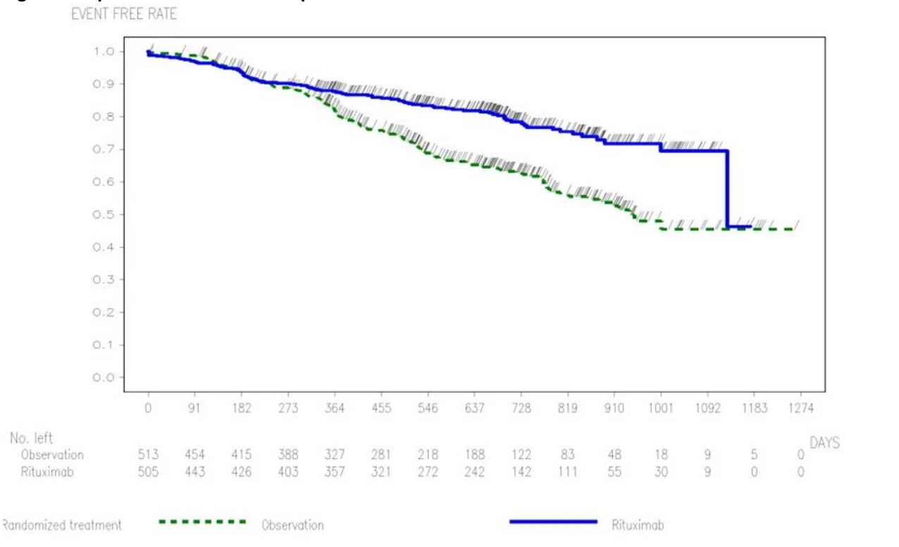
Rituximab maintenance treatment provided benefit in PFS in all subgroups tested: gender (male, female), age (<60 years, ≥ 60 years), FLIPI score (1, 2 or 3), induction therapy (R-CHOP, R-CVP) and regardless of the quality of response to induction treatment (CR or PR). The results of rituximab maintenance treatment in patients older than 75 years of age should be interpreted with caution due to the small number of patients in this subgroup.
The difference in overall survival between the two treatment arms was not conclusive. A longer follow-up is required to obtain mature overall survival results.
Relapsed/Refractory Follicular Non-Hodgkin's Lymphoma
Table 25 Relapsed/Refractory Follicular Non-Hodgkin's Lymphoma, Maintenance Therapy
| Induction Phase: Overview of Efficacy Results for CHOP vs R-CHOP | ||||
|---|---|---|---|---|
| Trial design | ||||
| Dosage | ||||
Number of study subjects | ||||
Mean age (Range) | ||||
| Gender | ||||
Results (50 months median observation time) | ||||
Prospective, open label, international, multicentre, Phase III trial | ||||
3)CHOP 4)R-CHOP | ||||
N= 231 N= 234 | ||||
54.1 (27-78) 54.1 (26-80) | ||||
Male: 118 (51%) Female: 113 (49%) Male: 107 (46%) Female: 127 (54%) | ||||
| CHOP | R-CHOP | RR1) | p-value (log-rank) | |
Primary Efficacy ORR2) CR2) PR2) | 74% 16% 58% | 87% 29% 58% | Na Na Na | 0.0003 0.0005 0.9449 |
Second. Efficacy OS (median) PFS (median) | NR 20.8 mo | NR 32.2 mo | 31% 36% | 0.0267 <0.0001 |
- 1. Estimates were calculated by hazard ratios
- 2. Last tumour response as assessed by the investigator. The “primary” statistical test for “response” was the trend test of CR versus PR versus non-response (p < 0.0001)
- 3. CHOP = cyclophosphamide (750 mg/m2 i.v., day 1), doxorubicin (50 mg/m2 i.v., day 1), vincristine (1.4 mg/m2 i.v., (max. 2 mg) day 1) and prednisone (100 mg orally, days 1-5, every 21 days for 6 cycles).
- 4. R-CHOP = Rituximab (375 mg/m2 i.v. infusion, on day 1 of each cycle for 6 cycles) plus CHOP chemotherapy.
Abbreviations: RR, risk reduction; NA, not available; NR, not reached; mo, months; ORR, overall response rate; CR, complete response; PR, partial response; OS, overall survival; PFS, progression free survival
Maintenance Phase: Overview of Efficacy Results Rituximab vs Observation (47.2 months median observation time) | |||||||||||||||||||||||||||||||||
|---|---|---|---|---|---|---|---|---|---|---|---|---|---|---|---|---|---|---|---|---|---|---|---|---|---|---|---|---|---|---|---|---|---|
| Demographics | Observation | Rituximab | |||||||||||||||||||||||||||||||
| Mean age (range) | 54.6 (27-80) | 53.3 (29-76) | |||||||||||||||||||||||||||||||
| Gender | Male: 83 (50%); Female: 84 (50%) | Male: 78 (47%); Female: 89 (53%) | |||||||||||||||||||||||||||||||
| Efficacy Analyses | Progression-Free Survival | Overall Survival | |||||||||||||||||||||||||||||||
Observation (N=167) | Rituximab (N=167) | Observation (N=167) | Rituximab (N=167) | ||||||||||||||||||||||||||||||
| Patients with event | 124 (74.3 %) | 95 (56.9 %) | 52 (31.1 %) | 37 (22.2 %) | |||||||||||||||||||||||||||||
| Patients without events1) | 43 (25.7 %) | 72 (43.1 %) | 115 (68.9 %) | 130 (77.8 %) | |||||||||||||||||||||||||||||
| Time to event (days) | |||||||||||||||||||||||||||||||||
| Median 2)* | 476.0 | 1304.0 | NR | NR | |||||||||||||||||||||||||||||
| 95% CI for Median 2) * | [375 ; 632] | [1072 ; 1605 -] | [- ; -] | [- ; -] | |||||||||||||||||||||||||||||
| 25% and 75%-ile | 203 ; 1623 | 432 ; - | 1287; - | 1885 - ; - | |||||||||||||||||||||||||||||
| Range3) | 20 to 2407 | 19 to 2429 | 127 to 2671 | 50 to 2688 | |||||||||||||||||||||||||||||
| p-value (Log-Rank Test) | <0.0001 | 0.0229 | |||||||||||||||||||||||||||||||
| Hazard Ratio | 0.49 | 0.61 | |||||||||||||||||||||||||||||||
| 95% CI | [0.37 ; 0.64] | [0.40 ; 0.94] | |||||||||||||||||||||||||||||||
| p-value (Wald Test) | < 0.0001 | 0.0243 | |||||||||||||||||||||||||||||||
| Month 12 | |||||||||||||||||||||||||||||||||
| Patients remaining at risk | 97 | 131 | 155 | 161 | |||||||||||||||||||||||||||||
| Event free rate | 0.59 | 0.78 | 0.93 | 0.96 | |||||||||||||||||||||||||||||
| 95% CI for rate | [0.51 ; 0.66] | [0.72 ; 0.85] | [0.90 ; 0.97] | [0.94 ; 0.99] | |||||||||||||||||||||||||||||
| Exploratory Analysis | Time to New Lymphoma Treatment or Death | Disease-Free Survival4) | |||||||||||||||||||||||||||||||
Observation (N=167) | Rituximab (N=167) | Observation (N=48) | Rituximab (N=49) | ||||||||||||||||||||||||||||||
| Patients with event | 112 (67.1 %) | 90 (53.9 %) | 36 (75.0 %) | 27 (55.1 %) | |||||||||||||||||||||||||||||
| Patients without events1) | 55 (32.9 %) | 77 (46.1 %) | 12 (25.0 %) | 22 (44.9 %) | |||||||||||||||||||||||||||||
| Time to event (days) | |||||||||||||||||||||||||||||||||
| Median 2) * | 659.0 | 1547.0 | 515.0 | 1591.0 | |||||||||||||||||||||||||||||
| 95% CI for Median 2) * | [568 ; 814] | [1143 ; 1750] | [450 ; 751] | [1120 ; -] | |||||||||||||||||||||||||||||
| 25% and 75%-ile | 326; 2062 - | 573 ; - | 331 ; 1408 | 564 ; - | |||||||||||||||||||||||||||||
| Range3) | 36 to 2407 | 27 to 2364 | 78 to 2144 | 76 to 2221 | |||||||||||||||||||||||||||||
| p-value (Log-Rank Test) | 0.0003 | 0.0014 | |||||||||||||||||||||||||||||||
| Hazard Ratio | 0.60 | 0.44 | |||||||||||||||||||||||||||||||
| 95% CI | [0.46; 0.80] | [0.26 ; 0.74] | |||||||||||||||||||||||||||||||
| p-value (Wald Test) | 0.0004 | 0.0018 | |||||||||||||||||||||||||||||||
| Month 12 | |||||||||||||||||||||||||||||||||
| Patients remaining at risk | 120 | 137 | 35 | 40 | |||||||||||||||||||||||||||||
| Event free rate | 0.72 | 0.82 | 0.75 | 0.82 | |||||||||||||||||||||||||||||
| 95% CI for rate | [0.66 ; 0.79] | [0.76 ; 0.88] | [0.62 ; 0.87] | [0.71 ; 0.92] | |||||||||||||||||||||||||||||
Abbreviations: NR, not reached | |||||||||||||||||||||||||||||||||
Follicular Non-Hodgkin's Lymphoma, Maintenance Therapy
In a prospective, open-label, international, multi-centre, Phase III trial, 465 patients with relapsed/refractory follicular NHL were randomized in a first step to induction therapy with either CHOP (cyclophosphamide, doxorubicin, vincristine, prednisone; n=231) or rituximab plus CHOP (R-CHOP, n=234). The two treatment groups were well-balanced with regard to baseline characteristics and disease status. The results are presented in Table 25. A total of 334 patients achieving a complete or partial remission following induction therapy were randomized in a second step to maintenance therapy with rituximab (n=167) or observation (n=167). Maintenance treatment with rituximab consisted of a single infusion of rituximab at 375 mg/m2 body surface area given every 3 months until disease progression or for a maximum period of two years.
The final efficacy analysis included all patients randomized to both parts of the study. After a median observation time of 50 months for patients randomized to the induction phase, R-CHOP significantly improved the outcome of patients with relapsed or refractory follicular NHL when compared to CHOP.
For patients randomized to the maintenance phase of the trial, the median observation time was 47.2 months from maintenance randomization. Maintenance treatment with rituximab led to a clinically relevant and statistically significant improvement in the primary endpoint, PFS, (time from maintenance randomization to relapse, disease progression or death) when compared to observation alone (p<0.0001 log-rank test). The median PFS was 42.9 months (range: 0.6 to 80.1 months) in the rituximab maintenance arm compared to 15.7 months (range: 0.6 to 79.4 months) in the observation arm. Using a cox regression analysis, the risk of experiencing progressive disease or death was reduced by 51% with maintenance treatment with rituximab when compared to observation (95% CI; 36 %-63 %). Kaplan-Meier estimated progression-free rates at 12 months were 78% in the rituximab maintenance group vs 59% in the observation group. An analysis of overall survival suggested a benefit of maintenance treatment with rituximab over observation (p=0.0229 log-rank test). The significance level for this analysis was set at 0.001.
The median time to new anti-lymphoma treatment was significantly longer with rituximab maintenance treatment than with observation (50.9 months (range 0.9 to 77.9 months) vs. 21.7 months (range 1.2 to 79.4 months), p=0.0003 log-rank test). The risk of starting a new treatment was reduced by 40% (95% CI; 20 %-54 %).
Table 26 Patients Starting New Lymphoma Treatment (NLT)/Reporting Disease Progression (PD)
| Observation (n=167) | Rituximab (n=167) | |
|---|---|---|
| Total Patients reporting NLT (n) | 85 (100%) | 56 (100%) |
| No PD reported before initiation of NLT | - | 2 (3.6%) |
| PD reported before initiation of NLT | 85 (100%) | 54 (96.4%) |
| PD reported during maintenance/observation phase | ||
| PD > 3 months before NLT | 27 (31.8%) | 12 (21.4%) |
| PD ≤ 3 months before NLT | 54 (63.5%) | 30 (53.6%) |
| PD reported after maintenance/observation phase (follow-up) | ||
| PD > 3 months before NLT | 1 (1.2%) | 4 (7.2%) |
| PD ≤ 3 months before NLT | 3 (3.5%) | 8 (14.3%) |
In patients achieving a CR/CRu (complete response unconfirmed) as best response during induction treatment, maintenance treatment with rituximab significantly prolonged the median disease free survival (DFS) compared to the observation group (52.3 (range 2.5 to 73.2 months) vs 16.9 months (range 2.6 to 70.7 months), p=0.0014) log-rank test. The risk of relapse in complete responders was reduced by 56 % (95% CI; 26 %-74 %).
The benefit of maintenance treatment with rituximab was confirmed in all subgroups analysed, regardless of induction regimen (CHOP or R-CHOP) or quality of response to induction treatment (CR or PR) (refer to Overview of Clinical Trials). Maintenance treatment with rituximab significantly prolonged median PFS in patients responding to CHOP induction therapy (median PFS 36.9 months (range 0.7 to 80.1 months) vs 11.6 months (range 0.7 to 67.5 months), p<0.0001). The risk of experiencing progressive disease or death was reduced by 64% with maintenance treatment with rituximab when compared to observation (95% CI; 46%-75%). Maintenance treatment with rituximab also prolonged median PFS in patients responding to R-CHOP induction (median PFS 51.6 months (range 0.6 to 77.9 months) vs 23.1 months (range 1.4 to 79.4 months), p=0.0273). The risk of experiencing progressive disease or death was reduced by 35% with maintenance treatment with rituximab when compared to observation (95% CI; 4 %-55%). Since subgroup analysis based on induction therapy was not prespecified in the protocol, the results should be interpreted with caution.
Maintenance treatment with rituximab provided consistent benefit in all subgroups tested [gender (male, female), age (≤ 60 years, > 60 years), stage (III, IV), WHO performance status (0 versus 1 or 2), B symptoms (absent, present), bone marrow involvement (no versus yes), IPI (0-2 versus 3-5), FLIPI score (0-1, versus 2 versus 3-5), number of extra-nodal sites (0-1 versus > 1), number of nodal sites (< 5 versus ≥ 5), number of previous regimens (1 versus 2), best response to prior therapy (CR/PR versus NC/PD), hemoglobin (< 12 g/dL versus ≥ 12 g/dL), ≤β2- microglobulin (< 3mg/L versus ≥ 3 mg/L), LDH (elevated, not elevated) except for the small subgroup of patients with bulky disease.
Table 27 Diffuse Large B-cell Non-Hodgkin’s Lymphoma
| Trial design | Dosage | Number of study subjects | Mean age (Range) | Gender | Results (24 months median follow-up) | ||||
|---|---|---|---|---|---|---|---|---|---|
| Randomized open-label, phase III trial | 1)CHOP | N = 197 | 68.9 (60- 80) | Male: 107 (54%) Female: 90 (46%) | 24 month survival rate | CHOP | R-CHOP | Risk Ratio | p-value (log-rank) |
| Event-free survival3) * | 37.3% | 57% | 0.58 | 0.0001 | |||||
| 2)R-CHOP | N=202 | 69.5 (59- 80) | Male: 92 (46%) Female: 110 (54%) | Overall survival3) * | 57.3% | 70.2% | 0.63 | 0.0072 | |
1CHOP = cyclophosphamide (750 mg/m2 i.v.), doxorubicin (50 mg/m2 i.v.), vincristine (1.4 mg/m2 up to a maximum of 2 mg on day 1), prednisone (40 mg/m2/day on days 1-5, every 3 weeks for 8 cycles). 2R-CHOP = Rituximab (375 mg/m2 i.v., every 3 weeks, on day 1 of the treatment cycle for 8 cycles) plus CHOP chemotherapy. 3Kaplan-Meier estimate. | |||||||||
Diffuse Large B-cell Non-Hodgkin’s Lymphoma
In a randomized, open-label trial, a total of 399 previously untreated elderly patients (age 60 to 80 years) with diffuse large B-cell lymphoma received standard CHOP chemotherapy (cyclophosphamide 750 mg/m2, doxorubicin 50 mg/m2, vincristine 1.4 mg/m2 up to a maximum of 2 mg on day 1, and prednisone 40 mg/m2/day on days 1-5) every 3 weeks for eight cycles, or rituximab 375 mg/m2 plus CHOP (R-CHOP). Rituximab was administered on the first day of the treatment cycle. In a planned interim analysis, a total of 328 patients (159 CHOP, 169 R-CHOP) were analyzed for efficacy. After a median follow up of approximately 12 months, R-CHOP led to a highly statistically significant increase in event free survival compared to CHOP (p = 0.0002), where events were death, relapse or progression of lymphoma, or institution of a new anti-lymphoma treatment; R-CHOP treatment reduced the risk of an event by 48%. Lower rates of disease progression during treatment and of relapse after complete response accounted for this difference. Overall survival was statistically significantly prolonged in the R-CHOP group compared to CHOP (p = 0.0055), with a 49% reduction in the risk of death. R-CHOP treatment was also associated with a statistically significant benefit, compared to CHOP, for complete response rate at the end of treatment (71% vs 59%; p = 0.0176), progression-free survival (p = 0.0001), and disease-free survival (p = 0.0048). The risk of disease progression was reduced by 54% and the risk of relapse after complete response by 51%. R-CHOP treatment benefited both low-risk and high-risk patients (age-adjusted International Prognostic Index score 0-1 and 2-3, respectively): the risk of an event was reduced by 69% in the low-risk group and 36% in the high-risk group.
An updated efficacy analysis including the total study population of 399 patients (197 CHOP, 202 RCHOP), with a median follow-up of 24 months, presented in Table 27, confirmed that R-CHOP significantly prolongs both event-free survival (p=0.0001) and overall survival (p=0.0072). R-CHOP treatment reduced the risk of an event by 42% and the risk of death by 37%. Kaplan Meier estimates of event-free survival at 24 months were 57.0% in the R-CHOP arm compared to 37.3% in the CHOP arm and of overall survival were 70.2% in the R-CHOP arm compared to 57.3% in the CHOP arm.
Chronic Lymphocytic Leukemia (previously untreated and previously treated patients)
In two open-label randomized Phase 3 trials, a total of 817 previously untreated patients and 552 previously treated patients with CLL were randomized to receive either FC chemotherapy (fludarabine 25mg/m2, cyclophosphamide 250 mg/m2, days 1-3) every 4 weeks for 6 cycles or rituximab in combination with FC (R-FC). Rituximab was administered at a dosage of 375 mg/m2 during the first cycle one day prior to chemotherapy and at a dosage of 500 mg/m2 on day 1 of each subsequent treatment cycle.
A total of 810 previously untreated patients (primary analysis: 403 R-FC, 407 FC; updated OS (final) analysis: 408 R-FC, 409 FC) and 552 previously treated patients (276 R-FC, 276 FC) were analyzed for efficacy.
Previously Untreated CLL
Table 28 Study ML17102 Treatment of Previously Untreated Chronic Lymphocytic Leukemia (CLL) Overview of Efficacy Results for Rituximab Plus FC vs. FC alone
| Efficacy Parameter | Primary Analysisa | Final Analysisb | ||
|---|---|---|---|---|
| Efficacy Parameter | Primary Analysisa | Final Analysisb | ||
| Efficacy Parameter | Primary Analysisa | Final Analysisb | ||
| Analyses at the time of primary PFS analysis (20.7 months median observation time) | Analyses at the time of final OS analysis (66.4 months median observation time) | |||
| Analyses at the time of primary PFS analysis (20.7 months median observation time) | Analyses at the time of final OS analysis (66.4 months median observation time) | |||
| Analyses at the time of primary PFS analysis (20.7 months median observation time) | Analyses at the time of final OS analysis (66.4 months median observation time) | |||
| FC N = 407 | R-FC N = 403 | FC R-FC N = 409 N = 408 | ||
| FC N = 407 | R-FC N = 403 | FC R-FC N = 409 N = 408 | ||
| FC N = 407 | R-FC N = 403 | FC R-FC N = 409 N = 408 | ||
| Progression-free Survival | ||||
| Median time to event (months) | 32.2 | 39.8 | 32.8 56.0 | |
| p value (log-rank test) | p < 0.0001 | p < 0.0001 | ||
| adjusted HR [95% CI], p value (Wald test) | 0.56 [0.43;0.72], p < 0.0001 | 0.57 [0.48;0.67], p < 0.0001 | ||
| Overall Survival | ||||
| Median time to event (months) | NR NR | 85.8 NR | ||
| p value (log-rank test) | p = 0.0427 | p = 0.0010 | ||
| adjusted HR [95% CI], p value (Wald test) | 0.64 [0.41;1.00], p = 0.0487 | 0.68 [0.54;0.86], p = 0.0015 | ||
| Event-free Survival | ||||
| Median time to event (months) | 31.1 39.8 | 31.2 54.7 | ||
| p value (log-rank test) | p < 0.0001 | p < 0.0001 | ||
| adjusted HR [95% CI], p value (Wald test) | 0.55 [0.43;0.70], p < 0.0001 | 0.57 [0.48;0.67], p < 0.0001 | ||
| End of Treatment Response Ratec | ||||
| Responders (CR + PR/nPR) | 72.7% | 86.1% | 72.4% | 85.8% |
| Patients with | ||||
| complete response (CR) | 17.2% | 36.0% | 16.9% | 36.0% |
| partial response (PR/nPR) | 55.5% | 50.1% | 55.5% | 49.8% |
| stable disease (SD) | 7.6% | 4.7% | 7.6% | 4.7% |
| progressive disease (PD) | 7.6% | 3.5% | 7.8% | 3.7% |
| missing | 12.0% 5.7% | 12.2% | 5.9% | |
| Disease-free Survivald | ||||
| Median time to event (months) | NR NR | 48.9 | 60.9 | |
| p value (log-rank test) | p = 0.7882 | p = 0.0523 | ||
| adjusted HR [95% CI], p value (Wald test) | 0.93 [0.44;1.96], p = 0.8566 | 0.73 [0.52;1.02], p = 0.0689 | ||
| Duration of Responsee | ||||
| Median time to event (months) | 34.7 40.2 | 36.2 56.4 | ||
| p value (log-rank test) | p = 0.0040 | p < 0.0001 | ||
| adjusted HR [95% CI], p value (Wald test) | 0.61 [0.43;0.85], p = 0.0036 | 0.58 [0.48;0.71], p < 0.0001 | ||
| Time to New Treatment | ||||
| Median time to event (months) | NR NR | 47.8 68.4 | ||
| p value (log-rank test) | p = 0.0052 | p < 0.0001 | ||
| adjusted HR [95% CI], p value (Wald test) | 0.65 [0.47;0.90], p = 0.0082 | 0.59 [0.49;0.72], p < 0.0001 | ||
NR: not reached; nPR: nodular partial response. Hazard ratios are from non-stratified (adjusted) analyses. 1 month = 30.4375 days.
- a Clinical cut-off July 04, 2007. Informed consent forms for seven patients (2 FC, 5 R-FC) were missing at the time of the primary analysis; hence, these patients were excluded from the analysis. Informed consent forms were later collected from those seven patients, and their data were added to the database ahead of the first updated analysis of efficacy.
- b Last patient visit October 31, 2011.
- c The response for one patient with PR at the time of the primary and updated analyses has changed to missing (and hence non-responder) at the time of this final analysis.
d Based on patients with confirmed CR (including late responders).
e Based on patients with confirmed response (CR, PR,nPR).
Table 29 Summary of Progression-Free Survival According to Binet Stage (ITT) Primary Analysis (20.7 Months Median Observation Time)
FC N = 407 | R-FC N = 403 | ||||||||||||
|---|---|---|---|---|---|---|---|---|---|---|---|---|---|
FC N = 407 | R-FC N = 403 | ||||||||||||
| Binet Stage A | |||||||||||||
| N | 22 | 18 | |||||||||||
| Progression Free Survival –Median (months) | 31.6 | Not Reached | |||||||||||
| Log Rank p-value | 0.0099 | ||||||||||||
| Hazard Ratio (95% CI) | 0.13 (0.03; 0.61) | ||||||||||||
| p-value (Wald test, not adjusted) | 0.0093 | ||||||||||||
| Binet Stage B | |||||||||||||
| N | 257 | 259 | |||||||||||
| Progression Free Survival –Median (months) | 32.3 | 43.3 | |||||||||||
| Log Rank p-value | < .0001 | ||||||||||||
| Hazard Ratio (95% CI) | 0.45 (0.32; 0.63) | ||||||||||||
| p-value (Wald test, not adjusted) | < 0.0001 | ||||||||||||
| Binet Stage C | |||||||||||||
| N | 126 | 125 | |||||||||||
| Progression Free Survival –Median (months) | 33.4 | 38.0 | |||||||||||
| Log Rank p-value | 0.4671 | ||||||||||||
| Hazard Ratio (95% CI) | 0.88 (0.58; 1.33) | ||||||||||||
| p-value (Wald test, not adjusted) | 0.5406 | ||||||||||||
Table 30 Summary of Progression-Free Survival According to Age (ITT) Primary Analysis (20.7 Months Median Observation Time)
FC N = 407 | R-FC N = 403 | ||||||||||||
|---|---|---|---|---|---|---|---|---|---|---|---|---|---|
| Age < 65 | |||||||||||||
| N | 288 | 279 | |||||||||||
| Progression Free Survival –Median (months) | 31.7 | 43.3 | |||||||||||
| Log Rank p-value | < .0001 | ||||||||||||
| Hazard Ratio (95% CI) | 0.54 (0.40;0.72) | ||||||||||||
| p-value (Wald test, not adjusted) | <.0001 | ||||||||||||
| Age ≥ 65 - ≤ 70 | |||||||||||||
| N | 94 | 91 | |||||||||||
| Progression Free Survival –Median (months) | 27.4 | 39.9 | |||||||||||
| Log Rank p-value | 0.0037 | ||||||||||||
| Hazard Ratio (95% CI) | 0.45 (0.26;0.78) | ||||||||||||
| p-value (Wald test, not adjusted) | 0.0046 | ||||||||||||
| Age > 70 | |||||||||||||
| N | 25 | 33 | |||||||||||
| Progression Free Survival –Median (months) | Not Reached | 38.0 | |||||||||||
| Log Rank p-value | 0.3787 | ||||||||||||
| Hazard Ratio (95% CI) | 1.61 (0.55; 4.74) | ||||||||||||
| p-value (Wald test, not adjusted) | 0.3832 | ||||||||||||
In the primary analysis of the study in previously untreated patients (see Table 28) the median PFS, calculated by applying the Kaplan-Meier method, was 39.8 months in the R-FC group and 32.2 months in the FC group (p < 0.0001, log-rank test). The primary analysis that led to the stopping of the study based on crossing the statistical boundary for PFS, showed an improvement of R-FC over FC for the secondary endpoint overall survival (p=0.0427). In updated overall survival results (final analysis) after a median of 64.4 months of observation, overall survival was significantly prolonged in the R-FC group compared with the FC group (p = 0.0010, log-rank test; adjusted HR 0.68 (95% CI [0.54, 0.86], p = 0.0015, Wald test). Although based on small numbers of patients, hazard ratios were greater than 1 (with wide confidence intervals) for the > 70 and ≥ 75 year age subgroups, and in the subgroup of patients who were diagnosed 6 to <12 months before entering the study. Due to the exploratory nature of subgroup analyses, these results need to be interpreted with caution. The benefit in terms of PFS was consistently observed in most patient subgroups analyzed according to disease risk at baseline, although it was not statistically significant in patients with Stage C disease or for patients > 70 years (see Tables 34 and 35).
Study ML17102 was initially open to all symptomatic patients in need of treatment, regardless of stage. From amendment #1 onwards, however, new patients in the lowest risk group (Binet A) were excluded from the study. A total of 40 patients (22 FC arm, 18 R-FC arm) had been enrolled at that time, which represents 5% of the overall intent-to-treat (ITT) population. Within the Binet A patients, patients who received R-FC had a better outcome compared to those who received FC. If Binet A patients were to be excluded from the ITT analysis of ML17102, the overall results of the remaining Binet B and C patients would be slightly lower to the current overall results, but, due to the small numbers, would not change any of the overall results and conclusions of the study.
In all subgroups analyzed according to Binet stage, the median PFS in the primary analysis was increased or not yet reached in Binet A for R-FC and the risk of disease progression or death [(Hazard Ratio (HR)] was decreased by the addition of rituximab to FC when compared to FC alone, although not statistically significantly decreased in patients with stage C disease. The effect was most pronounced in the group of patients with stage A disease, and least in patients in stage C disease.
The effect of rituximab when added to FC seems to be most pronounced with younger age. Due to the small size of the subgroup of patients over the age of 70 (FC n=25, R-FC n=33), no meaningful conclusion can be drawn for the effect rituximab might have in this age category.
180/403 (45%) of patients in the R-FC arm received Colony Stimulating Factors vs. 95/407 (23%) in the FC arm. A comparison with regards to the primary endpoint, PFS, yields a result favoring the R-FC arm: HR=0.59, 95% CI [0.43;0,81]. This outcome is similar to the overall study results. As is also true for the overall population, and as expected, in the subgroups more AEs were found in the R-FC arm compared to FC regardless if G-CSF was given or not.
Previously Treated CLL
Table 31 Treatment of Previously Treated6 Chronic Lymphocytic Leukemia (CLL)
Overview of Efficacy Results for Rituximab plus FC vs. FC Alone
| Trial design | Dosage | Number of study subjects | Mean age (Range) | Gender | Efficacy Results (25.3 months mean observation time) | ||||||||||||||||
|---|---|---|---|---|---|---|---|---|---|---|---|---|---|---|---|---|---|---|---|---|---|
| Analysis | Investigator-Assessed Results3) * | IRC Results3) * | |||||||||||||||||||
Random ized open label, Phase III trial | FC1) R-FC 2) | N = 276 N = 276 | 61.3 (35-81) 62.1 (35-83) | Male: 181 (66%) Female: 95 (34%) Male: 187 (68%) Female: 89 (32%) | FC | R-FC | Log rank pvalue | Hazard Ratio | FC | R-FC | Log rank pvalue | Hazard Ratio | |||||||||
Progressi on-free Survival (PFS) (months) | 20.6 (18.1; 24.0) 5) | 30.6 (26.0; 38.1) 5) | 0.0002 | 0.65 (0.51; 0.82) 5) | 21.7 (18.3; 24.1) 5) | 26.7 (22.0; 31.1) 5) | 0.0218 | 0.76 (0.60; 0.96) 5) | |||||||||||||
PFS with censorin g of new CLL treatme nt7) (months) | 22.5 (18.3; 29.0) 5) | 31.5 (26.2; 42.2) 5) | 0.0012 | 0.69 (0.53; 0.86) 5) | 22.6 (18.8; 25.2) 5) | 28.0 (22.9; 32.3) 5) | 0.0439 | 0.78 (0.61; 0.99) 5) | |||||||||||||
Overall Survival (months) | 51.9 (46.3; ...) 5) | NR (51.0; ...) 5) | 0.83 (0.59; 1.17) 5) | ||||||||||||||||||
Respons e rate4) (CR, nPR, PR) | 58.0% (51.9; 63.9% )5) | 69.9% (64.1; 75.3%) 5) | NA | 48.6 % (42.5; 54.6 %) 5) | 60.5% (54.5; 66.3%) 5) | NA | |||||||||||||||
- 1) FC = (fludarabine 25 mg/m2, cyclophosphamide 250 mg/m2, days 1-3) every 28 days for 6 cycles
- 2) R-FC = Rituximab (375 mg/m2 during the first cycle one day prior to chemotherapy and at a dosage of 500 mg/m2 on day 1 of each subsequent treatment cycle with FC chemotherapy.
- 3) Kaplan-Meier estimate.
- 4) Response rate is based on the Best Overall Response
- 5) 95% CI
- 6) Previous treatment included one of the following chemotherapy regimens: single agent chlorambucil +/- prednisone/ prednisolone, single agent fludarabine (or other nucleoside analogue), or alkylator containing combination therapy (e.g. CHOP/CVP)
- 7) These results are based on a sensitivity analysis with censoring of new CLL treatment before documented disease progression
NR: not reached. NA: not applicable.
Table 32 Summary of Progression Free Survival According to Age (ITT) as Assessed by IRC*
| Age Subgroup | N | HR (95% CI) | FC | R-FC | ||||||||||
|---|---|---|---|---|---|---|---|---|---|---|---|---|---|---|
| Patients (N) | Median PFS (months) | Patients (N) | Median PFS (months) | |||||||||||
| <65 | 317 | 0.61 [0.44;0.84] | 162 | 22.5 | 155 | 30.2 | ||||||||
| Age Subgroup | N | HR (95% CI) | FC | R-FC | ||||||||||
| Patients (N) | Median PFS (months) | Patients (N) | Median PFS (months) | |||||||||||
| ≥ 65 to ≤ 70 | 142 | 0.94 [0.60;1.47] | 68 | 23.3 | 74 | 26.1 | ||||||||
| > 70 | 93 | 1.10 [0.63;1.91] | 46 | 18.8 | 47 | 15.5 | ||||||||
- These results are based on exploratory analyses
Table 33 Summary of Progression-Free Survival According to Binet Stage (ITT) as Assessed by IRC*
| Binet Stage | N | HR (95% CI) | FC | R-FC | ||||||||||
|---|---|---|---|---|---|---|---|---|---|---|---|---|---|---|
| Patients (N) | Median PFS (months) | Patients (N) | Median PFS (months) | |||||||||||
| Binet A | 55 | 0.68 [0.29;1.57] | 31 | 22.8 | 24 | 51.0 | ||||||||
| Binet B | 326 | 0.79 [0.58;1.09] | 160 | 24.6 | 166 | 30.2 | ||||||||
| Binet C | 171 | 0.70 [0.47;1.03] | 85 | 15.8 | 86 | 21.3 | ||||||||
- These results are based on exploratory analyses
In the previously treated CLL study (see Table 31), the investigator-assessed median progression-free survival (primary endpoint) was 30.6 months in the R-FC group and 20.6 months in the FC group (p=0.0002, log-rank test). The risk of having a PFS event (progression or death, whichever occurred first) was statistically significantly decreased by 35% (HR = 0.65; 95% CI: [0.51, 0.82]; p=0.0002, Wald test) for patients in the R-FC arm compared to the FC arm (see Table 31). Forty-four percent of the patients in the FC arm, and 60% of those in the R-FC arm, were progression-free at two years using Kaplan-Meier estimates.
Based on Independent Review Committee (IRC) assessments, the median PFS was 21.7 months in the FC arm and 26.7 months in the R-FC arm (p = 0.0218, non-stratified Log-Rank test). The addition of rituximab to FC reduced the risk of disease progression or death by 24% (HR = 0.76; 95% CI [0.60, 0.96]; p = 0.0222, Wald test) compared to FC alone. Forty-three percent of patients in the FC arm and 54% of patients in the R-FC arm were progression-free at 2 years using Kaplan-Meier estimates. Please see Tables 37 and 38 for a summary of progression-free survival according to Age and Binet stage respectively, as assessed by IRC. These results are based on exploratory analyses.
In this open-label randomized trial, the discordance between investigators’ efficacy results and IRC’s assessments were due to differences in assessing disease status (progression or not) and in determining the time of progression. The discordance observed reflects the subjectivity of PFS assessment in open-labeled trials. The results should be interpreted cautiously.
OS benefit has not been demonstrated and follow-up is needed to draw meaningful conclusions about the treatment effect of R-FC compared to FC in terms of OS.
Rheumatoid Arthritis
The efficacy and safety of rituximab in alleviating the symptoms and signs of rheumatoid arthritis was demonstrated in three randomized, controlled, double-blind, multicenter studies.
Study 1 was a double blind comparative study which included 517 patients that had experienced an inadequate response or intolerance to one or more TNF inhibitor therapies. Eligible patients had severe active rheumatoid arthritis, diagnosed according to the criteria of the American College of Rheumatology (ACR). The primary endpoint was the percent of patients who achieved an ACR20 response at week 24. Patients received two 1000 mg IV infusions of rituximab, each following an IV infusion of 100 mg methylprednisone and separated by an interval of 15 days. Patients were also pre-medicated with acetaminophen and diphenhydramine before each infusion of rituximab. All patients received concomitant oral methotrexate (10 – 25 mg/week) and 60 mg oral prednisone on days 2-7 and 30 mg on days 8- 14 following the first infusion. Patients were followed beyond week 24 for long term endpoints, including radiographic assessment. During this time patient could receive further courses of rituximab (2 x 1000 mg + MTX) under an open label extension study protocol (see Radiographic Response). Retreatment frequency was determined by clinical evaluation, but no sooner than 16 weeks after the preceding course of rituximab.
Study 2 was a randomized, double-blind, double-dummy, controlled, 3 x 3 multifactorial study which compared two different dose levels of rituximab given with or without one of two per infusional corticosteroid regimens in combination with weekly methotrexate in patients with active rheumatoid arthritis which had not responded to treatment with 1 but no more than 5 other Disease-Modifying Anti-Rheumatic Drugs (DMARDs).
The comparator group in all three studies was weekly methotrexate (10-25 mg weekly).
Disease Activity Outcomes
In all three studies, rituximab 2 x 1000 mg significantly increased the proportion of patients achieving at least a 20% improvement in ACR score compared with patients treated with methotrexate alone (Table 34). The treatment effect was similar in patients independent of rheumatoid factor status, age, gender, body surface area, race, number of prior treatments or disease status.
ACR20 response rates at week 24 in RF negative patients were significantly higher in patients receiving rituximab + MTX (40%) compared to those receiving placebo + MTX (12%, p=0.0009), although lower than among rheumatoid factor positive patients (54%). In HACA positive patients, a total of 61/96 patients (63.4%) achieved at least an ACR20 response following their first treatment course. Mean change from original baseline DAS in HACA positive patients and HACA negative patients are -2.36 and -2.23 respectively.
The proportion of rituximab patients achieving an ACR20 response at week 24 in the US and non-US (including Canada) were 44% vs 61% respectively. ACR20 response in placebo patients was 18% in both regions. Treatment effect in favor of rituximab was statistically significant for both regions (p < 0.001).
Table 34 Cross-Study Comparison of ACR Responses at Week 24 (ITT Population)
| ACR Response | Placebo + MTX | Rituximab + MTX | |||||||
|---|---|---|---|---|---|---|---|---|---|
| Study 1 | N= 201 | N= 298 | |||||||
| ACR20 | 36 (18%) | 153 (51%)1 | |||||||
| ACR50 | 11 (5%) | 80 (27%)1 | |||||||
| ACR70 | 3 (1%) | 37 (12%)1 | |||||||
| Study 2 | N= 143 | N= 185 | |||||||
| ACR20 | 45 (31%) | 96 (52%)2 | |||||||
| ACR50 | 19 (13%) | 61 (33%)2 | |||||||
| ACR70 | 6 (4%) | 28 (15%)2 | |||||||
| Study 3 | N= 40 | N= 40 | |||||||
| ACR20 | 15 (38%) | 28 (70%)3 | |||||||
| ACR50 | 5 (13%) | 17 (43%)3 | |||||||
| ACR70 | 2 (5%) | 9 (23%)3 | |||||||
1p ≤ 0.0001; 2p ≤ 0.001; 3p <0.05
In study 3, the ACR20 response in patients treated with rituximab alone was 65% compared with 38% on methotrexate alone (p=0.025).
Clinically and statistically significant improvement was also noted on all individual components of the ACR response (tender and swollen joint counts, patient and physician global assessment, disability index scores (Health Assessment Questionnaire - HAQ), pain assessment and C- reactive protein (CRP - mg/dL).
Table 35 Components of ACR Response in Study 1
Rituximab + MTX (N=122) | Study 1 [RF(+) and RF (-) Patients] | |||||||||||||||||||||||||||||
|---|---|---|---|---|---|---|---|---|---|---|---|---|---|---|---|---|---|---|---|---|---|---|---|---|---|---|---|---|---|---|
| Placebo + MTX (N=201) | Rituximab + MTX (N=298) | |||||||||||||||||||||||||||||
Wk 0(SD) range | Wk 24 (SD) range | % mean Change (SD) range | Wk 0(SD) range | Wk 24 (SD) range | % mean change (SD) range | |||||||||||||||||||||||||
| Tender Joint | 32.9 (15.61) | 30.2 (18.99) | 7.2 (144.58) | 33.9 (15.23) | 19.5* | -41.8 (52.39) | ||||||||||||||||||||||||
| Count (68) | 1:68 | 0:68 | -100:1733.3 | 3:68 | (18.53) 0:68 | -100:264.7 | ||||||||||||||||||||||||
| Swollen | 22.9 (12.71) | 20.3 (13.44) | -5.6 (59.19) | 23.4 (11.87) | 13.0* | -43.0 (52.65) | ||||||||||||||||||||||||
| Joint Count (66) | 8:64 | 0:63 | -100:387.5 | 4:66 | (12.70) 0:64 | -100:366.7 | ||||||||||||||||||||||||
| Physician | 6.7 (1.629) | 6.1 (2.573) | -4.2 (47.23) | 6.9 (1.597) | 4.0* (2.573) | -40.8 (39.31) | ||||||||||||||||||||||||
| Global | 1.8:10 | 0.2:10 | -97.1:183.3 | 1.2:9.8 | 0:10 | -100:100 | ||||||||||||||||||||||||
| Assessmenta | ||||||||||||||||||||||||||||||
| Patient | 7.0 (2.006) | 6.4 (2.521) | -3.1 (44.01) | 6.9 (2.106) | 4.3** (2.752) | -25.4 | ||||||||||||||||||||||||
| Global Assessmenta | 0.9:10 | 0.3:10 | -95.9:240 | 0.1:10 | 0.0:10 | (117.90) -100:1300 | ||||||||||||||||||||||||
| Paina | 6.5 (2.132) | 6.2 (2.561) | 2.8 (55.61) | 6.4 (2.228) | 4.1**(2.711) | -23.8 | ||||||||||||||||||||||||
| 0.6:10 | 0.1:10 | -98.4:347.4 | 0.2:10 | 0.0:10 | (131.59) -100:2050 | |||||||||||||||||||||||||
| Disability | 1.9 (0.54) | 1.8 (0.64) | -2.0 (30.46) | 1.9 (0.58) | 1.4* (0.74) | -24.3 (34.92) | ||||||||||||||||||||||||
| Index (HAQ)b | 0.5:3.0 | 0.0:3.0 | -100:183.3 | 0.1:3.0 | 0.0:3.0 | -100:100 | ||||||||||||||||||||||||
| CRP | 3.8 (4.07) | 3.7 (4.12) | 80.0 | 3.7 (3.83) | 1.7* (2.45) | -36.3 (80.3) | ||||||||||||||||||||||||
| (mg/dL) | 0.2:22.7 | 0.2:23.9 | (452.94) | 0.2:23.7 | 0.2:22.2 | -99.1:550 | ||||||||||||||||||||||||
| -98.2:4800 | ||||||||||||||||||||||||||||||
a Visual Analogue Scale: 0=best, 10=worst
b Disability Index of the Health Assessment Questionnaire: 0=best, 3=worst
*p<0.0001, **p<0.005 rituximab + MTX minus Placebo + MTX stratified for rheumatoid factor, region and baseline ACR
Negative % change from baseline value indicates an improvement.
Patients treated with rituximab had a significantly greater reduction in disease activity score (DAS28) than patients treated with methotrexate alone. A good to moderate European League against Rheumatism (EULAR) response was achieved by significantly more patients treated with rituximab compared to patients treated with methotrexate alone (Table 36).
Treatment with rituximab + MTX (2 x 1 g) over multiple courses resulted in sustained improvements in the clinical signs and symptoms of RA and physical function for patients who remained on treatment.
In a study, using treatment to DAS28-ESR remission, where all patients initially received rituximab followed by retreatment with either rituximab or placebo, patients who received rituximab retreatment had improved efficacy compared with placebo -retreated subjects at Week 48 relative to baseline, as measured by ACR20 response (53.5% vs. 44.6%; p = 0.0195).
Table 36 Cross-Study Comparison of DAS and EULAR Responses at Week 24 (ITT Population)
| Placebo+MTX | Rituximab + MTX 2 × 1g | |
|---|---|---|
| Study 1 | (n = 201) | (n = 298) |
| Change in DAS28 [Mean (SD)] | -0.4 (1.2) | -1.9 (1.6)* |
| EULAR Response | ||
| None | 78% | 35% |
| Moderate | 20% | 50%* |
| Good | 2% | 15% |
| Study 2 | (n = 143) | (n = 185) |
| Mean change in DAS28 (SD) | –0.8 (1.4) | –2.0 (1.6) |
| EULAR response | ||
| None | 61% | 37% |
| Moderate | 35% | 40% |
| Good | 4% | 23% |
| Study 3 | ||
| Change in DAS [Mean (SD)] | -1.3 (1.2) | -2.6 (1.3) |
| EULAR response | ||
| None | 50% | 18% |
| Moderate | 45% | 63% |
| Good | 5% | 20% |
*p value < 0.0001. p values not calculated for studies 2 and 3.
Radiographic Response
In study 1 (WA17042) joint damage was assessed radiographically and expressed as changes in Genant-modified Total Sharp Score (TSS) and its components, the erosion score (ES) and the joint space narrowing (JSN) score. Rituximab + MTX slowed the progression of joint damage compared to placebo + MTX after 1 year as shown in Table 37.
Table 37 Mean Radiographic Change from Baseline to 104 Weeks
| Inadequate Response to TNF Antagonists | ||||
|---|---|---|---|---|
| Parameter | Rituximab 2 x 1000 mg + MTXb | Placebo + MTXc | Treatment Difference (Placebo – Rituximab) | 95% CI |
| Change during First Year | ||||
| TSS | 0.66 | 1.77 | 1.11 | (0.48, 1.76) |
| ES | 0.44 | 1.19 | 0.75 | (0.31, 1.19) |
| JSN Score | 0.22 | 0.58 | 0.36 | (0.10, 0.62) |
| Change during Second Year | ||||
| TSS | 0.48 | 1.04 | --- | --- |
| ES | 0.28 | 0.62 | --- | --- |
| JSN Score | 0.20 | 0.42 | --- | --- |
- a) Based on radiographic scoring following 104 weeks of observation.
- b) Patients received up to 2 years of treatment with Rituximab + MTX.
- c) Patients receiving Placebo + MTX. Patients receiving Placebo + MTX could have received retreatment with rituximab + MTX from Week 16 onward.
In RA Study 1 and its open-label extension, 70% of patients initially randomized to rituximab + MTX and 72% of patients initially randomized to placebo + MTX were evaluated radiographically at Year 2. As shown in Table 37, progression of joint damage in rituximab + MTX patients was further reduced in the second year of treatment.
Following 2 years of treatment with rituximab + MTX, 57% of patients had no progression of joint damage, defined as a change in TSS of zero or less compared to baseline. During the first year, 60% of rituximab + MTX treated patients had no progression from baseline to Week 56 compared to 46% of placebo + MTX treated patients. In their second year of treatment with rituximab + MTX, more patients had no progression from Week 56 to Week 104 than in the first year (68% vs. 60%). Additionally, 87% of the rituximab + MTX treated patients who had no progression in the first year also had no progression in the second year.
Physical Function and Quality of Life Outcomes
Rituximab-treated patients reported an improvement in all patient-reported outcomes (Health Assessment Questionnaire Disability Index (HAQ-DI), Functional Assessment of Chronic Illness Therapy-Fatigue (FACIT-F) and Short Form Health Survey (SF-36) questionnaires, (Tables 43 and 44). Significant reductions in disability index (HAQ-DI), fatigue (FACIT-F), and improvement in both the physical and mental health domains of the SF-36 were observed in patients treated with rituximab compared to patients treated with methotrexate alone.
Table 38 Short Form Health Survey (SF-36): Mean and Categorical Change from Baseline to Week 24
| Study 1 | |||||||||
|---|---|---|---|---|---|---|---|---|---|
| Placebo+MTX N=197# | Rituximab+MTX N=294# | ||||||||
| Mental Health | 1.3 (9.4) | 4.7 (11.8) | |||||||
| Mean change (SD) | |||||||||
| p-value* | 0.0002 | ||||||||
| Range | -28:46 | -24:60 | |||||||
| Improved | 40 (20%) | 111 (38%) | |||||||
| Unchanged | 128 (65%) | 144 (49%) | |||||||
| Worsened | 29 (15%) | 39 (13%) | |||||||
| p-value* | 0.0015 | ||||||||
| Physical Health | |||||||||
| Mean change (SD) | 0.9 (5.7) | 5.8 (8.5) | |||||||
| p-value* | <0.0001 | ||||||||
| Range | -24:23 | -29:31 | |||||||
| Improved | 25 (13%) | 141 (48%) | |||||||
| Unchanged | 158 (80%) | 136 (46%) | |||||||
| Worsened | 14 (7%) | 17 (6%) | |||||||
| p-value* | <0.0001 | ||||||||
*No test was performed on study 2 data
Mental Health Change Category: Change > 6.33 = improved, -6.33<= Change < 6.33 = unchanged, Change < -6.33 = worsened Physical Health Change Category: Change > 5.42 = improved, -5.42<=
Change < 5.42 = unchanged, Change < -5.42 = worsened
# Results based on Last Observation Carried Forward (LOCF). Number of patients that completed the survey at week 24 are 116 and 262 in the placebo and rituximab arm respectively.
Table 39 HAQ Responses at Week 24 in Study 1
| Week 24 response: Change from baseline | Placebo + MTX1 N= 201# mean (SD) | Rituximab +MTX1 N= 298# mean (SD) | p-value |
|---|---|---|---|
| HAQ2 | -0.1 (0.5) -2.0:1.4 (range) | -0.4 (0.6) -2.5:1.3 (range) | <0.0001 |
1 MTX (Methotrexate), 2 Health assessment questionnaire (HAQ)
# Results based on LOCF. Number of patients that completed the survey at week 24 are 120 and 273 in the placebo and rituximab arm respectively.
At week 24, in all three studies, the proportion of rituximab treated patients showing a clinically relevant improvement in HAQ-DI (defined as an individual total score decrease of >0.25) was higher than among patients receiving methotrexate alone.
Laboratory Evaluations
In protocols WA17042, WA16291 and WA17043 rheumatoid factor (RF) positive patients, marked decreases were observed in rheumatoid factor concentrations following treatment with rituximab (range 45-64%, Figure 2).
Figure 2 Percentage Change in Total RF Concentration Over Time in Study 1 (ITT Population, RF-Positive Patients)
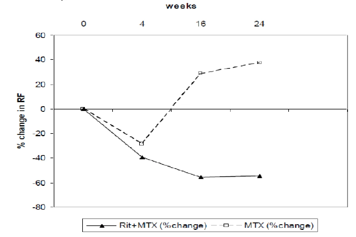
Plasma total immunoglobulin concentrations, total lymphocytes counts, and white cells generally remained within normal limits following treatment with rituximab, with the exception of a transient drop in white cells counts over the first four weeks following therapy. Titers of IgG antigen specific antibody to mumps, rubella, varicella, tetanus toxoid, influenza and streptococcus pneumococci remained stable over 24 weeks following exposure to rituximab in rheumatoid arthritis patients.
Effects of rituximab on a variety of biomarkers were evaluated in patients enrolled into Study 3 (WA16291). This substudy evaluated the impact of a single treatment course of rituximab on levels of biochemical markers, including markers of inflammation [Interleukin 6, C Reactive protein, Serum amyloid type A protein, Protein S100 isotypes A8 and A9], autoantibody (RF and anti-cyclic citrullinated peptide immunoglobulin) production and bone turnover [osteocalcin and procollagen 1 N terminal peptide (P1NP). Rituximab treatment, whether as monotherapy or in combination with methotrexate or cyclophosphamide reduced the levels of inflammatory markers significantly, relative to methotrexate alone, over the first 24 weeks of follow-up. Levels of markers of bone turnover, osteocalcin and P1NP, increased significantly in the rituximab groups compared to methotrexate alone.
120-minute infusion rate study (ML25641) in RA patients
In Study ML25641, a total of 351 patients with moderate-to-severe active rheumatoid arthritis (RA) who had an inadequate response to at least one TNF inhibitor and had no prior rituximab experience (N = 306) or had received one or two prior rituximab courses (N = 45) were evaluated in an open-label, multi-center, single-arm trial for the safety of 120-minute rituximab infusions. Patients with previous serious infusion-related reaction to any prior biologic therapy, including rituximab, or with clinically significant cardiovascular disease, were excluded from the study.
Eligible patients received 2 courses of rituximab treatment with 2 infusions of 1000 mg plus MTX treatment per course. The first course was administered on Day 1 (Infusion 1) and Day 15 (Infusion 2) and the second course six-months later on Day 168 (Infusion 3) and Day 182 (Infusion 4). Infusion 1 was administered over a 4.25-hour period. Infusion 2, 3, and 4 were administered over 120 minutes. Any patient experiencing a serious infusion-related reaction (IRR) with any infusion was withdrawn from the study. The main outcome measure was the incidence of infusion-related reactions during or within 24 hours after the 120 -minute infusion at Infusion 2.
The incidence of infusion-related reactions (IRRs) at Infusion 2 was 6.5% (95% CI [4.1%-9.7%]) and was consistent with the rate observed historically. For Infusion 2, the incidence of Grade 3 - 4 IRRs was 0.6% (95% CI [0.1%, 2.1%]) and there were no serious IRRs observed. For Infusion 3 and Infusion 4, the incidence of IRRs was 5.9% (95% CI [3.5%-9.3%]) and 0.7% (95% CI [0.1%-2.6%]), respectively. Data observed for Infusion 3 and 4 demonstrates a low incidence of IRRs, similar to the rate observed historically; no Grade 3 - 4 or serious IRRs were observed. Acute infusion-related reactions requiring dose modification (stopping, slowing, or interruption of the infusion) occurred in 12% and 3.9% of patients receiving Infusion 1 at standard infusion regimen and Infusion 2 at 120-minute faster infusion, respectively (see 8 Adverse Reactions-Clinical Trial Adverse Drug Reactions).
Granulomatosis with Polyangiitis (GPA, also known as Wegener's Granulomatosis) and Microscopic Polyangiitis (MPA)
The efficacy and safety of rituximab in patients with severely active Granulomatosis with Polyangiitis (GPA, also known as Wegener's Granulomatosis) and Microscopic Polyangiitis (MPA) was demonstrated in an active controlled, randomized, double-blind, multicentre non-inferiority study.
A total of 197 patients with severely, active GPA/MPA were enrolled and treated. Patients were 15 years of age or older, diagnosed with severely, active Granulomatosis with Polyangitis, also known as Wegener’s Granulomatosis (75% of patients) or Microscopic Polyangiitis (MPA) (24% of patients)
according to the Chapel Hill Consensus conference criteria (1% of patients had unknown GPA/MPA type). Of the 99 rituximab treated patients participating in the Phase III study, 3% were less than 18 years of age, 61% were between 18-64 years of age and 36% of patients were aged 65 years or older. Refer to Table 40 below for a summary of patient demographics and baseline disease characteristics.
Patients were randomized in a 1:1 ratio to receive either oral cyclophosphamide daily (2 mg/kg/day) for 3-6 months, followed by azathioprine or rituximab (375 mg/m2) once weekly for 4 weeks. Patients in both arms received 1000 mg of pulse intravenous (IV) methylprednisolone (or another equivalent-dose glucocorticoid) per day for 1 to 3 days, followed by oral prednisone (1 mg/kg/day, not exceeding 80 mg/day). Prednisone tapering was to be completed by 6 months from the start of study treatment.
Table 40 Baseline Demographic and Disease Characteristics in RAVE
Rituximab n = 99
Cyclophosphamide n = 98
All Patients n = 197
Age at screening (years)
Mean (SD)
54.0 (16.76)
51.5 (14.07)
52.8 (15.49)
Rituximab n = 99
Cyclophosphamide n = 98
All Patients n = 197
Range (min−max)
16−92
15−80
15−92
Sex (%) Male
46.5
54.1
50.3
Female
53.5
45.9
49.7
Primary Race (%)
White
91.9
94.9
93.4
Black or African American
3.0
3.1
3.0
Asian
1.0
0.0
0.5
Other
4.0
2.0
3.0
Ethnicity (%)
Not Hispanic or Latino
91.9
94.9
93.4
Hispanic or Latino
6.1
3.1
4.6
Unknown
2.0
2.0
2.0
ANCA-associated vasculitis type (%)
Wegener’s granulomatosis
73.7
75.5
74.6
Microscopic polyangiitis
24.2
24.5
24.4
Indeterminate
1.0
0
0.5
Missing
1.0
0
0.5
Newly diagnosed at enrollment (%)
48.5
49.0
48.7
BVAS/WG score, mean (SD) a
8.1 (2.82)
8.0 (3.41)
8.0 (3.12)
Creatinine clearance, mean (SD) (mL/min)
76.5 (46.27)
91.4 (49.24)
83.9 (48.23)
Creatinine clearance, median (mL/min)
67.61
87.47
73.81
Organ involvement (%) b
Renal
65.7
66.3
66.0
Hematuria
28.3
28.6
28.4
Red blood cell casts
37.4
35.7
36.5
Rise in creatinine > 30% or fall in creatinine clearance > 25%
34.3
36.7
35.5
Pulmonary
52.5
54.1
53.3
Alveolar hemorrhage
27.3
23.5
25.4
Endobronchial involvement
4.0
9.2
6.6
Nodules or cavities
18.2
27.6
22.8
Other lung infiltrate
25.3
21.4
23.4
Pleurisy
8.1
9.2
8.6
Respiratory failure
2.0
0
1.0
ANCA =anti-neutrophil cytoplasmic antibody; BVAS/WG =Birmingham Vasculitis Activity Score for Wegener’s granulomatosis; c-ANCA =cytoplasmic ANCA; MPO =myeloperoxidase; p-ANCA =perinuclear ANCA; PR3 =proteinase 3.
- a Scores for new/worse disease range from 0 to 67 (a higher score means more active disease). n =77 for Rituximab arm and n =67 for cyclophosphamide arm.
- b Tabulation includes new/worse disease and not persistent.
Complete Remission
The primary outcome measure was achievement of complete remission at 6 months defined as a Birmingham Vasculitis Activity Score for Wegener’s Granulomatosis (BVAS/WG) of 0, and off glucocorticoid therapy. The prespecified non-inferiority margin for the treatment difference was 20%. The study demonstrated non-inferiority of rituximab to cyclophosphamide for complete remission at 6 months (Table 41).
Table 41 Percentage of Patients Who Achieved Complete Remission at 6 Months (Intent to Treat Population)
| Rituximab (n=99) | Cyclophosphamide (n = 98) | Treatment Difference (Rituximab – Cyclophosphamide) | |
|---|---|---|---|
| Rate | 63.6% | 53.1% | 10.6% |
| 95.1%b CI | (54.1%, 73.2%) | (43.1%, 63.0%) | (−3.2%, 24.3%) a |
CI = confidence interval.
- a) Non-inferiority was demonstrated since the lower bound (− 3.2%) was higher than the pre determined non-inferiority margin (− 20%).
- b) The 95.1% confidence level reflects an additional 0.001 alpha to account for an interim efficacy analysis.
Retreatment with rituximab
The safety and efficacy of subsequent courses of rituximab in patients with GPA and MPA have not been determined (see 7 Warnings and Precautions and4 Dosage and Administration ).
16 Non-Clinical Toxicology
General Toxicology:
Immunohistology Studies with Human Tissues
The tissue reactivity of the chimeric mouse/human antibody rituximab was evaluated using a panel of 32 different human tissues fixed with acetone. The antibody was biotinylated to avoid background staining. No loss of immunoreactivity, as determined by FACS (fluorescence activated cell sorter) analysis using antigen-positive cells, was observed following biotinylation.
Biotinylated rituximab exhibited a highly restricted pattern of tissue reactivity, binding to antigen was found only on a subset of cells of lymphoid origin. Immunoreactivity was noted in the white pulp of the spleen, the lymphoid follicles of the tonsil, and in some, but not all, of the B lymphocytes present in the lymph node. Also, lymphoid cells present in other organs, e.g., large and small intestines and stomach, were immunoreactive with rituximab.
All simple epithelial cells, as well as the stratified epithelia and squamous epithelia of different organs, were found to be unreactive. Similarly, no reactivity was seen with neuroectodermal cells, including those in the brain, spinal cord and peripheral nerves. Mesenchymal elements, such as skeletal and smooth muscle cells, fibroblasts, endothelial cells, and polymorphonuclear inflammatory cells were found to be negative.
In Vitro Testing for Cross-Reactivity with Human Tissues: Rituximab Lot 0111
The human tissue specificity of biotinylated rituximab antibody Lot 0111 was evaluated using immunoperoxidase staining of formalin-fixed, normal adult human tissues obtained at autopsy. Biotinylated rituximab was used to avoid background reactivity caused by use of anti-human secondary reagents. CD20-positive (SB) and CD20-negative (HSB) human cell lines were used as controls, as was an irrelevant biotinylated mouse/human chimeric antibody termed S-004. The molar ratio of biotin-to-protein was approximately 10:1 for both antibodies; no loss of immunoreactivity was observed by flow cytometry using CD20-positive SB cells and the biotinylated rituximab antibody. Positive reactivity with staining intensity of 2+ to 3+ was observed with >90% of the CD20-positive control (SB) cells. No reactivity was observed with the CD20-negative cell line HSB.
The CD20 antigen exhibited a highly restricted pattern of distribution in the normal human tissues analyzed, and was mostly found on a subset of cells of lymphoid origin. Immunoreactivity was observed in the bone marrow, lymph node, peripheral blood B cells, white pulp of the spleen and in the lymphoid follicles of the tonsil. Some lymphoid nodules in other organ tissues, e.g., esophagus, kidney, small intestine, pancreas and stomach were also reactive.
All simple epithelial cells, and stratified epithelia and squamous epithelia of different organs were unreactive except for two specimens of large intestine with staining patterns of focal to diffuse. Reactivity was not seen in most neuroectodermal cells, including those of the brain and peripheral nerves; weak reactivity was observed in 30% of microglial cells present in 1 of 3 spinal cord specimens. Mesenchymal elements such as skeletal and smooth muscle cells, fibroblasts, and endothelial cells were unreactive.
Plasma Sample Analysis from Lot 0111 of Rituximab
Rituximab was evaluated in cynomolgus monkeys in a high-dose pathology/toxicology study designed to evaluate the safety of rituximab antibody Lot 0111 produced in suspension culture. Additionally, plasma samples from monkeys infused with this lot of rituximab antibody were analyzed for rituximab antibody levels as well as for the presence of anti-rituximab antibody: monkey anti-murine (MAMA) and monkey anti-rituximab (MACA). Groups 1 and 2, consisting of two animals each, received only vehicle; Groups 3 and 4, consisting of 6 animals each divided equally by sex, received rituximab (20 mg/kg). Groups 1 and 3 were dosed for four consecutive weeks; Groups 2 and 4 were dosed for eight consecutive weeks. Preliminary results from Groups 1 and 3 are available.
Plasma clearance study results indicate that high rituximab plasma levels (186 - 303 μg/mL) were achieved in all treated monkeys 24 hours after the first and second infusions. Plasma antibody levels achieved 24 hours after the third and fourth antibody injections were similar to those detected after the first two injections in three Group 3 monkeys. Further, concentrations persisted at significant levels for two weeks after the last infusion in these animals. In the other three Group 3 animals, rituximab levels were markedly reduced at both the 24 hours and seven day time points after the third and fourth infusions; results correlated with the production of a MAMA response.
As seen in previous monkey studies, marked B-cell depletion occurred in all animals after each of the four infusions of rituximab antibody. However, the level of B-cell depletion was more marked in three of the six monkeys on day 36.
Three of the six Group 3 monkeys produced anti-rituximab antibodies that were detected two weeks after the last antibody injection. Results are confirmed by the rapid recovery of B lymphocytes in the peripheral blood of the three animals at time points that correlate with the appearance of the potentially neutralizing anti-chimeric antibody responses. None of the other Group 3 monkeys showed an anti-rituximab immune response greater than 0.2 μg/mL on day 36. Results indicate that certain monkeys with competent immune systems may respond to multiple antibody exposures by producing significant amounts of neutralizing antibodies that alter the efficacy (depleting capability) of the antibody.
17 Supporting Product Monographs
RITUXAN® (Intravenous Infusion, 10 mg/mL), Submission Control Number: 271112, Product Monograph, Hoffmann-La Roche Ltd. (Jun 02, 2023).