


Product Monograph - Isovue 300 Injection
RECENT MAJOR LABEL CHANGES
Not applicable
TABLE OF CONTENTS
Sections or subsections that are not applicable at the time of authorization are not listed.
RECENT MAJOR LABEL CHANGES ........................................................................................... 2
TABLE OF CONTENTS ............................................................................................................. 2
PART I: HEALTH PROFESSIONAL INFORMATION ..................................................................... 5
1 INDICATIONS .............................................................................................................. 5
1.1 Pediatrics ................................................................................................................... 5
1.2 Geriatrics ................................................................................................................... 5
2 CONTRAINDICATIONS ................................................................................................. 6
3 SERIOUS WARNINGS AND PRECAUTIONS BOX ............................................................ 6
4 DOSAGE AND ADMINISTRATION ................................................................................. 6
4.1 Dosing Considerations .............................................................................................. 6
4.2 Recommended Dose and Dosage Adjustment ......................................................... 7
INTRAVASCULAR USE ......................................................................................................... 7
Peripheral Arteriography ................................................................................................... 7
Coronary Arteriography and Left Ventriculography ......................................................... 7
Pediatric Angiocardiography ............................................................................................. 8
Excretory Urography .......................................................................................................... 8
Peripheral Venography (Phlebography) ............................................................................ 9
Cerebral arteriography ....................................................................................................... 9
Computed Tomography ................................................................................................... 10
Intravenous Contrast Enhancement in Computed Tomography of the Head ............... 10
Intravenous Contrast Enhancement in Computed Tomography of the Body ............... 10
SUBARACHNOID USE ........................................................................................................ 11
4.4 Administration ........................................................................................................ 12
5 OVERDOSAGE ........................................................................................................... 13
6 DOSAGE FORMS, STRENGTHS, COMPOSITION AND PACKAGING ............................... 13
7 WARNINGS AND PRECAUTIONS ................................................................................ 14
General ............................................................................................................................. 14
Cardiovascular .................................................................................................................. 15
Contamination .................................................................................................................. 16
Endocrine and Metabolism .............................................................................................. 16
Hematologic...................................................................................................................... 17
Hepatic/Biliary/Pancreatic............................................................................................... 17
Neurologic ........................................................................................................................ 18
Renal ................................................................................................................................. 19
Sensitivity/Resistance ...................................................................................................... 20
7.1 Special Populations ................................................................................................. 21
7.1.1 Pregnant Women .............................................................................................. 21
7.1.2 Breast-feeding ................................................................................................... 21
7.1.3 Pediatrics ........................................................................................................... 21
7.1.4 Geriatrics ........................................................................................................... 21
8 ADVERSE REACTIONS................................................................................................ 22
8.1 Adverse Reaction Overview .................................................................................... 22
8.2 Clinical Trial Adverse Reactions .............................................................................. 23
8.5 Post-Market Adverse Reactions .............................................................................. 26
9 DRUG INTERACTIONS ............................................................................................... 26
9.1 Serious Drug Interactions ....................................................................................... 26
9.2 Drug Interactions overview..................................................................................... 27
9.3 Drug-Behavioural Interactions ................................................................................ 27
9.4 Drug-Drug Interactions ........................................................................................... 27
9.5 Drug-Food Interactions ........................................................................................... 28
9.6 Drug-Herb Interactions ........................................................................................... 28
9.7 Drug-Laboratory Test Interactions.......................................................................... 28
10 CLINICAL PHARMACOLOGY ....................................................................................... 28
10.1 Mechanism of Action ........................................................................................ 28
10.2 Pharmacodynamics ........................................................................................... 28
10.3 Pharmacokinetics .............................................................................................. 32
11 STORAGE, STABILITY AND DISPOSAL ......................................................................... 33
12 SPECIAL HANDLING INSTRUCTIONS ........................................................................... 33
PART II: SCIENTIFIC INFORMATION ...................................................................................... 34
13 PHARMACEUTICAL INFORMATION ........................................................................... 34
14 CLINICAL TRIALS ....................................................................................................... 35
14.1 Trial Design and Study Demographics .............................................................. 35
15 MICROBIOLOGY ....................................................................................................... 35
16 NON-CLINICAL TOXICOLOGY ..................................................................................... 36
PATIENT MEDICATION INFORMATION ................................................................................. 39
PART I: HEALTH PROFESSIONAL INFORMATION
1 INDICATIONS
ISOVUE® (iopamidol) Injection is indicated for:
Intravascular Procedures:
Adults
- Peripheral venography (phlebography) (ISOVUE-200 (41% w/v, 200 mg I/mL))
- Peripheral arteriography (ISOVUE-300 (61% w/v, 300 mg I/mL))
- Cerebral arteriography (ISOVUE-300 (61% w/v, 300 mg I/mL))
- Excretory urography (ISOVUE-300 (61% w/v, 300 mg I/mL))
- Intravenous contrast enhancement in computed tomography of the head (ISOVUE-300 (61% w/v, 300 mg I/mL))
- Intravenous contrast enhancement in computed tomography of the body (ISOVUE-300 (61% w/v, 300 mg I/mL), ISOVUE-370 (76% w/v, 370 mg I/mL))
- Coronary arteriography and left ventriculography (ISOVUE-370 (76% w/v, 370 mg I/mL))
Myelography:
Adults
- Lumbar myelography by lumbar injection (ISOVUE-200 (41% w/v, 200 mg I/mL))
1.1 Pediatrics
Pediatric Patients (0 to 18 years of age)
- Excretory urography (ISOVUE-300 (61% w/v, 300 mg I/mL))
- Intravenous contrast enhancement in computed tomography of the head (ISOVUE-300 (61% w/v, 300 mg I/mL))
- Intravenous contrast enhancement in computed tomography of the body (ISOVUE-300 (61% w/v, 300 mg I/mL))
Pediatric Patients (6 weeks to 18 years of age)
- Angiocardiography (ISOVUE-370 (76% w/v, 370 mg I/mL))
Pediatric Patients (2 to 18 years of age)
- Thoracolumbar myelography (ISOVUE-200 (41% w/v, 200 mg I/mL))
1.2 Geriatrics
Geriatrics: Evidence from clinical studies and experience suggests that use in the geriatric population may be associated with differences in safety or effectiveness (see 7 WARNINGS AND PRECAUTIONS).
2 CONTRAINDICATIONS
- ISOVUE (iopamidol injection) is contraindicated in patients with known hypersensitivity to the product and in patients with significant impairment of both renal and hepatic functions.
- Because of overdosage considerations, immediate repeat myelography in the event of technical failure is contraindicated (see Interval Recommendations under 4 DOSAGE AND ADMINISTRATION).
- Intrathecal administration of ISOVUE with corticosteroids is contraindicated.
- Direct intracisternal administration or gravitational displacement of a concentrated bolus of iopamidol into the intracranial subarachnoid spaces is contraindicated.
- Myelography should not be performed in the presence of significant local or systemic infection where bacteremia is likely when lumbar puncture is contraindicated.
3 SERIOUS WARNINGS AND PRECAUTIONS BOX
Serious Warnings and Precautions
|
4 DOSAGE AND ADMINISTRATION
4.1 Dosing Considerations
It is desirable that solutions of radiopaque diagnostic agents be at or close to body temperature when injected. As other sterile parenteral products, contrast media should not be transferred into other delivery systems except immediately prior to use.
Withdrawal of contrast agents from their containers should be accomplished under aseptic conditions with sterile syringes. The transferring of ISOVUE from the ISOVUE Multipack-300 (iopamidol injection, 300 mg I/mL) and ISOVUE Multipack-370 (iopamidol injection, 370 mg I/mL) pharmacy bulk package should be performed in a suitable work area, such as a laminar flow hood, using aseptic technique. The container closure may be penetrated only one time, utilizing a suitable transfer device.
Sterile techniques must be used with any subarachnoid and intravascular injections, and with catheters and guidewires and prolonged contact of the contrast medium with blood has to be avoided.
Parenteral drug products should be inspected visually for particulate matter and discoloration prior to administration. ISOVUE (iopamidol injection) solutions should be used only if clear and within the normal colorless to pale yellow range.
Patients should be well hydrated prior to and following administration of ISOVUE.
As with all radiopaque contrast agents, only the lowest dose of ISOVUE necessary to obtain adequate visualization should be used. A lower dose reduces the possibility of an adverse reaction. Most procedures do not require use of either a maximum dose or the highest available concentration of ISOVUE; the combination of dose and ISOVUE concentration to be used should be carefully individualized, and factors such as age, body size, size of the vessel and its blood flow rate, anticipated pathology and degree and extent of opacification required, structure(s) or area to be examined, disease processes affecting the patient, and equipment and technique to be employed should be considered.
If antihistamines or corticosteroids are to be used, they should not be mixed with the contrast medium because of chemical incompatibility.
4.2 Recommended Dose and Dosage Adjustment
Only the concentration recommended for the particular procedure to be performed should be used (see 3 SERIOUS WARNINGS AND PRECAUTIONS).
INTRAVASCULAR USE
Peripheral Arteriography
ISOVUE-300 (iopamidol injection, 300 mg I/mL) is recommended.
Adult Dosage:
For injection into the femoral artery, a single dose of 15-40 mL may be used; for the subclavian artery, 5 to 20 mL; for injection into the aorta for a distal runoff, 25 to 50 mL is the usual single dose. These doses may be repeated if indicated, however 15-20 minutes should be allowed to elapse between injections to allow for subsidence of hemodynamic disturbances, and the total procedural dose should be limited to the smallest volume necessary to obtain a diagnostic examination (see 7 WARNINGS AND PRECAUTIONS and 8 ADVERSE REACTIONS).
Administration:
The injection is usually made through a catheter introduced into the femoral artery with the tip placed to achieve lower aortic or aorto-iliac runoff for visualization of an individual femoral artery or its distribution throughout the lower limb. Pressure injection is usually employed. Visualization is similar to that achieved with ionic media of similar dose and concentration. Sedative premedication may be employed for the procedure, however, anesthesia is usually not considered necessary (see "Peripheral arteriography and venography" under 8 ADVERSE REACTIONS - INTRAVASCULAR).
Coronary Arteriography and Left Ventriculography
ISOVUE-370 (iopamidol injection, 370 mg I/mL) is recommended.
Adult Dosage:
The usual single dose for selective coronary artery injections is 2 to 10 mL; for left ventriculography, 35-50 mL. For nonselective opacification of multiple coronary arteries following injection at the aortic root, the usual single dose is 15-35 mL. These doses may be repeated if indicated, however 15-20 minutes should be allowed to elapse between injections to allow for subsidence of hemodynamic disturbances. The total procedural dose should be limited to the smallest volume necessary to obtain a diagnostic examination (see 7 WARNINGS AND PRECAUTIONS). Continuous ECG monitoring and close observation are essential (see "Coronary arteriography and left ventriculography" under 8 ADVERSE REACTIONS - INTRAVASCULAR).
Pediatric Angiocardiography
ISOVUE-370 (iopamidol injection, 370 mg I/mL) is recommended.
Pediatric Dosage:
Pediatric angiocardiography may be performed by injection into a large peripheral vein or by direct catheterization of the heart. The amount to be administered will depend on the number of injections required, the anatomical sites to be studied, the anticipated pathology, the size of the patient, diseases processes affecting the patient, and possibly other factors. In all cases, the minimum amount of contrast necessary to obtain an adequate imaging study should be used.
The following table serves as a guide for single dose injections:
| Age | mL |
6 weeks - 2 years 2 - 9 years 10 - 18 years | 10 - 15 15 - 30 20 - 50 |
The maximum cumulative doses recommended are as follows:
| Age | mL |
6 weeks - 2 years 2 - 4 years 5 - 9 years 10 - 18 years | 40 50 100 125 |
(see "Pediatric angiocardiography" under 8 ADVERSE REACTIONS - INTRAVASCULAR)
Excretory Urography
ISOVUE-300 (iopamidol injection, 300 mg I/mL) is recommended.
Adult Dosage:
The usual dose is 50 mL administered by intravenous injection. Doses up to 100 mL may be required in some patients.
Pediatric Dosage:
The dosage recommended is 1.0 to 3.0 mL/kg by intravenous injection. It should not be necessary to exceed a total dose of 30 grams of iodine.
Administration:
Preparatory dehydration is not recommended since it is unnecessary and may even be dangerous (see 7 WARNINGS AND PRECAUTIONS – General).
Peripheral Venography (Phlebography)
ISOVUE-200 (iopamidol injection, 200 mg I/mL) is recommended.
Adult Dosage:
The usual dose is 30 to 125 mL for the lower extremity. The combined total dose for multiple injections should not exceed 250 mL (see "Peripheral arteriography and venography" under 8 ADVERSE REACTIONS - INTRAVASCULAR).
Cerebral arteriography
ISOVUE-300 (iopamidol injection, 300 mg I/mL) is recommended.
Adult Dosage:
The usual single adult doses are as follows:
Common carotid artery Internal carotid artery External carotid artery Vertebral artery Aortic arch injection (four vessel studies) | 6 - 12 mL 5 - 10 mL 4- 8 mL 5 - 10 mL 20 - 50 mL |
These doses may be repeated if indicated; however, the total procedural dose should be limited to the smallest volume necessary to achieve a diagnostic examination. The total dose should not exceed 90 mL.
Administration:
Appropriate patient preparation is indicated. Cerebral arteriography should be undertaken with extreme care and special caution in patients with advanced vascular disease, increased intracranial pressure, slowed cerebral circulation, spasm, breached blood brain barrier, recent cerebral thrombosis, embolism or bleeding, and in patients in poor clinical condition (see 7 WARNINGS AND PRECAUTIONS). This may include suitable premedication. It is advisable to inject at rates approximately equal to the flow rate of the vessel being injected.
The incidence of adverse reactions appears to be related to the number of repeated injections, administration of doses higher than those recommended, and the method and technique of injection. Serious adverse reactions include stroke, hemiparesis, cortical blindness, hypotension, bradycardia, arrhythmias, convulsions, coma, death (see 7 WARNINGS AND PRECAUTIONS, and 8 ADVERSE REACTIONS – INTRAVASCULAR – “Cerebral arteriography").
Computed Tomography
Since an unenhanced scan may be sufficient for diagnosis, and the injection of contrast media may obscure certain lesions visible on the plain scan, contrast enhancement is usually performed only if the unenhanced scan has not provided or cannot be expected to provide sufficient information.
The decision to employ contrast enhancement should be carefully weighed, taking into consideration the patient's clinical condition, renal and cardiac reserve, and the status of the blood-brain-barrier since contrast enhancement may be associated with increased risk (see 7 WARNINGS AND PRECAUTIONS).
Experience with children 1 year of age and younger is limited.
Intravenous Contrast Enhancement in Computed Tomography of the Head
ISOVUE-300 (iopamidol injection, 300 mg I/mL) is recommended.
Adult dosage:
The usual adult dose is 50 to 100 mL by intravenous administration. In clinical studies, patients have safely received doses up to 150 mL.
Pediatric dosage:
The usual dose is 1.0 to 3.0 mL/kg by intravenous administration. It should not be necessary to exceed a total dose of 30 g of iodine.
Intravenous Contrast Enhancement in Computed Tomography of the Body
ISOVUE-300 (iopamidol injection, 300 mg I/mL) may be used for contrast enhancement of organs, tissues and larger blood vessels of the chest, abdomen and pelvis.
Adult dosage:
The usual adult dose is 100 - 150 mL administered by rapid intravenous bolus injection or infusion. Scanning is usually performed immediately after injection.
Equivalent doses of ISOVUE-370 (iopamidol injection, 370 mg I/mL) based on organically bound iodine content may also be used. (80 - 120 mL)
The total dose should not exceed 60 grams of iodine.
Pediatric dosage:
The usual dose is 1.0 to 3.0 mL/kg by intravenous administration. It should not be necessary to exceed a total dose of 30 g of iodine.
SUBARACHNOID USE
Only the recommended concentration should be used (see 3 SERIOUS WARNINGS AND PRECAUTIONS).
Myelography
Adult Dosage:
ISOVUE-200 (iopamidol injection, 200 mg I/mL), a solution that is approximately isotonic with the cerebrospinal fluid is recommended for examination of the lumbar region. The usual dose is 10-15 mL. A total dose in excess of 3000 mg iodine, and ISOVUE formulated to contain more than 200 mg I/mL should not be used intrathecally.
Pediatric Dosage:
ISOVUE-200 (iopamidol injection, 200 mg I/mL) is also recommended for thoracolumbar myelography in children over the age of two years. The minimum dose needed to perform a procedure should always be used. An ISOVUE formulation containing more than 200 mg I/mL should not be used intrathecally in children.
Pediatric Dosage for Lumbar, Thoracic Myelogram
| Age (years) | Dose ISOVUE-200 (mL) |
2 - 7 8 - 12 13 - 18 | 7 - 9 8 - 11 10 - 12 |
These doses, intended as a guideline, are based on age rather than weight because the brain and CSF capacity are independent of weight. Variations in dose will depend on such factors as height, suspected pathology, the patient's condition and technique used. Loss of contrast due to dispersion of the medium because of movement is less apt to occur in children, because of their shorter spinal cord.
Administration:
Anesthesia is not necessary, however in very young patients it may be considered for technical reasons. Premedication with sedatives or tranquilizers is usually not needed. In patients with a history of seizure activity who are not on anticonvulsant therapy, premedication with barbiturates or phenytoin should be considered.
Lumbar puncture is usually made between L3 and L4; if pathology is suspected at this level, the interspace immediately above or below may be selected.
Rate of Injection: To avoid excessive mixing with cerebrospinal fluid and consequent loss of contrast as well as premature cephalad dispersion, injection must be made slowly over one to two minutes; the needle may then be removed.
An interval of at least 5-7 days should be allowed before repeat examination. Aspiration of ISOVUE is unnecessary following intrathecal administration (see 10 CLINICAL PHARMACOLOGY).
Suggestions for Patient Management
Pre-procedure
- See 9 DRUG INTERACTIONS regarding discontinuation of neuroleptic agents.
- Maintain normal diet up to 2 hours before procedure.
- Ensure hydration - fluids up to time of procedure.
During Procedure
- Use minimum dose and concentration required for satisfactory contrast.
- Inject slowly over 1 to 2 minutes to avoid excessive mixing.
- Avoid abrupt or active patient movement which causes excessive mixing with CSF.
- Instruct patient to remain passive. Move patient slowly and only as necessary.
- To maintain as a bolus, move medium within the lumbosacral region very slowly under fluoroscopic control.
- In all positioning techniques, keep the patient's head elevated above highest level of spine.
- Do not lower head of table more than 15° during the procedure.
- Avoid intracranial entry of a bolus.
- Avoid early and high cephalad dispersion of the medium.
- At completion of the procedure, raise head of table steeply (45°) for about 2 minutes to restore medium to lower lumbar levels.
Post-procedure
- Raise head of stretcher to at least 30° before moving patient onto it.
- Movement onto stretcher, and off the stretcher to bed, should be done slowly with patient completely passive, maintaining head up position.
- Before moving patient onto bed, raise head of bed 30° to 45° and maintain the patient in this position under close observation for 12 to 24 hours.
- Advise patient to remain quiet in bed, in head up position.
- Obtain visitors cooperation in keeping the patient quiet and in head up position, especially in first few hours.
- Encourage oral fluids and diet as tolerated.
- Antinauseants of the phenothiazine class should not be administered to treat post procedural nausea or vomiting. Since persistent nausea and vomiting may result in dehydration, prompt consideration of volume replacement by intravenous fluid is recommended.
4.4 Administration
Please refer to 4.2 Recommended Dose and Dosage Adjustment
5 OVERDOSAGE
In myelography, even use of a recommended dose can produce severe CNS disturbances that are equivalent to overdosage if incorrect management of the patient during or immediately following the procedure permits inadvertent early intracranial entry of a large portion of the medium. It should be ensured that the patient is kept in the head up position.
Treatment of an overdose of an injectable radiopaque contrast medium is directed toward the support of all vital functions and prompt institution of specific therapy.
| For management of a suspected drug overdose, contact your regional poison control centre. |
6 DOSAGE FORMS, STRENGTHS, COMPOSITION AND PACKAGING
Table – Dosage Forms, Strengths, Composition and Packaging
ISOVUE-200 (iopamidol injection 41% w/v) provides 408 mg of iopamidol per mL (equivalent to 200 mg of organically bound iodine per mL) and approximately 0.029 mg (0.001 mEq) of sodium per mL. It is supplied in 20, 50, 100 and 200 mL vials.
ISOVUE-300 (iopamidol injection 61 % w/v) provides 612 mg of iopamidol per mL (equivalent to 300 mg of organically bound iodine per mL) and approximately 0.043 mg (0.002 mEq) of sodium per mL. It is supplied in 15, 50, 100 and 150 mL vials; and in 100mL and 150mL power injector syringes (Power injector syringes are supplied with connector tubing).
ISOVUE Multipack-300 (iopamidol injection 61 % w/v) provides 612 mg of iopamidol per mL (equivalent to 300 mg of organically bound iodine per mL) and approximately 0.043 mg (0.002 mEq) of sodium per mL. It is supplied as pharmacy bulk packages in 200 and 500 mL bottles.
| Route of Administration | Dosage Form / Strength/Composition |
| Non-medicinal Ingredients | |
| Subarachnoid, Intravascular | ISOVUE 200 (iopamidol injection 41% w/v) |
| Calcium disodium edetate (EDTA), hydrochloric acid, tromethamine and water for injection | |
| Intravascular | ISOVUE 300 (iopamidol injection 61 % w/v) |
| Intravascular | ISOVUE Multipack-300 (iopamidol injection 61% w/v) |
| Intravascular | ISOVUE 370 (iopamidol injection 76% w/v) |
| Intravascular | ISOVUE Multipack-370 (iopamidol injection 76% w/v) |
ISOVUE-370 (iopamidol injection 76% w/v) provides 755 mg of iopamidol per mL (equivalent to 370 mg of organically bound iodine per mL) and approximately 0.053 mg (0.002 mEq) of sodium per mL. It is supplied in 50, 100, 150 and 200 mL vials; and in 75 mL and 100mL power injector syringes (Power injector syringes are supplied with connector tubing).
ISOVUE Multipack-370 (iopamidol injection 76% w/v) provides 755 mg of iopamidol per mL (equivalent to 370 mg of organically bound iodine per mL) and approximately 0.053 mg (0.002 mEq) of sodium per mL. It is supplied as pharmacy bulk packages in 200 and 500 mL bottles.
7 WARNINGS AND PRECAUTIONS
Please see 3 SERIOUS WARNINGS AND PRECAUTIONS BOX
General
Since serious or fatal reactions can occur following the use of iodinated contrast media, diagnostic imaging procedures which involve the use of radiopaque contrast agents should be carried out only by physicians with the prerequisite training and with a thorough knowledge of the particular procedure to be performed. They should be thoroughly familiar with the emergency treatment of all adverse reactions to contrast media. Appropriate facilities, resuscitative drugs, equipment and personnel must be readily available in case a severe immediate or delayed reaction occurs (see 8 ADVERSE REACTIONS – TREATMENT OF ADVERSE REACTIONS TO CONTRAST MEDIA).
Caution is also advised in patients with severe cardiovascular disease, hyperthyroidism, concomitant renal and hepatic disease and in patients with endotoxemia or elevated body temperature.
Generally accepted contraindications, warnings, precautions and adverse reactions commonly related to the use of other radiopaque contrast media should be kept in mind during the administration of ISOVUE.
General anesthesia may be indicated in the performance of some procedures in selected patients; however, a higher incidence of adverse reactions has been reported with radiopaque media in anesthetized patients, which may be attributable to the inability of the patient to identify untoward symptoms, or to the hypotensive effect of anesthesia which can reduce cardiac output and increase the duration of exposure to the contrast agent.
Subarachnoid Use:
Intrathecal administration of corticosteroids with ISOVUE is contraindicated.
In rare cases following their use in myelography, water soluble iodinated contrast media have been associated with fatal, potentially fatal or extremely serious adverse reactions sometimes resulting in permanent damage. It is therefore imperative that the indication for myelography be well supported, the expected benefits be carefully weighed against the risks, the lowest diagnostic concentration and dose be used and proper patient management be carefully followed before, during and after the procedure.
Myelography should not be performed in the presence of significant local or systemic infection where bacteremia is likely or when lumbar puncture is contraindicated.
Because of overdosage considerations, immediate repeat myelography in the event of technical failure is contraindicated (see Interval Recommendations under 4 DOSAGE AND ADMINISTRATION).
Caution is advised in patients with severe cardiovascular disease, increased intracranial pressure or suspicion of intracranial tumor, abscess or hematoma; in patients with a history of convulsive disorder, chronic alcoholism, multiple sclerosis, multiple myeloma, sickle cell disease or with a history of bronchial asthma or other allergic manifestations; and in elderly patients. Particular attention must be given to the state of hydration of the patient, the concentration of ISOVUE used, the dose and the technique used.
Cardiovascular
Intravascular Use
Serious, rarely fatal, thromboembolic events causing myocardial infarction and stroke have been reported during angiographic procedures with non-ionic and also with ionic contrast media (see 7 WARNINGS AND PRECAUTIONS – Hematologic).
ISOVUE produces generally less of a circulatory osmotic load than conventional ionic contrast agents; however, it can produce significant hemodynamic disturbances, especially in patients with congestive heart failure or reduced cardiac reserve. Accordingly, the volume of injection should be kept to a minimum and patients should be observed for several hours following administration to detect delayed hemodynamic disturbances.
In angiographic procedures, the possibility of dislodging plaques or damaging, dissecting or perforating the vessel wall causing hemorrhage, thrombosis and injury to neighbouring organs should be borne in mind during catheter manipulations and contrast medium injection. Pulsation must be present in the artery to be injected. The presence of a vigorous, pulsatile flow should be established by a small "pilot" dose (about 2 mL) to locate the exact site of the needle or catheter tip to help prevent injection of the main dose into a branch of the aorta or intramurally. The inhibitory effects of non-ionic contrast media on mechanisms of hemostasis have been shown to be less than ionic contrast media at comparable concentrations. For this reason, meticulous angiographic procedures should always be followed: angiographic catheters should be flushed frequently, and prolonged contact of blood with contrast medium in syringes and catheters should be avoided.
Selective coronary arteriography should be performed only in selected patients in whom the expected benefits outweigh the procedural risk. Continuous monitoring of ECG and vital signs and adequate facilities for immediate resuscitation and cardioversion are mandatory. The risk in coronary arteriography is increased if the procedure is performed shortly after the occurrence of myocardial infarction. Angiography should be avoided whenever possible in patients with homocystinuria, because of the risk of inducing thrombosis and embolism. The inherent procedural risks of angiography in patients with compromised pulmonary function must be weighed against the necessity for performing the procedure (also see section on PEDIATRIC USE).
Extreme caution is advised in considering peripheral arteriography or venography in patients suspected of having thromboangiitis obliterans (Buerger's disease) since any procedure (even insertion of a needle or catheter) may induce a severe arterial or venous spasm. Caution is also advisable in patients with severe ischemia associated with ascending infection. Special care is required in patients with suspected thrombosis, thrombophlebitis, ischemic disease, local infection or a significantly obstructed vascular system. Extreme caution during injection of contrast media is necessary to avoid extravasation and fluoroscopy is recommended. This is especially important in patients with severe arterial or venous disease. In the presence of venous stasis, irrigation of the vein should be considered following venography. Occasional serious neurologic complications, including paraplegia have been reported in patients with aorto iliac or femoral artery bed obstruction, abdominal compression, hypotension, hypertension or with anesthesia and following injection of vasopressors when conventional ionic media were used.
Great care is necessary to avoid entry of a large concentrated bolus into an aortic branch. Mesenteric necrosis, acute pancreatitis, renal shut down, serious neurologic complications including spinal cord damage and hemiplegia, or quadriplegia have been reported following inadvertent injection of a large part of the aortic dose of contrast media into an aortic branch or arterial trunks providing spinal or cerebral artery branches.
When large individual doses are administered through multiple intra-arterial administrations, approximately 15-20 minutes should be permitted to elapse between injections to allow for subsidence of hemodynamic disturbances.
Following catheter procedures gentle pressure hemostasis is advised followed by immobilization of the limb for several hours to prevent hemorrhage from the site of arterial puncture.
Transient hypotension may occur after intraarterial (brachial) injection of contrast media.
Cerebral arteriography should be undertaken with extreme care. Special caution is required in elderly patients, patients in poor clinical condition, those with advanced arteriosclerosis, severe arterial hypertension, recent cerebral embolism or thrombosis, cardiac decompensation, subarachnoid hemorrhage and following a recent attack of migraine, if the examination is considered essential for the welfare of the patient. Since the contrast medium is injected into a sensitive area, the patient should be watched for possible untoward reactions.
Contamination
Sterile technique must be employed and recommended doses and concentrations must not be exceeded.
If non disposable equipment is used, scrupulous care should be taken to prevent residual contamination with traces of cleansing agents.
Endocrine and Metabolism
Pheochromocytoma
Administration of radiopaque materials to patients known or suspected of having pheochromocytoma should be performed with extreme caution. When, in the opinion of the physician, the possible benefits of contrast enhanced imaging outweighs the considered risks, the amount of radiopaque medium injected should be kept to an absolute minimum. The blood pressure should be assessed throughout the procedure and measures for treatment of a hypertensive crisis should be readily available. These patients should be monitored very closely when contrast agents are administered.
Thyroid Dysfunction
ISOVUE, like all other iodinated contrast media, may induce changes in thyroid function in some patients. Transient hyperthyroidism or hypothyroidism has been reported following iodinated contrast media administration to adult and pediatric patients.
Reports of thyroid storm occurring following the use of iodinated radiopaque diagnostic agents in patients with hyperthyroidism or an autonomously functioning thyroid nodule suggest that this additional risk be evaluated in such patients before the use of ISOVUE or any contrast medium.
Thyroid dysfunction in infants:
Decreased levels of thyroxine (T4) and triiodothyronine (T3) and increased Level of TSH were reported after exposure to ICM in infants, especially preterm infants, which remained for up to a few weeks or more than a month (see 8 ADVERSE REACTIONS). Some patients were treated for hypothyroidism. Hypothyroidism in infants may be harmful for growth and development, including mental development and may require treatment. Thyroid function in infants exposed to ICM should therefore be evaluated and monitored until thyroid function is normalized.
Hematologic
Contrast media may promote sickling in individuals who are homozygous for sickle cell disease.
Disseminated intravascular coagulation has been reported rarely with the use of iodinated contrast agents.
Intravascular Use:
Non-ionic iodinated contrast media inhibit blood coagulation less than ionic contrast media. Clotting has been reported in vivo and in vitro when blood remains in contact with syringes, catheters or tubes containing non-ionic contrast media.
Serious, rarely fatal, thromboembolic events causing myocardial infarction and stroke have been reported during angiographic procedures with non-ionic and also with ionic contrast media. Therefore, meticulous intravascular administration technique is necessary, particularly during angiographic procedures, to minimize thromboembolic events. Numerous factors, including length of procedure, number of injections, catheter and syringe material, underlying disease state, and concomitant medications may contribute to the development of thromboembolic events. For these reasons, meticulous angiographic techniques are recommended including close attention to keeping guidewires, catheters, and all angiographic equipment free of blood, use of manifold systems and/or three way stopcocks, frequent catheter flushing with heparinized saline solutions, and minimizing the length of the procedure. Non-ionic contrast media are not recommended as flush solutions. The use of plastic syringes in place of glass syringes has been reported to decrease but not eliminate the likelihood of in vitro clotting.
Intravascular injection of large doses of contrast media including ISOVUE may be associated with significant hemodynamic changes.
Hepatic/Biliary/Pancreatic
ISOVUE is contraindicated in patients with significant impairment of both renal and hepatic functions.
Caution must be exercised in patient with less significant impairment of both renal and hepatic functions. The amount of contrast medium administered to such patients should be kept to the absolute minimum and re-examinations should be delayed for one week.
Neurologic
Extreme care and special precaution are advised in patients with increased intracranial pressure, cerebral thrombosis or embolism, primary or metastatic cerebral lesions, subarachnoid hemorrhage, arterial spasm, transient ischemic attacks, and in any condition when the blood brain barrier is breached or the transit time of the contrast material through the cerebral vasculature is prolonged, since clinical deterioration, convulsions, and serious temporary or permanent neurological complications (including stroke, aphasia, cortical blindness, etc.) may occur following intravenous or intraarterial injection of relatively large doses of contrast media. Such patients and patients in clinically unstable or critical condition should undergo examinations with intravascular contrast media only if in the opinion of the physician the expected benefits outweigh the potential risks, and the dose should be kept to the absolute minimum.
Subarachnoid Use:
Intrathecal administration of corticosteroids with ISOVUE is contraindicated.
Rare extremely serious or fatal adverse reactions have been reported with the use of water-soluble iodinated contrast media in myelography. The expected benefits of myelography must be carefully weighed against the risks, and the lowest diagnostic concentration and dose must be used. Since severe adverse reactions including convulsions may be delayed following the use of ISOVUE, it should be used in hospitalized patients only and the patients should be kept under close surveillance for at least 24 hours. It appears that the propensity for seizure activity is related to the rate, concentration and amount of iopamidol reaching the brain cortex.
Caution is advised in patients with increased intracranial pressure or suspicion of intracranial tumor, abscess or hematoma; in patients with a history of convulsive disorder, or multiple sclerosis.
The incidence of significant adverse reactions is increased if the more cephalad segments of the spinal cord are exposed to the contrast material. The risk of convulsions is likely to be increased in patients taking phenothiazine derivatives.
Inadvertent intracranial entry of a large or concentrated bolus of the contrast medium, which increases the risks of neurotoxicity, must be prevented by careful patient management. Active patient movement which can cause rapid dispersion of the contrast medium and a consequent inadvertent rise in intracranial levels should be avoided. If such intracranial entry of iopamidol occurs, patients must be placed in upright position and prophylactic oral anticonvulsant treatment with diazepam or barbiturates for 24 to 48 hours should be seriously considered.
If bloody CSF is encountered, the possible benefits of a myelographic procedure should be considered in terms of the risk to the patient.
In patients with a history of epilepsy, myelography with iopamidol should only be performed when the use of a water soluble contrast agent for this procedure is considered essential. Extreme caution and close observation is advised. Patients receiving anticonvulsants should be maintained on this therapy. Should a seizure occur, intravenous diazepam or sodium phenobarbital is recommended (see 8 ADVERSE REACTIONS – TREATMENT OF ADVERSE REACTIONS TO CONTRAST MEDIA).
Myelography should not be performed or repeated within 5-7 days of a previous spinal puncture or myelography unless the increased risk is considered acceptable by the physician.
Computed tomography (CT) scans of the head performed within 24 hours following water soluble myelography may yield false results due to the permeation of the brain by the contrast medium from adjacent CSF spaces. If a CT scan of the brain is indicated, it should be performed before, or after a period of at least 24 hours following myelography.
Focal and generalized convulsive seizures have been reported after intrathecal use of water-soluble contrast agents including ISOVUE especially when higher than recommended doses were employed. In view of this the following should be avoided:
- Deviations from recommended neuroradiologic procedures or patient management.
- Use in patients with a history of epilepsy.
- Overdosage.
- Intracranial entry of a bolus or premature diffusion of a high concentration of the medium.
- Lowering of the head from its elevated position during the procedure, on the stretcher, and in bed.
- Excessive and particularly active patient movement or straining.
Renal
ISOVUE is contraindicated in patients with significant impairment of both renal and hepatic functions.
Renal function should be assessed before injecting ISOVUE. ISOVUE is cleared by glomerular filtration; patients with renal insufficiency have increased systemic exposure to ISOVUE as compared to patients with normal renal function. Exercise caution and use the lowest necessary dose of ISOVUE in patients with renal insufficiency. Before ISOVUE is administered, patients should be fully assessed and precautions must be taken in patients with renal impairment. Implementation of prevention strategies is considered to be the best approach to reducing development of contrast media-induced nephrotoxicity.
Adequately hydrate patients prior to and following ISOVUE administration in order to minimize the risk of contrast media-induced nephrotoxicity. Patients on dialysis, if without residual renal function, may receive ISOVUE for radiological procedures as iodinated contrast media are cleared by the dialysis process.
Acute renal insufficiency or failure may occur following ISOVUE administration, particularly in patients with pre-existing renal impairment, sepsis, hypertension, dehydration, advanced vascular disease, congestive heart disease, diabetes mellitus, multiple myeloma or other paraproteinacious diseases, patients on medications which alter renal function, and the elderly with age-related renal impairment.
Radiopaque diagnostic contrast agents are potentially hazardous in patients with multiple myeloma or other paraproteinemias because of the potential of causing protein precipitation and consequent fatal renal failure. Although neither the contrast agent nor dehydration has been proven separately to be the cause of anuria in myelomatous patients, it has been speculated that the combination of both may be causative. The risk in myelomatous patients is not an absolute contraindication; however special precautions including maintenance of normal hydration and close monitoring are required.
Elderly patients may present a greater than normal risk. The need for the imaging procedure in these patients should be evaluated carefully. Special attention must be paid to dose and concentration of the contrast medium, hydration and technique used. In patients with normal hepatic and renal function most of the iopamidol dose is excreted by the kidneys with less being excreted by the liver (see 10 CLINICAL PHARMACOLOGY). In renal impairment, excretion of iopamidol occurs more slowly than usual. In patients with severe renal insufficiency, a higher proportion of the drug is excreted by the liver into the bile. Excretion by this route into the bile occurs at a much slower rate and opacification of the gallbladder may occur. Therefore, caution must be exercised in patients with severely impaired renal function. The amount of contrast medium administered to such patients, or those with combined renal and hepatic disease or significant oliguria should be kept to the absolute minimum and re-examinations should be delayed for one week.
Intravascular Use:
Contrast media-induced nephrotoxicity, presenting as transient impairment of renal function, may occur after intravascular ISOVUE administration. Patients with pre-existing renal impairment, diabetes mellitus, sepsis, hypotension, dehydration, cardiovascular disease, elderly patients, and patients with multiple myeloma, hypertension, patients on medications which alter renal function and patients with hyperuricemia, are at increased risk of this condition. Patients with both renal impairment and diabetes are at the highest risk for contrast media-induced nephrotoxicity.
Sensitivity/Resistance
ISOVUE (iopamidol injection) is contraindicated in patients with hypersensitivity to iopamidol or to any ingredient in the formulation, including any non-medicinal ingredient, or component of the container. The reported incidences of adverse reactions to contrast media are twice as high in patients with a history of allergy than in the general population. Patients with a history of previous reactions to a contrast medium or iodine are three times more susceptible than other patients. Most adverse reactions to injectable contrast agents appear within one to 30 minutes after the start of injection, but delayed reactions also do occur.
Before ISOVUE (iopamidol injection) is administered, the patient should be questioned for a history of previous reaction to a contrast medium, a known sensitivity to iodine per se, or known clinical hypersensitivity (bronchial asthma, hay fever, food allergies, or other hypersensitivity manifestations). Although a history of allergy or hypersensitivity may imply a greater than usual risk, it does not arbitrarily contraindicate the use of the medium. The risk benefit ratio should be carefully evaluated in such patients and premedication with antihistamines or corticosteroids to avoid or minimize possible allergic reactions may be considered. Recent reports indicate that such pretreatment does not prevent serious life-threatening reactions but may reduce both their incidence and severity. Antihistamines or corticosteroids should not be mixed in the same syringe with the contrast medium because of chemical incompatibility.
The intravenous injection of a test dose of 0.5 to 1 mL of contrast agent before injection of the full dose has been employed in an attempt to predict severe or fatal adverse reactions. The preponderance of recent scientific literature, however, now demonstrates that this provocative test procedure is not reliably predictive of serious or fatal reactions. Severe reactions and fatalities have occurred with the test dose alone, with the full dose after a non-reactive test dose, and with or without a history of allergy. No conclusive relationship between severe or fatal reactions and antigen-antibody reactions or other manifestations of allergy has been established. A history of allergy may be more useful in predicting reactions, and warrants special attention when administering the drug. Since delayed severe reactions can occur the patient should be kept under observation following injection.
7.1 Special Populations
7.1.1 Pregnant Women
There are no studies on the use of ISOVUE in pregnant women. At doses similar to those recommended in humans, no definite teratogenic effects were observed in rats and rabbits. It is not known whether iopamidol crosses the placental barrier or reaches fetal tissues, although many injectable contrast agents are known to do so in humans. ISOVUE should not be used during pregnancy unless the potential benefit to the mother clearly justifies the potential risk to the fetus.
Radiologic procedures involve some risk to the fetus from exposure to ionizing radiation.
7.1.2 Breast-feeding
It is not known to what extent iopamidol is excreted in human milk. It has been shown that many injectable contrast agents are excreted unchanged in human milk. Although it has not been established that serious adverse reactions occur in nursing infants, mothers should stop nursing for at least 48 hours following the administration of ISOVUE.
7.1.3 Pediatrics
Safety and efficacy have been established in pediatric angiocardiography, excretory urography and lumbar myelography. Pediatric patients at higher risk of experiencing adverse events during contrast administration may include those having asthma, a sensitivity to medication and/or allergens, cyanotic heart disease, congestive heart failure, a serum creatinine greater than 1.5 mg/dL or those less than 12 months of age.
The inherent procedural risks of angiography in cyanotic infants must be weighed against the necessity for performing this procedure. In pediatric angiocardiography, a dose of 10 mL to 20 mL of contrast media may be hazardous in infants weighing less than 7 kg. This risk is probably significantly increased if these infants have preexisting right heart strain, right heart failure and effectively decreased or obliterated pulmonary vascular beds.
Experience with computed tomography in children 1 year of age and younger is limited.
Decreased levels of thyroxine (T4) and triiodothyronine (T3) and increased Level of TSH were reported after exposure to ICM in infants, especially preterm infants, which remained for up to a few weeks or more than a month (see 8 ADVERSE REACTIONS). Some patients were treated for hypothyroidism. Hypothyroidism in infants may be harmful for growth and development, including mental development and may require treatment. Thyroid function in infants exposed to ICM should therefore be evaluated and monitored until thyroid function is normalized (see 7 WARNINGS AND PRECAUTIONS – Thyroid Dysfunction in Infants).
7.1.4 Geriatrics
Elderly patients may present a greater than normal risk. The need for the imaging procedure in these patients should be evaluated carefully. Special attention must be paid to dose and concentration of the contrast medium, hydration and technique used (see 7 WARNINGS AND PRECAUTIONS – Renal).
8 ADVERSE REACTIONS
8.1 Adverse Reaction Overview
Adverse reactions following the use of ISOVUE (iopamidol injection) are usually of mild to moderate severity, however, serious, life threatening and fatal adverse effects have been associated with both the subarachnoid and intravascular use of ISOVUE.
The same degree of careful patient observation for adverse reactions as with the use of conventional ionic contrast media should be strictly followed. Adequate equipment and appropriate personnel should be readily available in case a serious reaction should occur.
It should be kept in mind, that although most adverse reactions occur soon after the administration of the contrast medium, some adverse reactions may be delayed and could be of a long lasting nature.
Adverse reactions may also be caused by the technique (catheter manipulation, etc.) of the procedure. Such adverse reactions include extravasation with pain and severe tissue damage, perforation and dissection of blood vessels with bleeding, tamponade, hematomas, thrombosis, thrombophlebitis, dislodgement of atheromatous plaques or thrombi with embolization, subintimal injection, vascular spasm, rupture of a blood vessel, and injury to nerves and other vital tissues.
TREATMENT OF ADVERSE REACTIONS TO CONTRAST MEDIA
Contrast media should be injected only by physicians thoroughly familiar with the emergency treatment of all adverse reactions to contrast media. The assistance of other trained personnel such as cardiologists, internists and anesthetists, etc. is required in the management of severe reactions.
A guideline for the treatment of adverse reactions is presented below. This outline is not intended to, be a complete manual on the treatment of adverse reactions to contrast media or on cardiopulmonary resuscitation. The physician should refer to the appropriate texts on the subject.
It is also realized that institutions or individual practitioners will already have appropriate systems in effect and that circumstances may dictate the use of additional or different measures.
Minor Allergic Reactions:
When treatment is considered necessary, the intravenous or intramuscular administration of an antihistamine such as Diphenhydramine HCl 25-50 mg is generally sufficient (contraindicated in epileptics). The resulting drowsiness makes it imperative to ensure that out-patients neither drive nor go home unaccompanied.
Major or Life Threatening Reactions:
A major reaction may be manifested by signs and symptoms of cardiovascular collapse, severe respiratory difficulty and nervous system dysfunction. Convulsions, coma and cardiorespiratory arrest may ensue.
The following measures should be considered:
- 1. Start emergency therapy immediately - carefully monitoring vital signs.
- 2. Have emergency resuscitation team summoned - do not leave patient unattended.
- 3. Ensure patient airway - guard against aspiration.
- 4. Commence artificial respiration if patient is not breathing.
- 5. Administer oxygen if necessary.
- 6. Start external cardiac massage in the event of cardiac arrest.
- 7. Establish route for i.v. medication by starting infusion of appropriate solution (e.g. 5% Dextrose in Water).
- 8. Judiciously administer specific drug therapy as indicated by the type and severity of the reaction. Careful monitoring is mandatory to detect adverse reactions of all drugs administered.
Acute Allergic-Anaphylactic Reactions:
Soluble Hydrocortisone 500-1000 mg i.v.
AND/OR
Epinephrine Injection USP 1:1000 solution, 0.2-0.4 mL subcutaneously. In the presence of anoxia this may cause ventricular fibrillation. Caution is required in patients on adrenergic beta-blockers (see 9 DRUG INTERACTIONS). In extreme emergency 0.1 mL per minute, appropriately diluted, may be given intravenously until desired effect is obtained. Do not exceed 0.4 mL.
Cardiac Arrest:
Epinephrine Injection USP 1:1000 solution, 0.1-0.2 mL, appropriately diluted, may be given intracardially.
Hypotension:
Monitor blood pressure carefully.
Phenylephrine HCl 0.1-0.5 mg appropriately diluted slowly i.v. or by slow infusion.
OR
Norepinephrine 4 mL of 0.2% solution in 1000 mL of 5% Dextrose by slow drip infusion.
Acidosis:
Sodium bicarbonate 5%; 50 mL i.v. every 10 minutes as needed to combat post-arrest acidosis.
Sinus Bradycardia:
Atropine 0.4-0.6 mg i.v. May also reverse 2nd or 3rd degree block.
Convulsions:
Pentobarbital Sodium 50 mg in fractional doses slowly i.v. (contraindicated if cyanosis is present).
OR
Diazepam 5-10 mg slowly i.v., titrating the dose to the response of the patient.
Defibrillation, administration of antiarrhythmics and additional emergency measures and drugs may be required.
Transfer patient to intensive care unit when feasible for further monitoring and treatment.
8.2 Clinical Trial Adverse Reactions
Clinical trials are conducted under very specific conditions. The adverse reaction rates observed in the clinical trials; therefore, may not reflect the rates observed in practice and should not be compared to the rates in the clinical trials of another drug. Adverse reaction information from clinical trials may be useful in identifying and approximating rates of adverse drug reactions in real-world use.
Intravascular Use:
In a prospective clinical study involving patients undergoing an intravenous procedure, 169,284 patients received high osmolar ionic contrast media. The overall incidence of adverse reactions was approximately 13 in 100 in these patients. The incidence of severe reactions was approximately 20 in 10,000. A severe reaction was defined as one or any combination of the following symptoms that required some form of treatment: dyspnea, sudden drop in blood pressure, cardiac arrest, and loss of consciousness. Another group of 168,363 patients received non-ionic low osmolar contrast media, mostly iopamidol. The overall incidence of adverse reactions was approximately 3 in 100 patients and that of severe reactions was approximately 4 in 10,000.
The following adverse reactions have been associated with the intravascular use of ISOVUE:
Minor adverse reactions, such as a sensation of warmth, pain, burning sensation, flushing, nausea and vomiting, taste alterations are frequently associated with intravascular injections of ISOVUE, although their incidence is generally less than that seen with comparable injections of monomeric ionic contrast media.
More serious, potentially life-threatening adverse effects include:
Cardiovascular system: arrhythmias, atrial fibrillation, ventricular fibrillation, atrial and ventricular tachycardia, premature beats, severe bradycardia or tachycardia, bigeminy, bundle branch block, asystole, cardiac arrest, death, severe hypertension, hypotension, hypotensive shock, circulatory collapse, cardiogenic shock, congestive heart failure, pulmonary edema, cyanosis, angina, myocardial ischemia, myocardial infarction, vasovagal reaction, arterial spasm, thrombosis, ECG changes.
Central nervous system: stroke, hemiparesis, hemiplegia, stupor, somnolence, coma, cerebral edema, convulsions, cardio-respiratory arrest, apnea, aphasia, dysphasia, difficulty speaking, slurred speech, vision abnormalities, blurred vision, visual field defect, nystagmus, photoma, diplopia, transient or persistent blindness, sixth nerve palsy, psychotic reaction, delirium, impairment of memory, difficulty in coordination, transient ischemic attack, tremor, decreased reflexes, EEG changes.
Allergic-anaphylactoid: erythema, rash, urticaria, nasal congestion, rhinitis, conjunctivitis, laryngospasm, laryngeal edema, edema of glottis with airway obstruction, wheezing, bronchospasm, asthmatic attack, anaphylactic shock.
Urinary tract: albuminuria, hematuria, oliguria, renal failure, anuria, urinary retention, difficulty voiding, urinary incontinence, osmotic nephrosis.
Other reactions: pruritus, headache, fever, chills, sweating, sneezing, grimacing, tingling of lips, tongue, extremities, faintness, pallor, dizziness, tinnitus, tremor, lacrimation, diarrhea, anorexia, coughing, malaise, vertigo, rigors, salivary gland swelling ("iodism"), thyroid storm.
Transient changes in some laboratory parameters are not uncommon.
The incidence of serious adverse reactions with special procedures varies with the sensitivity of the organ to be examined and is also dependent on the technique, the volume, concentration, and number of injections administered, as well as on the presence and seriousness of pre-existing disease. Thus, the incidence of serious adverse reactions is generally highest with cerebral arteriography and coronary arteriography. The following adverse reactions deserve special mention in connection with specific imaging procedures:
Coronary arteriography and left ventriculography:
Serious cardiac arrhythmias, including ventricular tachycardia, ventricular fibrillation, bradycardia, heart block, ECG changes, cardiac arrest, hypotension, shock, chest pain, myocardial ischemia, coronary thrombosis, myocardial infarction, stroke and death. Complications due to the procedure include dissection and perforation of coronary arteries with tamponade, hemorrhage, thrombosis, dislodgement of atherosclerotic plaques, sinus arrest.
Pediatric angiocardiography:
In a clinical trial with 76 pediatric patients undergoing angiocardiography, two adverse reactions (2.6%) both remotely attributed to ISOVUE were reported. Both patients were less than 2 years of age, both had cyanotic heart disease with underlying right ventricular abnormalities and abnormal pulmonary circulation. In one patient preexisting cyanosis was transiently intensified following ISOVUE administration. In the second patient preexisting decreased peripheral perfusion was intensified for 24 hours following the examination (see 7 WARNINGS AND PRECAUTIONS for information on high risk nature of these patients).
Cerebral arteriography:
Seizures, thrombosis, embolism of cerebral vessels, cerebral infarct, transient or persistent hemiparesis, blindness and other disturbances in vision, speech and some cranial nerve functions, EEG changes, delirium, coma, arrhythmias, circulatory collapse and cardio-respiratory arrest. Permanent defects are possible.
Peripheral arteriography and venography:
Adverse reactions observed during peripheral arteriography and venography may be due to injection of the contrast medium or to trauma during the procedure. Reported adverse reactions include soreness in extremities, transient arterial spasm, perforation of vessels, extravasation with tissue damage, hemorrhage, hematoma formation, injury to nerves and other structures in close proximity to the vessels, thrombosis, embolism, gangrene, thrombophlebitis, dissecting aneurysm, arteriovenous fistula, dislodgement of atheromatous plaques, subintimal injection, dissection of vessels. Hypotension may occur especially after intraarterial injection of contrast media.
Intraarterial digital subtraction angiography:
Adverse effects may occur as a result of catheter manipulation in various areas of the vascular system or the administration of the contrast medium. Renal failure, thromboembolism, hemiparesis, visual disturbances, aphasia, convulsions, coma, loss of consciousness have been described.
Intravenous Contrast Enhancement in Computed Tomography:
Following the injection of relatively large doses of contrast media used in this procedure, transient or permanent neurological changes have been reported.
Subarachnoid Use:
Following subarachnoid administration of ISOVUE, as with other currently used non-ionic contrast media, the most important adverse reactions involve the central nervous system. The incidence of such adverse reactions increases when the more cephalad segments of the spinal cord are exposed to the contrast material. The concentration and amount of the contrast medium also appear to have a direct relationship to the frequency and severity of such adverse effects.
Adverse reactions known to occur with the subarachnoid use of other non-ionic iodinated contrast media may also follow the use of ISOVUE. Most adverse reactions occur several hours following the procedure necessitating close and prolonged observation.
The most serious adverse reactions involving the CNS are: convulsions, toxic encephalitis and myelitis, and aseptic meningitis with severe disturbance of central and peripheral nervous system function. The reported adverse effects include: grand mal seizures with cerebral edema, cerebral hemorrhage, meningeal hemorrhage, coma and death. Early onset of seizures (less than two hours) is indicative of early substantial intracranial entry of contrast material. Spinal convulsions, hyperreflexia, hypertonia, myoclonus, fasciculation, hyperkinesia, transverse myelitis, quadriplegia, hemiplegia, spastic paraparesis, cauda equina syndrome, urinary retention, fecal incontinence, sensory and motor defects, transient or persistent cortical blindness, amblyopia, diplopia, unilateral or bilateral loss of vision, oculomotor weakness, sixth nerve palsy, nystagmus, hearing loss, dysphasia, dysarthria, paralysis, flaccidity, areflexia, muscle weakness, nerve root pain, peripheral neuropathy; toxic brain syndrome, confusion, hallucination, disorientation, depersonalization, psychosis, decreased concentration, memory dysfunction, amnesia, apprehension, anxiety, agitation, depression, stupor, somnolence, EEG changes have occurred. Aseptic meningitis with fever, meningismus, neck stiffness, photophobia, oculomotor signs and mental confusion is usually preceded by pronounced headaches, nausea and vomiting; lumbar punctures reveal high WBC and protein and low glucose level with absence of organisms.
The most frequently reported adverse reactions are headache, nausea, vomiting and musculoskeletal pain. These reactions are usually delayed and disappear within 24 hours. Headaches may be severe and persist for days or weeks. Backache, neck pain and stiffness, numbness, paresthesias, leg pain, radicular irritation, muscle cramps, muscle weakness, sciatica, meningeal irritation, neuralgia, dizziness, fainting, lightheadedness, hot flushes, restlessness, tremor, tinnitus, sweating, anorexia, pyrexia are not uncommon.
Other reactions include hypotension, hypertension, bradycardia, tachycardia, arrhythmias, vasovagal reactions, urticaria, bronchospasm, respiratory distress, anaphylactic shock, cardiorespiratory arrest, chest pain, nasal congestion, chills, Guillain-Barré syndrome, alterations in vital signs, EEG, ECG and laboratory parameters.
Although not reported with ISOVUE, the possible occurrence of adhesive arachnoiditis following the use of water soluble contrast media cannot be excluded.
8.5 Post-Market Adverse Reactions
Endocrine Disorders
Thyroid function tests indicative of hypothyroidism or transient thyroid suppression have been uncommonly reported following iodinated contrast media administration to adult and pediatric patients, including infants. Some patients were treated for hypothyroidism.
9 DRUG INTERACTIONS
9.1 Serious Drug Interactions
- Intrathecal administration of corticosteroids with iopamidol is contraindicated
9.2 Drug Interactions overview
- Other drugs should not be admixed with iopamidol
- Oral cholecystographic agents should not be used during the 48 hours preceding injection of contrast media due to renal toxicity in patients with liver dysfunction (see 9.4 Drug-Drug Interactions)
- Drugs lowering the seizure threshold should be discontinued 48 hours before myelography and not be resumed for at least 24 hours after myelography (see 9.4 Drug-Drug Interactions)
- Vasopressors administration requires extreme caution due to potentiation of neurological effects (see 9.4 Drug-Drug Interactions)
- Biguanides (Metformin) administration should be discontinued 48 hours prior to non-urgent contrast injections or at the time of the contrast medium examination and withheld for 48 hours after the administration of contrast medium and reinstated only after adequate renal function remains stable (less than 25% increase compared to baseline creatinine) (see 9.4 Drug-Drug Interactions).
- Serum iodine levels will be elevated following administration of ISOVUE (see 9.7 Drug-Laboratory Test Interactions)
9.3 Drug-Behavioural Interactions
Interactions with behaviour have not been established.
9.4 Drug-Drug Interactions
A number of medications may lower the seizure threshold (phenothiazine derivatives, including those used for their antihistaminic properties; tricyclic antidepressants; monoamine oxidase (MAO) inhibitors; central nervous system (CNS) stimulants; analeptics; antipsychotic agents). Such agents should be discontinued at least 48 hours before myelography; should not be used to control nausea and vomiting during or after myelography; and should not be resumed for at least 24 hours post procedure.
If injection of a contrast medium is indicated following the administration of vasopressors, extreme caution is advised since marked potentiation of neurological effects can occur.
There have been reports in the literature indicating that patients on adrenergic beta blockers may be more prone to severe adverse reactions to contrast media. At the same time treatment of allergic anaphylactoid reactions in these patients is more difficult. Epinephrine should be administered with caution since it may not exhibit its usual effects. On the one hand, larger doses may be needed to overcome the bronchospasm, while on the other, these doses can be associated with excessive alpha adrenergic stimulation with consequent hypertension, reflex bradycardia and heart block and possible potentiation of bronchospasm. Alternatives to the use of large doses of epinephrine include use of vigorous supportive care such as with fluids and the use of beta agonists including parenteral salbutamol or isoproterenol to overcome bronchospasm and norepinephrine to overcome hypotension.
Renal toxicity has been reported in a few patients with liver dysfunction who received oral cholecystographic agents followed by parenterally administered contrast agents. Parenteral injection of contrast media should, therefore, be postponed for about 48 hours in patients who have been administered a cholecystographic contrast agent.
Other drugs should not be admixed with iopamidol.
Biguanides (Metformin): In patients with acute kidney failure or severe chronic kidney disease biguanides elimination can be reduced leading to accumulation and the development of lactic acidosis. As the application of ISOVUE can lead to renal impairment or an aggravation of renal impairment, patients, especially those with prior renal impairment, treated with metformin may be at an increased risk of developing lactic acidosis. As a precaution, biguanides should be discontinued 48 hours prior to non-urgent contrast injections or at the time of the contrast medium examination and withheld for 48 hours after the administration of contrast medium and reinstated only after adequate renal function remains stable (less than 25% increase compared to baseline creatinine).
9.5 Drug-Food Interactions
Interactions with food have not been established.
9.6 Drug-Herb Interactions
Interactions with herbal products have not been established.
9.7 Drug-Laboratory Test Interactions
Transient changes may occur in red cell and leucocyte counts, serum calcium, serum creatinine, serum glutamic oxaloacetic transaminase (SGOT) and uric acid in urine; transient albuminuria may also occur. These findings have not been associated with clinical manifestations.
Serum iodine levels will be elevated following administration of ISOVUE. Therefore, the results of protein bound iodine (PBI) and radioactive iodine uptake studies will not accurately reflect thyroid function for several weeks following administration. However, thyroid function tests not depending on iodine estimations, e.g., triiodothyronine (T3) resin uptake and total or free thyroxine (T4) assays are less likely to be affected. Such tests which are required and might be affected by the contrast medium should be performed prior to its administration.
10 CLINICAL PHARMACOLOGY
10.1 Mechanism of Action
lopamidol is a water-soluble, iodinated, nephrotropic, low osmolar X-ray contrast agent.
Intravascular injection of ISOVUE (iopamidol injection) opacifies those vessels in the path of flow of the contrast medium, permitting radiographic visualization of the vasculature of internal structures and extremities of the body until significant hemodilution occurs.
10.2 Pharmacodynamics
Intravascular Use:
Following intravenous contrast medium administration, the increase in density (increased X Ray absorption) in non neural tissue is dependent on the presence of iodine in the vascular and extravascular (extracellular) compartments. This is related to the rate and amount of contrast material administered, blood flow, vascularity, capillary permeability, extravascular diffusion, and renal filtration.
Peak iodine blood levels occur immediately following rapid intravenous administration, then fall rapidly as the contrast material is diluted in the plasma volume and diffuses from the vascular into the extravascular spaces. Equilibration between plasma and extravascular iodine concentration occurs within a few minutes.
Contrast enhancement (increase in the difference in density between adjacent tissues) is the result of differential vascular and extravascular iodine concentration between normal and abnormal tissues, which may accentuate inherent differences in preexistent tissue density. With contrast enhancement a pathological lesion may show increased or decreased density compared to the surrounding normal tissue. Some lesions however will remain or become isodense and thus undetectable by attempted contrast enhancement. Contrast enhancement in most cases is greatest immediately after bolus injection.
Iopamidol can be visualized in the renal parenchyma within 30-60 seconds following rapid intravenous administration. Opacification of the calyces and pelves in patients with normal renal function becomes apparent within 1 to 3 minutes, with optimum contrast occurring between 5 and 15 minutes. In patients with renal impairment, contrast visualization may be delayed or may not occur. Because of its lower osmolality, the diuresis following ISOVUE administration will be less than that associated with the administration of comparable doses of diatrizoate meglumine and diatrizoate sodium; this may result in a relative increase in iodine concentration in the renal collecting system.
Experience with ISOVUE suggests there is generally less discomfort (e.g., pain or warmth or both) with peripheral arteriography than seen with conventional ionic contrast agents.
In clinical studies, when ISOVUE was administered intravascularly, significant transient changes in vital signs and hemodynamic parameters did occur, but were generally smaller than with conventional ionic contrast media.
In vitro studies with animal blood showed that many radio opaque contrast agents, including iopamidol, may produce a slight aggregation of platelets, a mild reduction in erythrocyte sedimentation rate and a slight depression of plasma coagulation factors including prothrombin time, partial thromboplastin time, and slight fibrinogen depletion.
No evidence of in vivo complement activation has been found in normal subjects.
Iopamidol does not cross the intact blood brain barrier to any significant extent in rabbits following intravascular administration.
In vitro, iopamidol causes histamine release from rat mast cells and does not cause hemolysis of human erythrocytes.
Subarachnoid Use:
Following lumbar subarachnoid injection, conventional radiography will continue to provide good diagnostic contrast for at least 30 minutes. At about one hour, a diagnostic degree of contrast will usually not be available due to diffusion throughout the CSF and transfer to the general circulation.
Following lumbar subarachnoid injection of iopamidol, irrespective of patient positioning, upward diffusion takes place through the CSF into the intracranial subarachnoid cisterns and the cerebral ventricles, eventually permeating the cerebral cortex.
ANIMAL PHARMACOLOGY:
In ten radiographic studies (ventriculography, myelography, cisternoventriculography, urography, angiography, arteriography, aortography, and arthrography) in rats, rabbits or dogs, excellent visualization was observed with single doses of iopamidol.
Intravenous administration of 4 mL/kg iopamidol (400 mg I/mL) to four anesthetized cats produced transient decreases in mean arterial blood pressure (11%), left ventricular pressure (7%), dP/dt (16%), and heart rate (18%). These changes were significantly less, both in severity and duration, than those seen with 4 mL/kg doses of the two reference agents, iothalamate meglumine and sodium (400 mg I/mL); and diatrizoate meglumine and sodium (370 mg I/mL). A transient increase in respiratory rate (+30%) was observed and there were no changes in the ECG recordings. Similar changes were seen with the two reference agents.
In anesthetized dogs (4 ♂’s, 4 ♀’s) intracarotid injections of 1 and 2 mL/kg of iopamidol (400 mg I/mL) caused a reduction of diastolic pressure of 19 and 25%, a reduction of heart rate of 24 and 34%, maximum asystole of 0.64 and 0.93 seconds duration and an increase of respiratory rate of 168 and 164% respectively. In a similar group of dogs, the same doses given intraventricularly produced a transient lowering of mean arterial blood pressure of 4 and 10%, a transient 3 and 5% reduction in LVP, a reduction in dP/dt (0.8 and 0.5%) and a 16 and 35% increase in respiratory rate, respectively.
In a study to determine its effects on peripheral vascular tone using the perfused hind limb of dogs (n=4), 1 mL of iopamidol (370 mg I/mL) caused an 11% reduction of the perfusion pressure, whereas metrizamide and iothalamate meglumine and sodium at the same dose and iodine concentration caused about a 25% reduction.
Ten New Zealand rabbits were administered an aortic injection of 1 mL iopamidol (400 mg I/mL). Another group of ten received a similar injection of iothalamate sodium at the same concentration. Changes, respectively, of +8.7% and – 6.2% in clearance of para-aminohippuric acid (PAH) and –7.7% and – 23.1% in creatinine clearance were observed.
In a study to determine the effects of intracarotid injections of contrast agents on the blood-brain barrier of anesthetized rabbits (n= 12/agent), iopamidol (30 mL at 400 mg I/mL) had no effect on the permeability of cerebral vessels, as evidenced by the absence of staining of the cerebral tissue following subsequent i.v. perfusion with a solution of trypan blue. With iothalamate meglumine (30 mL at 282 mg I/mL), staining was seen in the cerebral tissue in 7 of the 12 treated animals.
In six anesthetized rabbits, administration of 3 mL/kg of iopamidol (400 mg I/mL) into the right atrium, caused a 28% increase in pulmonary arterial pressure and a 17% increase in mean aortic blood pressure.
Intracerebral administration of 0.05 mL of 370 mg I/mL iopamidol to groups of 24 ♀’s and 24 ♂’s caused a 29% reduction in spontaneous motility (motor activity) in male mice but not in females. In rats, given 0.24 mL/kg of 300 mg I/mL iopamidol intracerebrally, motor coordination (rotating drum method) was reduced in 2 of 12 females and 4 of 12 males.
Iopamidol (0.2 mL/kg at 300 mg I/mL) increased intravesical pressure and reduced spontaneous bladder motility in 3 of 8 cats following injection into the lumbar subarachnoid space.
A single dose of iopamidol, 0.125 mL/kg of a 400 mg I/mL solution (equivalent to 50 mg I/kg), was administered intracisternally by suboccipital puncture of the cisterna magna in 4 anesthetized beagle dogs. Samples of CSF, obtained in a similar manner, and serum were taken at various time intervals during the 144 hour test period. Urine was collected every 24 hours. Iopamidol was rapidly cleared from the CSF and serum after injection. Mean concentrations in CSF of iopamidol were 11,500 ± 4000 μg/mL at 6 hr, 1220 ±1,150 μg/mL at 24 hr, 70.9 ± 33.1 μg/mL at 72 hr, and 11.2 ± 3.4 μg/mL at 144 hr after dosing. Mean concentrations of iopamidol in serum reached a peak of 54.1 ± 23.8 μg/mL at 4 hr after dosing and had declined to 5.55 ± 1.94 μg/mL at 24 hr and to 0.14 ± 0.12 μg /mL at 144 hr after dosing. Iopamidol was excreted rapidly by the kidneys. At 24, 72 and 144 hr after dosing the cumulative percent of the dose excreted in the urine was 46.2 ± 2.8%, 72.1 ± 3.2%, and 75.0 ± 1.2% respectively.
After intravenous administration of 50 and 200 mgI/kg iopamidol (400 mg I/mL) to two groups of 5 rabbits and 2 dogs, iopamidol was excreted almost exclusively by the kidneys (88 and 93% in rabbits; 99 and 97% in dogs). Biliary excretion was less than one percent in both species.
Three rabbits and one dog received single intravenous injections of 200 mg I/kg (0.5 mL/kg of 400 mg I/mL). Chromatographic analysis of samples of blood, urine and bile taken at 0.5, 1, 2, 3, 4, 5, 6 and 7 hours after dosing revealed no metabolites.
Each of three anesthetized beagle dogs received i.v. iopamidol doses equivalent to 0.5, 2 and 8 g I/kg, 60 minutes apart at a rate of 1 mL per second (1.35, 5.4 and 21.6 mL/kg of a 370 mg I/mL solution respectively). Transient ECG changes seen after the 2 and 8 g I/kg doses included increases in PQ, QT and QRS durations, increases in R and S amplitudes and decreases in Q amplitudes. After 8 g I/kg transient decreases followed by increases in P amplitude were observed, as was a very slight transient depression of the ST segment and T-wave inversion. Transient dose-related changes observed after each dose included increases in respiratory rate, urine volume, urinary sodium and potassium excretion, decreases in total serum protein, urine osmolality and pre-ejection period (an index of myocardial performance). Transient changes observed only after the two higher doses included increases in serum osmolality and decreases in serum sodium and potassium, hematocrit and arterial pressure. Heart rate decreased briefly and then increased after these doses. Glomerular filtration rate increased after 0.5 and 2 g I/kg, but not in a dose dependent fashion after the highest dose. Transient dose-related increases in renal blood flow were seen after each dose. The changes in hematocrit, serum electrolytes, osmolality and total protein were thought to be related to the large volume and high concentration of the injection solution. Similarly, some of the ECG findings were considered an effect of the rapid injection of large volumes of solution.
In in vitro experiments:
- Iopamidol (37 mg I/mL of blood) did not cause hemolysis of human erythrocytes or erythrocyte aggregation when added to human whole blood.
- Erythrocyte sedimentation rate (ESR) was reduced by 1±8.5 and 11 ± 5% in citrated rabbit blood containing 0.38 and 3.8 mg I/mL of iopamidol. Iopamidol (3.8) mg I/mL added to citrated dog blood reduced ESR by 10%.
- Platelet aggregation was induced by approximately 8 and 20% following the addition 0.1 and 0.3 mL of a 300 mg I/mL solution to rat blood.
- Fibrinogen levels were reduced and prothrombin and partial thromboplastin times increased in a dose-dependent manner following the addition of 15, 27 and 50 mg I/mL to rat and dog plasma.
- Iopamidol (3 mL at 400 mg I/mL) caused a positive inotropic response (37%) and a negative chronotropic response (14%) in the isolated rabbit heart preparation.
- Iopamidol (150 mg I/mL) caused histamine release (48%) from rat mast cells.
- Samples of rabbit and dog plasma and human serum albumin (2 mL) in dialysis tubes were dialyzed against 10 mL of phosphate buffer (pH 7.3) solutions containing up to 2 mg of iopamidol 400 mgI/mL solution. No binding to plasma proteins occurred.
- Samples (1 mL) of up to 30 micrograms of iopamidol (400 mg I/mL) were added to 5 mL of rabbit or dog cerebrospinal fluid and tested for protein binding by molecular filtration. No binding was observed.
10.3 Pharmacokinetics
Absorption:
Intravascular Use:
Following intravascular injection, iopamidol is immediately diluted in the circulating plasma.
Subarachnoid Use:
After intrathecal administration, iopamidol appears in the plasma within one hour after injection and virtually all of the drug reaches the systemic circulation within 24 hours.
Distribution:
Calculations of apparent volume of distribution at steady state indicate that iopamidol is distributed between the circulating blood volume and other extracellular fluid compartments. There appears to be no significant deposition of iopamidol in tissues. In in vitro studies, iopamidol showed no binding to serum, plasma or cerebrospinal fluid proteins.
Metabolism:
No significant metabolism, deiodination, or biotransformation of iopamidol occurs.
Elimination
Intravascular Use:
The pharmacokinetics of intravenously administered iopamidol in normal subjects conform to an open two compartment model with first order elimination (a rapid alpha phase for drug distribution and a slow beta phase for drug elimination). The elimination serum or plasma half life is approximately two hours and is not dose dependent.
Iopamidol is excreted unchanged mainly by glomerular filtration but tubular excretion may also play a role. In the absence of renal dysfunction, the cumulative urinary excretion for iopamidol, expressed as a percentage of administered intravenous dose, is approximately 35 to 40 percent at 60 minutes, 80 to 90 percent at 8 hours, and 90 percent or more in the 72 to 96 hour period after administration. In normal subjects, approximately one percent or less of the administered dose appears in cumulative 72 to 96 hour fecal specimens. In patients with impaired renal function, the elimination half life is prolonged and depends on the degree of impairment. In such patients, biliary excretion increases significantly.
Subarachnoid Use:
Iopamidol is excreted mainly through the kidneys after intrathecal injection and is essentially undetectable in the plasma 48 hours later. Sixty six percent (range 29 100%) of the administered dose is recovered unchanged in urine in 48 hours in adults.
11 STORAGE, STABILITY AND DISPOSAL
The preparation should be stored at room temperature, protected from light. It should be visually inspected and used only if clear and within the normal colorless to pale yellow range. Discard unused portion. In the event that crystallization of the medium has occurred, it may be clarified by bringing the temperature of the contents of the vial within the range of maximum solubility of iopamidol (between 10°C and 20°C); crystals will not dissolve at higher temperatures. Shake the vial gently for 2 to 3 minutes or until the crystals redissolve. Do not use the solution if crystals persist.
12 SPECIAL HANDLING INSTRUCTIONS
Directions for proper use of ISOVUE Multipack-300 (iopamidol injection, 300 mg I/mL) and ISOVUE Multipack-370 (iopamidol injection, 370 mg I/mL)
The transferring of ISOVUE from the pharmacy bulk package should be performed in a suitable work area, such as a laminar flow hood, utilizing aseptic technique.
The container closure may be penetrated only one time, utilizing a suitable transfer device.
The withdrawal of container contents should be accomplished without delay. However, should this not be possible, a maximum time of 4 hours from initial closure is permitted to complete fluid transfer operation.
Temperature of container after the closure has been entered should not exceed 25°C.
PART II: SCIENTIFIC INFORMATION
13 PHARMACEUTICAL INFORMATION
Drug Substance
Proper name: iopamidol
Chemical name: (S)-N,N'-bis[2-hydroxy-l-(hydroxymethyl)ethyl]-2,4,6-triiodo-5-
lactamidoisophthalamide.
Molecular formula and molecular mass: C17H22I3N3O8, 777.09
Structural formula:
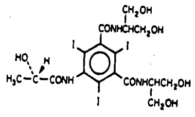
Physicochemical properties: White, odorless crystalline powder. Very soluble in water and methanol. Practically insoluble in chloroform.
The osmolality of iopamidol somewhat exceeds that of cerebrospinal fluid (approximately 301 mOsm/kg water) and of plasma (approximately 285 mOsm/kg water) but is substantially lower than that of conventional ionic contrast agents of equivalent iodine concentration.
| Parameter | Iopamidol Concentration | ||
| 41% | 61% | 76% | |
| Concentration mgI/mL | 200 | 300 | 370 |
| Osmolality @ 37ºC (mOsm/kg water) | 413 | 616 | 796 |
Viscosity (cP) @ 37ºC @ 20ºC | 2.0 3.3 | 4.7 8.8 | 9.4 20.9 |
| Specific Gravity @ 37ºC | 1.216 | 1.328 | 1.405 |
Product Characteristics:
Composition: ISOVUE (iopamidol) is a sterile solution of iopamidol in water for injection. The solution also contains tromethamine (0.1%) as a buffer and calcium disodium edetate (EDTA) (range 0.017-0.048%) as a sequestering agent but contains no preservative. The pH has been adjusted to 6.5-7.5 with hydrochloric acid.
14 CLINICAL TRIALS
14.1 Trial Design and Study Demographics
ISOVUE (iopamidol) has been evaluated in a clinical trial with 76 pediatric patients undergoing angiocardiography.
In a prospective clinical study involving patients undergoing an intravenous procedure, 169,284 patients received high osmolar ionic contrast media.
Another group of 168,363 patients received non-ionic low osmolar contrast media, mostly iopamidol.
15 MICROBIOLOGY
No microbiological information is required for this drug product.
16 NON-CLINICAL TOXICOLOGY
General Toxicology:
Acute Toxicity
| Route of Administration | Species | Number of Animals | Sex / Number Per Group | LD50 (g/kg) | Toxic Signs | ||||
| Iopamidol | iodine | ||||||||
| Intravenous | mouse rat rabbit dog | 40 32 18 20 | (4♀, 4♂) (4♀, 4♂) (3♀, 3♂) (2♀, 2♂) | 44.5 28.2 19.6 34.7 | 21.8 13.8 9.6 17.0 | Dyspnea, frequent and laborious turning, and tonic and clonic convulsions. Symptoms observed within a few hours of injection. Absent at 24 hours. Profound prostration and death. Deaths occurred within 24 hours in mice; within 1 hour in rats; after 5 minutes in rabbits and between 1-3 days in dogs | |||
| Intraperitoneal | mouse adult rats newborn rats young rats | 32 32 120 56 | (4♀, 4♂) (4♀, 4♂) (10♀, 10♂) (5♀, 5♂) | 28.0 24.3 72.5 29.4 | 13.7 11.9 35.5 14.4 | Reduced spontaneous motility, loss of grip strength, convulsions, loss of muscle tone, weight loss, bradypnea, dyspnea, collapse, prostration and death. Symptoms several hours after dosing to 24 hours. Deaths after ½ to 4 days in adult animals, 1 hours to 5 days in neonates; and 5 hours to 3 days in young rats. | |||
| Oral | mouse rat | 8 8 | (4♀, 4♂) (4♀, 4♂) | >49 >49 | >24 >24 | No deaths or overt signs of toxicity | |||
| Intracerebral | mouse rat | n/a n/a | (4♀, 4♂) (4♀, 4♂) | 3.02 >0.24 | 1.48 >0.12 | Prostration, dyspnea and tonoclonic convulsions (3-4 hours). Deaths within 48 hours. Prostration, dyspnea and reduction of motility. No deaths. | |||
| Intracisternal | Rat rabbit | n/a n/a | (4♀, 4♂) (4♀, 4♂) | >0.24 > 0.82 | >0.12 >0.40 | Prostration and dyspnea after 2 hours. No symptoms after 24 hours. No deaths. Dyspnea, tonoclonic convulsions, tonic stretching and loss of righting reflex (3-4 hours). | |||
n/a = not available
Subacute Toxicity
| Route of Administration | Species | Number of Animals | Daily Dose (mL/kg) | Duration of Study (days) | Toxic Signs |
| Intravenous | rat | 60 (30♀, 30♂) | 2.5, 5.0,7.5 (400 mg I/mL solution) | 28 | No body-weight changes and no clinical signs of toxicity. Reduction in urine specific gravity, reduction in hemoglobin, increase in partial thromboplastin time, decrease or increase in plasma fibrinogen, decrease in serum albumin, albumin globulin ratio, BUN, total bilirubin, SGOT, SGPT, phosphorus and increase or decrease in chloride concentration, thyroid activity. |
| Intravenous | rat | 60 (30♀, 30♂) | 4,8,16 (40 mg I/mL solution) | 28 | Blue coloration of the skin, diuresis, diarrhea, mechanical vein damage, reduced food intake, decreased body-weight, less efficient utilization of food, excretion of a smaller volume of a more concentrated and acidic urine, increase in SGPT values, increase in liver, kidneys and thyroid weights, degenerative changes in liver and kidney, thyroid activation. |
| Intravenous | dog | 18 (9♀, 9♂) | 2.5, 5.0, 10.0 (400 mg I/mL solution) | 28 | No body-weight changes. Occasional emesis, increase in urine specific gravity, elevation of SGPT, increase or decrease of plasma chloride, renal lesions (swelling and dissociation of the tubular epithelium, presence of pyknotic nuclei and lympho-histoid granulomas), thyroid activation. |
| Intrathecal | rabbit | 30 (15♀, 15♂) | 0.17, 0.42, 1.0 (300 mg I/mL solution) | 28 | No body-weight changes and no clinical signs of toxicity. Increase in bilirubin and total protein, hyperglycemia, increase or decrease in SGPT values, decrease in concentration of potassium and increase in concentration of calcium, increase in prothrombin time, increase in kidney weights, decrease in spleen weight, CSF: increase in glucose and GOT, decrease in magnesium, increase or decrease in chloride, decrease in potassium concentrations, increase in WBC count. |
| Intracisternal | dog | 18 (9♀, 9♂) | 0.13, 0.30, 0.83 (300 mg I/mL solution) | 28 | No body-weight changes and no clinical signs of toxicity. Increase in partial thromboplastin time and plasma prothrombin time, decrease in concentration of plasma potassium, presence of granulomas in the kidneys, edema and ischemia of the medulla oblongata, CSF: increase in total protein, white blood cells and GOT. |
Genotoxicity:
In the ames test, iopamidol at doses of 10-1000 μg/test plate did not induce a significant number of reversions in the five strains of Salmonella typhimurum used, whether in the absence or presence of mouse liver microsomal enzymes.
At a concentration of 1000 ppm, iopamidol did not induce a significant increase of genetic conversions in Saccharomyces cerevisiae, when tested in vitro in the presence or absence of mouse liver microsomal enzymes.
Iopamidol did not induce significant increases of mutation rates in the yeast strain Schizosaccharomyces pombe either in vitro (in the presence or absence of mouse liver microsomal enzymes) at a concentration of 1000ppm, or in vivo (mouse) at a dose of 5g/kg.
Reproductive and Developmental Toxicology:
In one study, iopamidol (400 mg I/mL solution) was administered intravenously to groups of 12 male rats in single daily doses of 1.5, 3.7, or 10 mL/kg for 9 weeks before being paired with females that had been dosed similarly for 2 weeks. After mating, dosing was continued until Day 13 of gestation or until the day of delivery. No effects on gonadal function, estrous cycle, mating behaviour, conception, early gestation, organogenesis, late gestation, parturition, lactation and growth of offspring and subsequent fertility were found. The pregnancy index (percent of pregnant females delivering live fetuses) was 100% in controls and 92.9, 100 and 87.5% in the respective dose groups. The percent of viable young at 4 days after delivery was 96.7% for controls and 91.9, 86.9 and 82.7% respectively for the treated groups.
In another study, iopamidol (400 mg I/mL solution), was administered intravenously to groups of 15 pregnant rats from day 6 through 16 of gestation at 0, 1.5, 3.75, and 10 mL/kg. No teratogenic effects were observed in the offspring obtained by cesarian section at day 21. Resorptions were less numerous in the low and middle dose groups (0.3/litter) than in controls (0.4/litter) and slightly higher in the high dose group (0.5/litter). Post-implantation losses followed a similar pattern (2.7, 2.4 and 10.3% loss vs 5% in controls). The mean number of live fetuses per litter at the low and middle doses was higher than in controls (11.9 and 12.7 vs 11.6). Mean litter weights were 3.9g for the low dose, 3.7g for the intermediate dose and 3.8g for controls. At the high dose, there were 10.9 live fetuses/litter and mean fetal weight of 3.6g.
In rabbits, iopamidol (400 mg I/mL) was given intravenously to groups of 15 pregnant females at daily doses of 0.7, 2.0 or 5.0 mL/kg from day 6 through 18 of gestation. A control group received similar injections of saline. At day 30, fetuses were delivered by cesarian section and external, internal and skeletal morphology was examined. One mid-dose rabbit showed multiple malformations and another had ascites and a cyst of the liver. The incidence of 2 malformations in 282 fetuses was considered less than the spontaneous rate in this species.
Groups of 20 pregnant rats received iopamidol (370 mg I/mL) intravenously in single daily doses of 0, 2.8, 5.6 and 11.1 mL/kg from Day 15 of gestation through Day 21 of lactation. There was no evidence of detrimental effects on the test animals based on indices of gestation, length of gestation, parturition, lactation, maternal mortality and litter size, as well as appearance, behaviour, growth and viability of offspring. Only the mean number of live fetuses at 4 days postpartum was slightly reduced in the high dose group (7.7±6.8) compared to controls (9.8 ± 2.2).