


Product Monograph - Methotrexate Sodium Injection USP
ACTION AND CLINICAL PHARMACOLOGY
Methotrexate (MTX), the 3-amino N10- methyl analog of folic acid, is a potent folate antagonist, and is cell cycle specific in the S-phase. MTX blocks the reduction of dihydrofolate to the active tetrahydrofolate (FH4) which functions as a cofactor in one-carbon (C1) transfer reactions (carrier), crucial for the synthesis of purine nucleotides and thymidylate. MTX competes with folate, and acts as an antimetabolite by relatively irreversible intracellular binding to the enzyme dihydrofolate reductase
(DHFR).
Inhibition of the formation of FH4 produces an acute intracellular deficiency of folate coenzymes and a vast accumulation of the toxic substrate FH2 polyglutamates. The C1 carrier reactions cease, subsequently interrupting the synthesis of DNA and RNA. Polyglutamate derivatives, increasing as a function of drug concentration and time of exposure, occur through extensive intracellular metabolism of MTX, extending the cytotoxicity of MTX to the cells in the S-phase. Protein synthesis is also inhibited, since reduced folates are cofactors in the conversion of glycine to serine, and homocysteine to methionine. Decreased ability to synthesize MTX polyglutamates, DHFR gene amplification leading to overproduction of the target enzyme, decreased or impaired membrane transport of MTX into cells, and decreased affinity for the MTX binding site due to production of altered forms of DHFR, are the four biochemical manifestations implicated in the mechanism of resistance or decreased sensitivity of tumor cells to MTX.
Drug concentrations in excess of 10-8M may be required to inhibit DNA synthesis in resistant cells.
Due to limited lipid solubility of MTX, and folic acid as well as many of its analogs being very polar, diffusion across the blood-brain barrier is poor; however intrathecal administration of MTX passes significantly into the systemic circulation. MTX is readily absorbed from the gastrointestinal tract at oral doses of < 20 mg/m2; however, larger doses are usually administered intravenously. I.V. infusions of 15 to 30 g/m2 MTX are required to achieve CSF drug levels that approximate those of 12 mg intrathecal
MTX therapy. MTX concentrations in CSF are only about 3% of those in the systemic circulation at steady state.9,10,17,45,82
PHARMACOKINETICS41,5,52,60,70,22,27,49,54,13,67,42,43,45,75
Absorption: Low doses of twice weekly 30 mg/m2 oral MTX were found equally as effective as equimolar doses of parenteral MTX. Peak plasma concentrations after oral MTX vary from
1-10 :M, while high-dose (HD) MTX infusions in excess of 1000 mg/m2 vary from 0.1-1 mM. After lumbar injection of 12 mg/m2 (prophylaxis) MTX, there was an initial rapid redistribution of the drug, followed by a biphasic decay curve with half-lives of 4.5 -14 hours. A mean peak MTX concentration of
2 X 10⁻⁷ M was found 3 to 12 hours after intrathecal injection, which disappeared biexponentially with half-lives of 5.5-24 hours. Intravenous injection of Methotrexate Sodium Injection USP (MTX)
disappears from plasma in a triphasic fashion; namely, the rapid distributive phase (t1/2% = 2-8 min.), the second or renal clearance phase (t1/2$ = 0.9-2 hours), and the final or gradual MTX cellular release phase
(t1/2( = 5.3-11 hours). Plasma levels are higher after the first 12 hours of a 24-hour infusion, than by bolus administration. The systemic bioavailability for oral and intramuscular administration83 was reported as
36±10% and 93±14%, respectively.
Distribution: Plasma protein binding is reported to be approximately 35-50%, with the major binding component being albumin. Levels of MTX over 50:mol/L serum result in saturation of protein binding capacity, leading to increased quantities of unbound drug. Presence of other organic acids may also reduce plasma protein binding capacity, resulting in increased amounts of extracellular MTX. MTX is rapidly distributed into liver, kidneys, skin, intestinal mucosa, and bile. Distribution into brain, fat, and skeletal muscle is minimal. Bile may contain up to 20% of the administered dose. MTX distribution into interstitial fluid spaces (CSF, pleural and peritoneal cavities) occurs slowly by passive transport;
however, if such spaces are expanded by ascites or pleural effusion, they may act as reservoirs, slowly releasing MTX back to plasma, resulting in increased drug exposure time and toxicity. Small quantities of
MTX are found in breast milk 44 and saliva 41.
Metabolism and Elimination: Systemic metabolism seems to play a minor role; however,
7-hydroxymethotrexate, reported to be potentially nephrotoxic, and 2.4-diamino-N10-methylpteroic acid, believed to be formed during enterohepatic metabolism, are metabolites accumulating after HD MTX.
Intestinal bacterial carbopeptidases metabolize MTX to 2.4-diamino-n10-methylpteroic acid by cleaving glutamate residues.
I.V. MTX Sodium Injection USP is predominantly eliminated through renal excretion through both active tubular secretion and glomerular filtration. Within 8-12 hours (i.v.), 45%, and within 48 hours approximately 90% of intact MTX is found in urine. Fecal excretion, probably through the biliary tract, does not exceed 1-2%. Renal clearance (n=22) after i.v. 30 mg/m2 MTX was calculated to the mean value of 78 ± 4.9 mL/min, and the pattern of urinary excretion paralleled plasma concentration decay.41
Children:18,25 Urinary excretion was faster in young children at all doses. Younger patients tolerated HD
MTX (5-250 mg/Kg) with citrovorum factor (CF) better than older patients. Extensive intra and interpatient variability in both children and adults must be considered.
INDICATIONS AND CLINICAL USE
Methotrexate Sodium Injection USP(MTX) is used in the treatment of neoplastic diseases:
- Choriocarcinoma, chorioadenoma destruens and/or hydatidiform mole: MTX alone,50a,40,58 or MTX combination chemotherapy.72,39
Intermediate or high grade Non-Hodgkin's Lymphoma (NHL): MTX as part of ProMACE-
CytaBOM84, ProMACE-MOPP84, or Magrath protocol85 (Schedule and dose variations of
ACOMLA+Pred for patients aged 2-35 years).
- Breast Cancer: MTX as part of CMF86.
- Acute Lymphoblastic Leukemia (ALL): MTX maintenance.47,55,56,35
- Head and Neck Cancer: MTX as part of combination chemotherapy.
- Gastric Cancer: FAM vs. high-dose MTX as part of FAMTX88.
- Metastasis of unknown origin (MUO Syndrome).
- Osteogenic Sarcoma: high-dose adjuvant MTX/CFR90.
- Bladder Cancer (advanced): MTX as part of M-VAC91.
- Leptomeningeal spread of malignancies (carcinomatosis, leukemia, lymphoma): MTX as single treatment or alternating with Ara-C and/or hydrocortisone92.
CONTRAINDICATIONS
Methotrexate Sodium Injection USP (MTX) is contraindicated in:
- 1.
Pregnancy: MTX has caused fetal deaths and congenital abnormalities,76,81 and small quantities of
MTX are found in breast milk.44
- 2. Presence of preexisting blood dyscrasia, such as bone marrow hypoplasia, leukopenia,81 thrombocytopenia,81 and anemia.81
- 3. Renal disease.16
- 4. Liver disease including cirrhosis, recent or active hepatitis and fibrosis.38,81
- 5. Active infectious disease.81
- 6. Active peptic ulcer.81
- 7. Ulcerative colitis.20
WARNINGS
METHOTREXATE SODIUM INJECTION USP (MTX) IS A POTENT CHEMOTHERAPEUTIC
AGENT WHICH SHOULD BE USED ONLY BY PHYSICIANS WHO ARE FAMILIAR WITH
THE DRUG, ITS ACTION, AND ITS SIDE EFFECTS.
MTX should be used with caution in patients with impaired bone marrow function65,53 and previous or concomitant wide-field radiotherapy. Methotrexate given concomitantly with radiotherapy may increase the risk of soft tissue necrosis and osteonecrosis.99
Patients with significant pleural effusion or ascites should have their fluid removed prior to therapy, in order to avoid third space accumulation of MTX and increased toxicity due to slow release of MTX from third space reservoirs.
Routine liver biopsies prior to MTX therapy are not indicated; however, liver function tests (LFTs)
should be determined prior to the initiation of therapy with MTX, and they should be regularly monitored throughout therapy.
The MTX toxicity for bone marrow and gut epithelium in humans is determined by a plasma concentration threshold of MTX > 2 x 10⁻⁸M, and a critical time threshold of about 42 hours, implicating the following:
- With HD-MTX (MTX > 1 g/m2), serum drug levels must be monitored.
- Drug levels exceeding the time threshold of about 42 hours may indicate significant toxicity.
Toxicity can be minimized by appropriate administration of folinic acid rescue
(leucovorin calcium CFR).
- It is imperative when using HD-MTX that urine be alkalinized, to prevent intratubular crystallization of MTX and its 7-hydroxy metabolite.61,93
Preservative-free MTX must be used for intrathecal, intraventricular, intraarterial and high dose MTX administration to eliminate the possibility of preservative toxicity.
At high and/or prolonged doses, MTX may be hepatotoxic,20 reported as atrophy, necrosis, cirrhosis, fatty degeneration, and periportal fibrosis. LFTs prior to and during therapy must be determined, since changes may occur without previous signs of gastrointestinal or hematologic toxicity.
Interruption due to toxicity of Methotrexate Sodium Injection USP (MTX) therapy is indicated in the following situations: ulcerative stomatitis,81 severe diarrhea, hemorrhagic enteritis, hepatic cirrhosis or fibrosis, impaired liver and/or renal function and inhibition of the hematopoietic system, and significant diffusional insufficiency of the lungs from MTX.
MTX may produce depression of the bone marrow, anemia, leukemia, thrombocytopenia, and bleeding.65,53
In women of childbearing potential and men capable of inducing conception, adequate nonhormonal contraception is strongly recommended during MTX therapy and for a period of at least 12 weeks after therapy since the risk for genetic abnormalities and teratogenesis may persist and possibly delay the new growth of normal germinal cells.65
MTX in the treatment of psoriasis must be restricted to only the recalcitrant and disabling form, diagnosed as such following dermatologic consultation. Deaths, probably the result of hypersensitivity reactions, have been reported in this therapy group.64,65
PRECAUTIONS
General: Methotrexate Sodium Injection USP (MTX) has a high potential for usually dose-related toxicity,65 and physicians must, therefore, be familiar with the drug.
Patients receiving MTX must be properly monitored for signs and symptoms of toxicity, since most adverse reactions are reversible if detected early enough. Reduction or discontinuance of the drug is required, and appropriate corrective measures have to be initiated under these circumstances. If necessary, this could include the use of leucovorin calcium and/or acute, intermittent hemodialysis with a high-flux dialyzer 100 (see SYMPTOMS AND TREATMENT OF OVERDOSAGE). General caution as well as alertness to possible recurrence of toxicity is required, if Methotrexate Sodium Injection
USP (MTX) is to be restarted. MTX chemotherapy requires pretreatment and periodic hematologic evaluations, to control possible MTX - related hematopoietic suppressive effects.
MTX administration should be discontinued with the occurrence of an unexpected large drop in white blood cell count, and appropriate therapy be instituted.
MTX should not, or only be used with utmost caution in patients with malignant disease in the presence of preexisting bone marrow hypoplasia leukopenia, thrombocytopenia or anemia.74
The concomitant use of non-steroidal anti-inflammatory drugs, including aspirin, may potentiate MTX toxicity, and may lead to severe adverse reactions including death.20,73
MTX therapy-induced occurrence of possibly severe leukopenia may lead to bacterial infection and nadir fever. In such case, MTX therapy should be interrupted and appropriate antibiotic treatment be instituted.
Care is necessary, when packed red cells are given to patients on extended (24-hour) MTX infusions.
Accumulation of MTX in red blood cells, and its slow elimination from these cells leads to increased drug toxicity.79a
The possible immunosuppressant action of MTX should be taken into consideration, when considering this drug for treatment in patients where immune responses are important or essential.65
Because MTX is primarily excreted by the kidney, impaired renal function can lead to drug accumulation with resultant toxicity and/or additional renal damage.74 Renal disease is, therefore, a contraindication to
MTX therapy.16 Monitoring of renal parameters during therapy is strongly recommended.
Use in the Elderly: Because of possibly reduced folate stores, reduced renal and/or hepatic function in this patient population, caution must be observed. Monitoring of renal and hepatic function is of utmost importance.
Use in children: Caution should be used in neonates and infants, due to reduced renal and hepatic function. Monitoring these functions are of utmost importance.
Use in obstetrics: (See WARNINGS). Menstrual function impairment has been reported.76,81
Nursing Mothers: MTX is excreted in breast milk.44 Therefore, breast-feeding is contraindicated. Patients with Special Diseases and Conditions: MTX must be used with extreme caution, in immunosuppressed patients in the presence of infections, live virus vaccination, peptic ulcer, ulcerative colitis, and general debilitation. For young children and geriatric patients, see appropriate sections.
Drug Interactions95,96: Serious (life-threatening) drug interactions have been reported in patients receiving MTX and various NSAIDs,20,73 including Aspirin96, due to reduced clearance.
Concurrent administration of intrathecal MTX with acyclovir may result in neurological abnormalities;
use with caution.
An increased risk of hepatotoxicity has been reported when the retinoic acid analog etretinate81 and other potentially hepatotoxic agents (e.g. leflunomide, azathioprine, sulfasalazine, retinoids) are given concurrently with methotrexate. Therefore, patients receiving concomitant therapy with methotrexate and other potentially hepatotoxic agents should be closely monitored for possible increased risk of hepatotoxicity.
Oral neomycin may decrease absorption of oral MTX.96 Polymyxin B, Nystatin and vancomycin may also decrease the absorption of MTX96. Kanamycin96 may increase the absorption of MTX, possibly requiring reduction of the MTX dose. Probenecid60,95,96 appears to inhibit the active renal tubular secretion of MTX, markedly increasing serum MTX concentrations. If this combination has to be used, monitoring serum MTX levels is a priority. Nephrotoxic drugs such as aminoglycosides95, amphotericin
B and cyclosporin could theoretically increase MTX toxicity by decreasing its elimination. Drugs known to bind to plasma proteins such as salicylates, sulfonamides, tetracyclines, chloramphenicol,
PABA, and phenytoin may displace MTX from plasma protein, resulting in increased toxicity.
Concurrent administration of these drugs is not recommended;5,22,24,81,95,96 however, if used in combination, monitoring of serum MTX levels is a priority.
Allopurinol concurrent with MTX may increase possible MTX-induced bone marrow depression.34
MTX-concurrent anticoagulants (coumarin-or indanediones-derivative). MTX may increase anticoagulant activity and/or increase the risk of hemorrhage as a result of decreased hepatic synthesis of procoagulant factors, and interference with platelet formation.
The concomitant use of hepatotoxic drugs and alcohol must be avoided.65,81
Vitamin preparations containing folic acid or its derivatives may alter responses to MTX.74
Concurrent administration of high cumulative cisplatin dosage with MTX significantly diminished MTX excretion. Increased folinic acid doses were required to prevent serious toxicity.19
MTX has been reported to slow the biotransformation of concurrent cyclophosphamide.57
Theophylline concentrations should be monitored in patients concurrently receiving low-dose MTX, to avoid the possibility of theophylline toxicity94.
Methotrexate given concomitantly with radiotherapy may increase the risk of soft tissue necrosis and osteonecrosis.99
With all drug-drug and drug-disease interactions, patient monitoring during therapy must be stressed.
Laboratory Tests: Renal and hepatic function, as well as blood elements should be assessed by history, physical examination, and laboratory tests such as CBC, urinalysis, serum creatinine, SGOT, and chest X-ray. These tests are to be made before MTX treatment, periodically during MTX therapy, and before reinstituting MTX therapy after interruption (toxicity or rest period).
Information for the patient: Patients should be fully informed as to the risk of fatal or severe toxic reactions associated with MTX therapy, to be administered under the physicians constant supervision.
Use of alcoholic beverages during MTX therapy must be denied. Any use or intended use of prescription or non-prescription drugs must be reported. In women of childbearing potential, and men capable of inducing conception, adequate nonhormonal contraception is strongly recommended during MTX therapy, and for a period of at least 12 weeks after therapy. Oral and/or dental hygiene must be enforced.
ADVERSE REACTIONS
Many side effects of antineoplastic therapy are unavoidable since they represent the drug's pharmacologic action. Early signs of Methotrexate Sodium Injection USP (MTX) toxicity include leucopenia, thrombocytopenia, anemia, mouth ulceration, stomatitis, diarrhea, nausea/vomiting, abdominal distress81 and adverse reactions of lungs to MTX. Also reported were increased fatigue, chills, fever, dizziness, and decreased resistance to infection. In general, incidence and severity of side effects may be considered dose-related.65,68,81
Skin: Erythematous rashes, pruritus, urticaria, photosensitivity, depigmentation, alopecia, ecchymosis, telangiectasia, acne, and furunculosis. Rarely, painful psoriatic plaque erosions may appear.101 Lesions of psoriasis may be aggravated by MTX-concomitant exposure to UV radiation.65 The incidence of loss of hair is less frequent with MTX than with combination chemotherapy.
Blood: Bone marrow depression, leukopenia, thrombocytopenia, anemia, hypogammaglobulinemia, hemorrhage from various sites, and septicemia.53,65,76 NOTE: With leukopenia and thrombocytopenia, the nadir of the leukocyte and platelet counts occurs after 7-10 days, with recovery 7 days later.
Gastrointestinal: Gingivitis, pharyngitis, stomatitis, anorexia, nausea/vomiting, diarrhea, hematemesis, melena, G.I. ulceration including bleeding and in some cases intestinal perforation, enteritis, hepatic toxicity with acute liver atrophy, necrosis, fatty degeneration, periportal fibrosis or cirrhosis.53,76,81 CNS: After intrathecal administration with MTX, cases of arachnoiditis with headaches, drowsiness, blurred vision, aphasia, hemiparesis, cranial nerve paresis and convulsions have been reported as well as cases with Guillain-Barre syndrome and necrotizing encephalitis.3,14,40,65,76
Urogenital System: Renal failure, azotemia, hyperuricemia, cystitis, hematuria, cutaneous vasculitis, severe nephropathy, defective oogenesis or spermatogenesis, transient oligospermia, menstrual dysfunction, infertility, abortion, and fetal defects.65
Other Reactions: related to or attributed to the use of MTX, such as pneumonitis,46,81 asthma, metabolic changes, precipitating diabetes, soft tissue necrosis99, osteonecrosis99, osteoporotic effects63 and sudden death have been reported. Although, not completely explained as yet, the sudden death would appear to point towards the possibility of hypersensitivity reactions.
SYMPTOMS AND TREATMENT OF OVERDOSAGE
Dosage of MTX should be discontinued or reduced as soon as ulceration, bleeding, diarrhea, or marked depression of the hematopoietic system becomes apparent.30 Leucovorin (calcium leucovorin, folinic acid, citrovorum factor),12,23,30 because of its ability to bypass the effects of Methotrexate Sodium Injection USP
(MTX), neutralizes the immediate toxic effects of MTX. Leucovorin calcium is administered as "rescue"
during high-dose HD-MTX or in MTX overdosage situations, to prevent the associated hematologic and gastrointestinal toxicity.
HD-MTX therapy should only be initiated, when leucovorin calcium is physically present and ready to be administered. In general, when overdose is apparent or suspected, a leucovorin calcium dose, within 1 hour of event, capable of producing a blood concentration $ the MTX blood concentration, is required.30 Fifteen to 25 mg/m2 leucovorin produce peak plasma concentrations of . 1 X 10⁻⁶M.
Leucovorin, in general, is continued until plasma MTX concentrations have reached non-toxic (<5 x 10-
8M) levels. Calcium leucovorin maybe administered by i.v. infusion in doses up to 75 mg within 12 hours, followed by 12 mg i.m. every six hours for 4 doses.10,12 Where average doses of MTX appear to have an adverse effect, 2-4 mL (6-12 mg) of leucovorin calcium may be given i.m. every 6 hours for four doses.48
Use of leucovorin calcium after an hour's delay is much less effective.
Effective clearance of methotrexate has been reported with acute, intermittent hemodialysis using a high-flux dialyzer.100
DOSAGE AND ADMINISTRATION
ANTINEOPLASTIC CHEMOTHERAPY:
General: Methotrexate Sodium Injection USP(MTX) may be given by i.v., i.m., intrathecal, intraventricular (best via an Ommaya Reservoir) or intra-arterial route. It is recommended that initial treatment take place in a hospital setting. As guideline, a ratio of 1:30 (range 1:20 to 1:40 by age and body build) is suggested for the conversion of mg/Kg body weight to mg/m2 body surface area. For intrathecal, intraventricular (via Ommaya Reservoir), intraarterial and high dose Methotrexate (more than
1 gm of MTX), preservative free MTX should be utilized. It is important that physicians familiarize themselves with constantly new advances in the role of low-dose, medium-dose, and high-dose MTX with and without CFR as part of combination chemotherapy regimen.
Choriocarcinoma, chorioadenoma destruens and/or hydatidiform mole: MTX is administered i.m. in doses of 15 to 30 mg/qd x 5. Such courses are usually repeated 3 to 5 times with rest periods of one or more weeks between courses, as required, or until toxic symptoms subside. The effectiveness of therapy is ordinarily evaluated through 24-hour quantitative analysis of urinary human chorionic gonadotropin
(hCG), which should return to normal or <0.50 IU/24h. This, usually occurs after the third or fourth course, to be followed by a complete resolution of measurable lesions in 4-6 weeks. Thereafter, one or two additional courses of MTX after normalization of hCG are recommended. Before each course of
MTX or combination regimen, careful clinical assessment is essential.4,40,48,50a,72 Since hydatidiform mole may precede choriocarcinoma, prophylactic chemotherapy with MTX is recommended. Chorioadenoma destruens is considered to be an invasive form of hydatidiform mole. MTX dosage is similar to that recommended for choriocarcinoma.
Intermediate or high grade NHL84,85: ProMACE-MOPP(P-M) and ProMACE-CytaBOM(P-C) as alternating half-cycle regimen have high CR rates. P-C, because of its unacceptable incidence of pneumocystis toxicity has in all patients been supplemented with prophylactic trimethoprim-sulfamethoxazole (co-trimoxazole, 1 double-strength tablet b.i.d.). This interaction may cause a >60% increase in systemic exposure to MTX in children.27 P-M consists of 650 mg/m2 (day 1) i.v.
cyclophosphamide, 120 mg/m2 (day 1) i.v. etoposide, and 25 mg/m2 (day 1) i.v. doxorubicin. On days 1 -
14 60 mg/m2 p.o. prednisone q.d. 6 mg/m2 (day 8) i.v. mechlorethamine, 1.4 mg/m2 (day 8) i.v.
vincristine. On days 8-14, 100 mg/m2 p.o. procarbazine q.d., 500 mg/m2(day 15) i.v. 12-hour infusion of
MTX, and on day 15 and every 6 hours for four doses 50 mg/m2 i.v., leucovorin rescue. No therapy was given on days 16-28. New cycle on day 29. P-C uses the same drugs on day 1 as P-M, followed by 300 mg/m2 (day 8) i.v. cytarabine, 5U/m2 (day 8) i.v. bleomycin, 1.4 mg/m2 (day 8) i.v. vincristine, 120 mg/m2
(day 8) i.v. MTX, and 25 mg/m2 p.o. leucovorin rescue every 6 hours for 4 doses beginning on day 9. The next cycle begins on day 22 for at least 6 cycles, 2 cycles beyond a complete remission (CR). The
Magrath Protocol (77-04) uses effective pulses of therapy, separated by as brief periods as possible. In each cycle, a prolonged MTX infusion was started close to the nadir of myelosuppression, resulting from the administration of cyclophosphamide and doxorubicin, and the subsequent cycle was begun as soon as bone marrow recovery had occurred. After the first 6 cycles, a new cycle was started every 28 days. 58 of
65 patients, aged 2 - 35 years with either undifferentiated (incl. Burkitt's) or lymphoblastic lymphomas, achieved CR (89%). The estimated survival (3 years) was 60%. Approximately 75% of all cycles were delivered at full drug dosage. Modification due to severe stomatitis was most frequently necessitated by
MTX and doxorubicin, and prednisone/prednisolone.85
Breast Cancer:86 A 21-day i.v. CMF adjuvant chemotherapy regimen, namely, 600 mg/m2 cyclophosphamide (every 21 days), 40 mg/m2 MTX (every 21 days), and 600 mg/m2 5-FU (every 21 days), shows that using the treatment program as described, one can administer a high rate of more than
85% doses of concurrent adjuvant chemotherapy to patients receiving irradiation for breast conservation management. Leukopenia occurs more often in this patient group, than in patients who have had modified radical mastectomy and undergo adjuvant chemotherapy. No life-threatening toxicity, or toxicity to skin and lungs was reported; however, short-lived ST-T abnormalities may develop from unavoidable small amount of radiation scatter to the heart.
ALL: Acute lymphoblastic leukemia in children and young adults is the most responsive to maintenance
MTX chemotherapy. In adult patients, clinical remission is more difficult to obtain and early relapse is common.1,2,7,12,18,25,29,35,39,47,55,56,59 MTX alone, or intrathecal MTX in combination with cranial radiation therapy , 6-MP and antifolic agents, appear to be a choice for securing CNS prophylaxis and interim maintenance of drug-induced remissions. Maintenance therapy is initiated with i.m. or p.o. doses of 30 mg/m2 MTX twice weekly, or as 2.5 mg/Kg intravenously every 14 days.
AGL: Acute granulocytic leukemia is rare in children, but common in adults; it responds poorly to chemotherapy, with short remissions and frequent relapses. Resistance to multiple chemotherapy develops rapidly.
Head and Neck Cancer: The following, are suggested combination therapy regimen containing MTX:
BMC equivalent to 10 U i.m. bleomycin (days 1,7,and 15), 40 mg/m2 i.m. MTX (days 1 and 15), and 50 mg/m2 i.v. cisplatin (day 4). Repeat the cycle every 21 days. CABO87 equivalent to 50 mg/m2 i.v. cisplatin
(day 4), 40 mg/m2 i.v. MTX (days 1 and 15), 10 U i.v. bleomycin (days 1,8, and 15), and 1 mg/m2 i.v.
vincristine (days 1,8, and 15). Courses are given every 3 weeks. VCR may be discontinued after 6 doses.
After 3 courses, weekly MTX maintenance is given.
Gastric Cancer: The doses of chemotherapy for FAMTX88, a regimen based upon biochemical modulation of fluorouracil with high remission rates in advanced gastric cancer, are: 1.5 g/m2 i.v. 5-fluorouracil (1 hour after the MTX infusion), 1.5 g/m2 i.v. MTX (on day 1), 15 mg/m2 p.o. leucovorin (24 hours after MTX) every 6 hours for 48 hours, and 30 mg/m2 i.v. doxorubicin (day 15). Repeat treatment every 4 weeks. This treatment was found better and less toxic than FAM (5-FU, adriamycin, and mitomycin).
Metastasis of Unknown Origin (MUO Syndrome): MTX as part of palliative CMF89. CMF, in a randomized trial of multiple-drug chemotherapy for metastatic adenocarcinoma of unknown primary, is described as 500 mg/m2 i.v. cyclophosphamide, 40 mg/m2 i.v. methotrexate, and 600 mg/m2 i.v. 5-FU on day 1 and then every 21 days. The vast majority of MUO syndrome-affected patients are in the median age range of 56-60 years, with males and females equally affected. Prognosis for most patients with
MUO syndrome is dismal.
Osteogenic Sarcoma:90 T-10, selected as preoperative chemotherapy for osteogenic sarcoma, and to be continued if greater than 90% of primary tumor revealed necrosis, is identified as 8-12 g/m2 i.v. MTX
(day 1,8,15,64,71,99,106. Delete after 12 or 16 doses), and 10-15 mg p.o. leucovorin every 6 hours for 10 doses starting 20 hours after each MTX dose. Other drugs in the T-10 regimen are 15 U/m2 i.v. bleomycin
(days 43 and 44), 600 mg/m2 i.v. cyclophosphamide (days 43 and 44), 600:g/m2 i.v. dactinomycin (days
43 and 44), and 30 mg/m2 i.v. doxorubicin (days 78-80).
Note: "Prior to MTX, the urine is alkalinized with i.v. sodium bicarbonate. Oral sodium bicarbonate was also given for 3 days following the MTX infusion at the dose of 2-3 mEq/Kg/24 hrs in divided doses."
On day 29, resection or amputation, and on day 116, endoprosthesis is performed.
Bladder Cancer (advanced): MTX as part of M-VAC91, identified as 30 mg/m2 i.v. MTX (day 1),
3 mg/m2 i.v. vinblastine (day 2), 30 mg/m2 i.v. doxorubicin (day 2), 70 mg/m2 i.v. cisplatin (day 2) after vigorous hydration, and concluded with repeating vinblastine and MTX on days 15 and 22. Cycles are repeated every 28 days, even if an interim dose is withheld because of myelosuppression or mucositis. Leptomeningeal spread of malignancies: (e.g.: carcinomatosis, leukemia, lymphoma). Single-agent
MTX treatment, or alternating treatment with Ara-C and /or hydrocortisone, may be given intrathecally
(eg: children92) as prophylactic or active chemotherapy. Usually, MTX is injected into the lumbar subarachnoid space in a dose of 6-12 mg/m2 twice weekly, until the CSF is free of leukemic cells; then,
MTX is given monthly. In case of prophylactic treatment, a total of 5 doses are given with or without cranial radiation therapy.
NOTE: Intrathecal MTX may be associated with acute toxicity, usually occurring after the 2nd to 4th dose, and chronic toxicity, most commonly seen with prior or concurrent radiation therapy. For patients whose lives would be jeopardized by myelosuppression, several low p.o. doses of 3-6 mg/m2 leucovorin every 6 hours should be administered 24 hours after the intrathecal MTX injection. When Ara - C is used intrathecally, doses range between 4.5 and 73 mg/m2 every 3 to 7 days. In adults, a dose of 50 mg/m2 Ara-
C yields peak CSF levels of 1 mM. Ara-C is reasonably well tolerated in reference92, intrathecal CNS-prophylactic therapy consisted of a maximum of 15 mg/m2 MTX, 15 mg/m2 hydrocortisone sodium succinate, and 30 mg/m2 cytosine arabinoside (Ara-C). These 3 drugs, in separate syringes, were injected sequentially during the same intrathecal treatment; the treatments being given weekly during the first month of remission, and once ever 2nd month thereafter, up to one year.
NOTE: For intrathecal injection, all drugs must be preservative-free.
Mycosis fungoides (MF): The classic presentation of this subset of low-grade T-cell lymphomas is its chronicity. Despite remarkable responsiveness to initial and salvage treatments, there is a great probability for relentless recurrence of MF. Patients with MF usually succumb to either end-organ failure or infection. The T-Cell Lymphoma skin classification for MF reads as T1=Limited plaques (<10% body surface area), T2=Generalized plaques (>10% body surface area), T3=Cutaneous tumors, and
T4=Generalized erythroderma. T1-T4 respond well to total-skin electron beam therapy (EBT) with or without topical treatment with PUVA (0.4-0.6 mg/Kg p.o. 8-methoxypsoralen followed after 2 hours by
UVA), HN2-mustard, topical 5-FU, topical BCNU (carmustine), topical retinoids or to topical combination therapy. Systemic therapy for MF produces higher CR rates, but is not curative. Systemic therapy includes 30 mg/m2 i.v. MTX once weekly, or 240 mg/m2 i.v. MTX with CFR. Polychemotherapy regimen, such as MOPP, CVP, CHOP or CAPO do not include MTX.
NOTE: When dealing with Non-Hodgkin's lymphoma (NHL), it is necessary prior to planning chemotherapy, to identify the type of lymphoma (Rappaport Classification) as nodular (N), or diffuse (D);
and have each subclassified as well, e.g. differentiated lymphocytic (NWDL or DWDL) etc. The term lymphosarcoma for the bulk of NHL has been abandoned. In addition, to facilitate effective chemotherapy regimen for NHL, the Working Formulation Classification (NCI: Cancer 1982, 49(10): 2112-2135)
should be consulted.
Lymphomas: All cases of Burkitt's lymphoma (BL) exhibit B-cell markers. The staging system for non-endemic (American) and endemic (African) BL78 is A=single extra-abdominal site, B=multiple extra-abdominal sites, C=intra-abdominal tumor, D=intra-abdominal tumor with involvement of multiple extra-abdominal sites, and AR=intra-abdominal tumor with >90% of tumor surgically resected. COM, COMP, and CHOMP are MTX-containing chemotherapy regimen used against non-endemic (American) BL.
CHOMP=1200 mg/m2 i.v. cyclophosphamide (day 1) 1.4 mg/m2 i.v. vincristine (day 1), 40 mg/m2 i.v.
doxorubicin, 40 mg/m2 i.v. prednisone (days 1-5),and 2.7 g/m2 i.v. MTX over 42 hours with CRF on day
- 10. Additional, intrathecal therapy has been found effective.
"IT" cycle I: Ara-C 30 mg/m2 (day 1-3, and 7), max. 12.5 mg/m2 MTX (day 10), 6-8 hours after commencement of systemic MTX infusion.
"IT" cycle II - III: Ara-C 30 mg/m2 (days 1 and 2), max. 12.5 mg/m2 MTX (day 3,10) 6-8 hours after commencement of systemic MTX infusion
"IT" cycle IV-VI (Max. 6 cycles for patients with stage A,B, and AR "BL"): Ara-C 45 mg/m2 (day 1), max. 12.5 mg/m2 MTX (day 10).
MTX-containing, abbreviated chemotherapy regimen used in NHL: Single-agent HDMTX as pulse infusions of 3-30 mg/Kg with and without CFR in ANN Arbor modified stage II, IV, and IVL childhood
NHL were effective (8/10 CR, 2/10PR)23 ACOMLA (MTX as 120 mg/m2 i.v.), COMLA (MTX as 120 mg/m2 i.v.), F-MACHOP (MTX as 500 mg/m2 i.v. cont. infusion), IMVP-16 (MTX as 30 mg/m2 i.m.),
LNH-80 (MTX as 15 mg IT), MACOP-B (MTX as 400 mg/m2 i.v.), M-BACOD (MTX as 3000 mg/m2 i.v.), m-BACOD(MTX as 200 mg/m2 i.v.), ProMACE-CytaBOM (MTX as 120 mg/m2 i.v.)84,
ProMACE-MOPP (MTX as 500 mg/m2 i.v. in flexitherapy)84, and the Magrath protocol85.
PSORIASIS CHEMOTHERAPY:53,64,66
NOTE: Methotrexate Sodium Injection USP (MTX) must only be used in patients with severe, recalcitrant and disabling disease that is inadequately controlled by other forms of treatment, including topical retinoids. Diagnosis must be confirmed through dermatologic consultation. MTX MUST ONLY BE USED BY PHYSICIANS WHO ARE FAMILIAR WITH MTX, ITS
ACTION, AND ITS SIDE EFFECTS. Due to the potency of, and the risks involved with MTX, the patient should be fully informed, and be under constant supervision of the physician.
Examples for MTX antipsoriasis therapy are: Psoriatic erythroderma, psoriatic arthritis, acute, localized, and extensive psoriasis.
Specific dosage recommendations (average 70 kg adult): An initial test dose of 5 to 10 mg MTX to detect any unusual sensitivity. Single oral doses range from 7.5 to 30 mg/wk with additional gradual increments of 2.5 to 5 mg/wk. Some patients may need a maximum 37.5 mg/wk. The range of single intramuscular or single rapid (push) intravenous dosage is 7.5 to 50 mg/wk, with some patients needing titration to maximum 75 mg/wk.
Slow intravenous drips of MTX should never be used. Divided oral doses are 2.5 to 5.0 mg at 12 hour intervals for three doses each week. Gradual increments of 2.5 mg/wk, not to exceed 30 mg/wk. CFR is used against toxicity. The goal is to achieve adequate control at the lowest possible MTX dose and the longest rest period between dosages.
The use of MTX may permit the return to conventional topical therapy, a course that should be encouraged.
Section WARNINGS and PRECAUTIONS must be consulted.
High-dose Methotrexate Sodium injection USP (HD-MTX)1,8,10,23,48,56,72 must be followed by CFR
(citrovorum factor rescue, calcium leucovorin or folinic acid rescue); therefore, CFR must physically be present and ready to be used, to avoid severe MTX toxicity with possibly fatal outcome. Administration of CRF should be consecutive to, rather than simultaneous with MTX, as not to interfere with the antineoplastic effects of MTX. Alkalinization of urine should be considered.
Parenteral products, prior to administration, need to be visually inspected for discoloration and for particulate matter.
SPECIAL INSTRUCTIONS:
METHOTREXATE SODIUM INJECTION USP (MTX) IS A POTENT DRUG THAT SHOULD
ONLY BE USED BY PHYSICIANS EXPERIENCED WITH CANCER CHEMOTHERAPEUTIC
DRUGS.
Safe handling of methotrexate sodium:
Work-practice guidelines11 for personnel dealing with cytotoxic antineoplastic preparations must be respected to minimize unnecessary exposure to MTX in physicians, nurses, pharmacists, health care workers, pharmaceutical technicians and patients. Protective wear includes unpowdered surgical latex gloves, to be changed frequently,50b 79b a protective lint-free, closed-front disposable gown with long sleeves and plastic cuffs, and a plastic face shield or splash goggles, should a biological safety cabinet #II
(vertical-flow containment hood) not be available.79b
Personnel, regularly handling these agents should have frequent hematologic examinations (CBC), and frequently be screened for urine mutagens. NOTE: If accidental methotrexate contamination of the eye occurs, the affected eye should immediately and thoroughly be washed with water, to prevent severe irritation and possible corneal ulceration.
Cytotoxic waste disposal of Methotrexate Sodium Injection USP (MTX) must be considered as
"hazardous waste"97, and all disposable materials having been in contact with the agent must either be deposited into a 2-mm thick polypropylene hospital trash bag (properly labelled), or otherwise be segregated and incinerated at above 1000°C.79b Sealed containers may explode. Spills:
Cleaning up immediately, and decontaminating areas of spills and breakages by experienced and well-protected personnel is of the utmost importance. Contaminated areas including hood interiors must have warning labels posted. Spill Kits, clearly labelled, must be easily accessible. It is suggested that spill kits include replacement hood filters, a respirator, chemical splash goggles, at least 2 pairs of gloves, at least 2 sheets (12" X 12") of absorbent materials, 250 mL and 1 L spill control pillows, an small scoop to collect glass fragments, and at least 2 large 2-mm thick polypropylene hospital trash bags, or other cytotoxic drug waste-disposal bags. Absorbents should be incinerable.79b
PHARMACEUTICAL INFORMATION
DRUG SUBSTANCE:
Proper Name: METHOTREXATE
Chemical Name: L-(+)-N-[p-[[(2,4-diamino-6 pteridinyl)methyl] methylamino]-glutamic acid.
Structural Formula: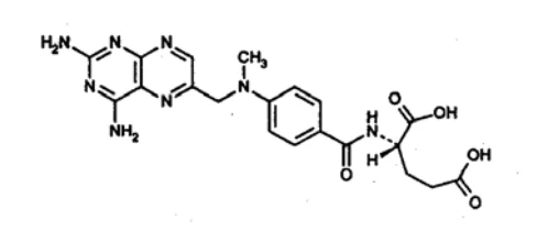
Molecular Formula: C20 H22 N8 O5
Molecular Weight: 454.44
PHYSICAL PROPERTIES
Physical form: Yellow to orange-brown crystalline powder.
Solubility: Soluble in mineral acids, dilute solutions of alkali and carbonates. Practically insoluble in water, alcohol, chloroform and ether.
pH values: 8.3 to 8.6 of a 0.25% aqueous solution.
pKa values: 2.15 and 3.8
COMPOSITION:
Each mL of Methotrexate Sodium Injection USP contains:
25 mg methotrexate, 4.9 mg sodium chloride and 4.4 mg sodium hydroxide in water for injection.
May also contain HCl for pH adjustment.
STABILITY AND STORAGE RECOMMENDATIONS:
Methotrexate Sodium Injection USP should be stored at 15-25oC, and be protected from light.
PARENTERAL PRODUCTS:
NOTE: Methotrexate sodium in contact with aluminum surfaces should be avoided, since there is a potential for formation of orange crystals after 36 hours at 24°C with protection from light.98
Parenteral products prior to administration, need to be visually inspected for discoloration and for particulate matter. All dosage forms represent a clear yellow solution.
Pharmacy Bulk Vials:
The contents are intended for use in a hospital, or a pharmacy with a recognized intravenous admixture program, and are restricted to the preparation of admixtures for infusion or, through a sterile transfer device for filling of empty sterile syringes. After employing a single puncture in a protected work area
(see SPECIAL INSTRUCTIONS), the unused portion should be safely discarded within 8 hours of withdrawal. Preservative-free solutions should be diluted immediately prior to use, and any unused portion be discarded. If stored for 24 hours at room temperature, and protected from light, a diluted solution of Methotrexate Sodium Injection USP (MTX) maintains approximately 90% of its label. Care must be taken to minimize the potential for introduction of microorganisms during manipulation in the hospital environment. It should be noted that when dispensed, the container or syringe (holding the individual dose prepared for administration to the patient) be enclosed in an over wrap bearing the statement: DO NOT REMOVE COVERING UNTIL MOMENT OF INJECTION.
AVAILABILITY OF DOSAGE FORMS
Methotrexate Sodium Injection USP is available as a 25 mg/ mL methotrexate sterile solution in 2 mL and 20 mL vials. All dosage forms are sterile, preservative-free and isotonic. The vials are single use vials. The 20 mL vial is a pharmacy bulk vial.
PRECLINICAL PHARMACOLOGY
Cellular transport: To prevent a cell from replicating, MTX (formerly amethopterin) must first cross the cell membrane. It has been shown that MTX uptake is an active process, and that this drug enters cells predominantly by using the C1 carrier system, responsible for transport of reduced folates. Once inside the cell, MTX rapidly binds to dihydrofolate reductase (DHFR).8a,13,67
Antitumor activity: The following relationship between the number of L-4946 (generalized leukemia)
cells inoculated (i.p.) in AK mice (n=10/experiment) and 100 days curability after 3 mg/Kg i.p. MTX was demonstrated:
| Leukemic cells | Control (days) | Life span (days) | Cure rate (%) |
1200000 120000 12000 1200 120 .12 | 13.4 16.4 | 30.8 - 35.3 39.6 | 0 8 82 96 100 100 |
In 8 other experiments in mice (n=10/experiment), see following table, the relationship between i.p. MTX dosage and increase in life span (days) against L-4946 leukemia was demonstrated as follows:32,33,69
| Exp. # | Control | 5.0 mg/Kg | 3.0 mg/Kg | 1.5 mg/Kg | 0.75 mg/Kg |
| 1 2 3 4 5 6 7 8 | 14.0 14.1 13.6 14.1 12.9 13.9 13.5 14.2 | 38.9 44.8* | 30.0 31.0 32.3 31.0 29.5 32.4 30.2 35.0 | 27.0 26.0 24.5 24.9 23.3 24.5 23.2 28.5 | 16.1 20.5 16.6 19.2 18.6 19.4 21.0 |
| Average | 13.8 | 41.9 | 31.4 | 25.2 | 18.7 |
*One of 10 mice survived 100 days Metabolism:76,62,58,54
Absorption: Plasma levels following i.v. administration of 0.2 mg/Kg 3H-methotrexate to one female beagle dog, and 0.3 mg/Kg 3H-methotrexate to each of two adult rhesus monkeys were similar, with a plasma t1/2 of 75 to 90 minutes. The volume of distribution in all 3 animals approximated total body fluid.37 The blood concentration of methotrexate in mice fell rapidly during the first 30 minutes following i.v. administration, being reduced to trace amounts in 3 to 4 hours. About 6% of the injected dose was found in the liver and kidneys 24 hours after injection.71
During 30 minutes i.v. infusion of 10 mg/Kg methotrexate into 4 New Zealand White rabbits, the plasma concentrations of methotrexate rose rapidly with a mean peak concentration (at time 0) of 48.1 :g/mL, and decreased poly-exponentially post infusion with a mean apparent terminal t1/2 of 3.26 ± 0.756 hours.
Thirty minutes after infusion, the mean AUC was 2360 ± 536 :g X min/mL.80
Distribution: In young adult male or female monkeys (n=19 %&) the following tissue uptake (:g/g) of
(3H)-methotrexate, after intraventricular 6 mg/m2 (1.69 mg/animal) (3H)-methotrexate in artificial CSF injection, was demonstrated:49
| Tissue | 20 min | 70 min | 130 min | 12 hrs | 24 hrs | 48 hrs |
Liver Spleen Kidney Intestine Duodenum Pancreas lung heart Cervical spinal cord Thoracic spinal cord Lumbar spinal cord Total Brain + spinal cord | 0.13 0.01 0.12 0.03 0.02 0.02 0.03 0.01 11.37 4.86 2.31 3.38 | 0.67 0.05 0.98 0.08 0.16 0.10 0.11 0.10 12.4 11.6 9.18 4.51 | 1.46 0.07 0.74 0.14 0.021 0.21 0.27 0.09 8.46 7.92 5.64 4.39 | 0.69 0.08 0.23 0.25 0.13 0.07 0.08 0.06 2.44 1.90 2.25 1.15 | 0.24 0.09 0.22 0.11 0.06 0.03 0.05 0.03 0.47 0.41 0.49 0.53 | 0.041 0.066 0.050 0.032 0.027 0.024 0.023 0.015 0.20 0.09 0.15 0.28 |
| Brain + spinal cord (intact methotrexate :g/mg) | 3.38 | 4.52 | 4.38 | 0.99 | 0.35 | 0.14 |
Conversion of methotrexate to non-methotrexate products was detected in the CSF between 12 and 24 hours after injection. Using radioimmunoassay, tissue distribution of MTX in rats (n=4/parameter), receiving either 30 mg/Kg i.v. bolus MTX or 30 mg/Kg drip infusion MTX, was determined at 60 and at
240 minutes after administration, respectively (ng/g tissue):
| Tissue54 | Bolus | Drip | ||
| 60 min. | 240 min. | 60 min. | 240 min. | |
Serum Kidney Gut Lung Liver Testis Muscle Fat Brain | 5094 7553 3307 1152 5704 937 200 1045 1865 | 334 3848 275 249 277 77 180 144 197 | 6570 13069 8712 2894 5483 1952 1715 1134 938 | 207 4266 279 116 1715 436 593 361 362 |
Elimination:62,37 The cumulative excretion in normal mice after 15 mg/Kg i.p. (3H)-MTX was at 4 hrs=54%, 8 HRS=56%, and at 24 hrs 61% in urine, and 35% in feces (96% total).
The cumulative excretion in normal and L-1210 tumor-bearing rats for 4,8,24, and 48 hours, respectively, was 31% and 34%, 32% and 38%, 37% and 50%, and 39% and 51% in urine. The respective fecal excretion for 24 hours was 29% and 41%, with total excretion, respectively, accounting for 82% and
84%. MTX is incompletely absorbed from the G.I. tract.
| Cumulative % Excretion of 3H-methotrexate | |||
| Parameter | Dog, bile fistula (0.5 mg/Kg i.v.) | Monkey, bile fistula (0.03 mg/Kg i.v.) | Monkey intact (0.3 mg/Kg i.v.) |
Urine 1 hr 6 hr 24 hr 48 hr Bile 1 hr 6 hr 24 hr Feces | 31 65 70 -- 2 7 -- -- | 46 62 73 74 <1 5 16 1 | 32 44 50 54 ------ -- |
| Total | 77% | 91% | 67% |
TOXICOLOGY
Among the LD50-tested rats and mice, only exceptional animals died within 72 hours after methotrexate
(MTX) administration; the majority of all deaths occurred between 3 and 7 days.
LD50 p.o. MTX 108±45 mg/Kg rats
LD50 i.p. MTX 6-25 mg/Kg rats
LD50 i.p./day x 5 MTX 5.6±1.7 mg/Kg rats
LD50 i.p. MTX 94±9 mg/Kg mice (&%)
LD50 i.p./day x 5 MTX 9.7±1.5 mg/Kg mice (&%)
LD50 i.p. MTX 95±12 mg/Kg mice (&)
LD50 i.p./day x 5 MTX 15±4 mg/Kg mice (&)
LD50 i.p. MTX 93±19 mg/Kg mice (%)
LD50 i.p./day x 5 MTX 6.5±1.6 mg/Kg mice (%)
In rats, the sex differences were similar to mice. dogs were usually sacrificed when severely intoxicated, and no attempt was made to establish an LD50. Of 2 dogs receiving single i.v. doses of 50 mg/Kg methotrexate (4-amino-N10-methyl- pteroylglutamic acid), 1 was dead on the third day, the other moribund after 5 days. Severe intoxication or death occurred in 3 of 11 dogs in 5 to 6 days after receiving i.m. 1.0 mg/Kg/day MTX, in 4 of 11 dogs in 10 to 11 days, after receiving i.m. 0.2 mg/Kg/day, and in the last 4 of 11 dogs in 14 to 37 days after receiving i.m. 0.05 mg/Kg/day MTX. After single intoxication doses in mice, rats, and dogs, no changes were noted during the first 24 hours, except a moderate weight loss in mice receiving the largest doses. During the second or third day, anorexia, diarrhoea, weight loss, lethargy, and weakness became apparent. These signs increased in severity until time of death. Feces in dogs became grossly bloody. Terminally, all animals had lost an average 20% of their body weight. The course of intoxication in animals receiving repeated doses was similar to that described; however, more delayed in onset.26
Toxicity data from mouse, rat and hamster, dog and monkey, and humans were quantitatively compared for the relationship between maximum tolerated dose (MTD) in dog, rhesus monkey, and man, versus
LD10 in mice, rats and hamster.
Amethopterin (i.p.)
(1-5 days)
LD10 Hamster 25 mg/Kg = 103 mg/m2
LD10 Swiss mouse 3.2 mg/Kg = 9.5 mg/m2
LD10 BDF1 mouse 5.2 mg/Kg = 16.0 mg/m2
LD10 rat 0.58 mg/Kg = 3.1 mg/m2
MTD monkey 3 mg/Kg = 35.0 mg/m2
MTD dog 0.12 mg/Kg = 2.0 mg/m2
MTD man 0.41 mg/Kg = 15.0 mg/m2
The investigators concluded that the experimental test systems used to evaluate the toxicities of potential anticancer drugs, correlate remarkably closely with the results in man.28
| Effect of Folic Acid (FA) and Dihydrofolate Reductase (DHFA) on Amethopterin (MTX) Toxicity in the Mouse. (For LD50 determinations: 6 groups of 6 mice each) | |||||
| Experiment I | Experiment II | ||||
| Treatment | Time (hr) | MTX LD50 (mg/Kg) | Treatment | Time (hr) | MTX LD50 (mg/Kg) |
none DHFA (25 mg/Kg) MTX (100 mg/Kg) MTX (100 mg/Kg) DHAF (25 mg/Kg) | -----1 -24 -24 -1 | 200 450 <30 180 ---- | none FA (25 mg/Kg) DHFA (25 mg/Kg) FA (25 mg/Kg) DHFA (25 mg/Kg) | -----1 -1 +1 +1 | 250 350 630 180 360 |
When rats were treated chronically with i.v. 3.4 to 10 mg/Kg MTX over 5 to 9 weeks, or initially i.p. 0.1 mg/kg/day x 5, a doubling of the dose every third week to a maximum 0.4 mg/Kg/day, the marrow appeared to be hypocellular. The thymus showed marked atrophy. There was significant anemia, and neutropenia leading to various forms of infections, as well as colonic ulceration in rats and dogs.
Carcinogenicity/Mutagenicity: Carcinogenicity studies in mice, rats, and hamsters were inconclusive.
Methotrexate was found to be mutagenic.
Reproduction and Teratology: In rats, receiving treatment with MTX for longer than 4 months, there was impaired spermatogenesis and seminiferous tubular atrophy in the testes. Intraperitoneal administration of 0.2 and 0.3 mg/Kg MTX to rats on day 9 of pregnancy was embryolethal in 63 and
84%, respectively. 19.2 mg/Kg i.v. MTX caused developmental toxicity in virgin rabbits; this toxicity was ameliorated by 75 mg/Kg leucovorin.6,21 In rats 0.2 and 0.3 mg/Kg MTX were teratogenic in 35% and
75%, respectively.
BIBLIOGRAPHY
- 1. Abramovitch M, Ochs J, Pui Ch-O et al. Efficacy of high-dose methotrexate in childhood acute lymphocytic leukemia: analysis by contemporary risk classification. Blood 1988;71(4):866-869.
- 2.
Acute Leukemia Group B. Acute lymphocytic leukemia in children. JAMA 1969;207(5):923-
928.
- 3. Asada Y, Kohga S, Sumiyoshi A et al. Disseminated necrotizing encephalopathy induced by methotrexate therapy alone. Acta Pathol Jpn 1988;38(10):1305-1312.
- 4. Ayhan A, Ergeneli MH, Yüze K et al. Effects of prophylactic chemotherapy for postmolar trophoblastic disease in patients with complete hydatidiform mole. Int J Gynecol Obstet 1990;32:39-41.
- 5.
Balis FM. Pharmacokinetic drug interactions of commonly used anticancer drugs. Clin
Pharmacokinet 1986;11:223-235.
- 6. Bantle, JA, Fort DJ et al. Further validation of FETAX: evaluation of the developmental toxicity of five known mammalian teratogens and non-teratogens. Drug Chem Toxicol 1990;13(4):267-282.
- 7.
Bell BA, Whitehead VM. Chemotherapy of childhood acute lymphoblastic leukemia. Dev
Pharmacol Ther 1986;9:145-170.
8a. Bertino JR. Towards improved selectivity in cancer chemotherapy. Cancer Res
1979;39(Feb):293-294.
8b. Bertino JR(Symposium ed.) Sequential methotrexate and 5-fluorouracil in the management of neoplastic disease. Semin Oncol 1983;10(suppl.2):1-38.
- 9.
Black DJ, Livingston RB. Antineoplastic drugs in 1990. A review (part I-II). Drugs 1990;39(4-
5):495-498, 664-673.
- 10.
Bleyer WA. The Clinical pharmacology of methotrexate. New applications of an old drug.
Cancer 1978;41(1):36-51.
- 11. Canadian Society of Hospital Pharmacists (CSHP): IX: Guidelines for handling and disposal of hazardous pharmaceuticals (including cytotoxic drugs). CSHP Ottawa, Canada 1991, pp. 1-27
- 12. Capizzi RL, De Conti RC et al. Methotrexate therapy of head and neck cancer; improvement of therapeutic index by the use of leucovorin "rescue". Cancer Res 1970;30(June):1782-1788.
- 13.
Chabner BA, Clendeninn N et al. Biochemistry of methotrexate. Progr Cancer Res Ther
1986;33:1-38.
- 14. Chessells JM, Cox TCS, Kendall B et al. Neurotoxicity in lymphoblastic leukemia: comparison of oral and intramuscular methotrexate and two doses of radiation. Arch Dis Childhood 1990;65:416-422.
- 15.
Cheung Y-W, Vishnuvajjala BR et al. Stability of cytarabine, methotrexate sodium and hydrocortisone sodium succinate admixtures.
Am J Hosp Pharm 1984;41(9):1802-1806.
- 16. Condit T, Chanes RE et al. Renal toxicity of methotrexate. Cancer 1969;23(1):126-131.
- 17.
Condit PT, Mead JAR. Further observations on the site of action of amethopterin. Biochem
Pharmacol 1963;12:94-96.
- 18. Craft AW, Rankin A, Aherne W. Methotrexate absorption in children with acute lymphoblastic leukemia. Cancer Treat Rep 1981;65 (Suppl.1):77-81.
- 19. Crom WR, Teresi ME et al. The intrapatient effect of cisplatin on the pharmacokinetics of high-dose methotrexate. Drug Intell Clin Pharm 1985;19:467.
- 20.
Daley HM, Scott GL et al. Methotrexate toxicity precipitated by azapropazone. Br J Dermatol
1986;114:733-735.
- 21. DeSesso JM, Goeringer GC. Amelioration by leucovorin of methotrexate developmental toxicity in rabbits. Teratology 1991;43:210-215.
- 22. Dixon RL, Henderson ES et al. Plasma protein binding of methotrexate and its displacement by various drugs. Fed Proc 1965;24:454, #1807
- 23. Djerassi I, Kim JS. Methotrexate and citrovorum factor rescue in the management of childhood lymphosarcoma and reticulum cell sarcoma (Non-Hodgkin's Lymphomas). Prolonged unmaintained remissions. Cancer 1976;38(3):1043-1051.
- 24.
Evans WE, Christensen ML. Drug interactions with methotrexate. A review. St. Jude Children's
Research Hospital, Memphis Tenn. Lederle Labs. Brochure (616-5) 1985;Aug:1-6.
- 25. Evans WE, Stewart CF, Hutson PR et al. Disposition of intermediate-dose methotrexate in children with acute lymphocytic leukemia. Drug Intell Clin Pharm 1982;16:839-842.
- 26. Ferguson FC Jr, Thiersch JB et al. The action of 4-amino-N10 methylpteroylglutamic acid in mice, rats, and dogs. J Pharmacol Exp Ther 1950;98:293-299.
- 27. Ferrazzini G, Klein J et al. Interaction between trimethoprim-sulfamethoxazole and methotrexate in children with leukemia. J Pediatr 1990;117(Nov):823-826.
- 28. Freireich EJ, Gehan EA, Rall DP et al. Quantitative comparison of toxicity of anticancer agents in mouse, rat, hamster, dog, monkey, and man. Cancer Chemother Rep 1966;50(4):219-244.
- 29.
Gardner NH. Acute lymphoblastic leukemia: problems, antileukemic agents and management. J
Am Med Wom Assoc (JAMWA) 1967;22:21-27.
- 30. Gauthier E, Gimonet JF, Piedbois P et al. Efficacité de l'hémodialyse dans un cas d'intoxication aiguë par le methotrexate. Presse Méd 1990;19(44):2023-2025.
- 31.
Goldin A. Studies with high-dose methotrexate. Historical background. Cancer Treat Rep
1978;62(2):307-312.
- 32.
Goldin A, Humphreys St R et al. Prolongation of the life span of mice with advanced leukemia
(L 1210) by treatment with halogenated derivatives of amethopterin. JNCI 1959;22(4):811-823.
- 33. Goldin A, Venditti JM et al. Comparison of relative effectiveness of folic acid congeners against advanced leukemia in mice. JNCI 1957(6):1133-1135.
- 34.
Grindey GB, Moran RG. Effects of allopurinol on therapeutic efficacy of methotrexate. Cancer
Res 1975;35(Jul):1702-1705.
- 35. Haaxma-Reiche H, Daenen S. Acute lymphoblastic leukemia in adults: results of intravascular maintenance chemotherapy for central nervous system prophylaxis and treatment. Eur J Cancer Clin Oncol 1988;24(4):615-620.
- 36.
Hansen J, Keilgard B et al. Kinetics of degradation of methotrexate in aqueous solution. Int J
Pharmaceut 1983;16:141-152.
- 37. Henderson ES, Adamson RH et al. The metabolic fate of tritiated methotrexate. I. Absorption, excretion, and distribution in mice, rats, dogs and monkeys. Cancer Res 1965;25 (Aug):1008-1017.
- 38. Hersh EM, Wong VG et al. Hepatotoxic effects of methotrexate. Cancer 1966;19(4):600-606.
- 39. Hertz R, Ross GT et al. Chemotherapy in women with trophoblastic disease: choriocarcinoma, chorioadenoma destruens and complicated hydatidiform mole. Ann NY Acad Sci 1964;114:881-885.
- 40. Homesley HD, Blessing JA, Schlaerth J et al. Rapid escalation of weekly intramuscular methotrexate for nonmetastatic gestational trophoblastic disease: a gynecologic oncology group study. Gynecol Oncol 1990;39:305-308.
- 41.
Huffman DH, Wan SH, Azarnoff DL et al. Pharmacokinetics of methotrexate. Clin Pharmacol
Ther 1973;14:572-579.
- 42. Jacobs SA et al. 7-hydroxy methotrexate as a urinary metabolite in human subjects and rhesus monkeys receiving high-dose methotrexate. J Clin Invest 1976;57:534-538.
- 43. Jacobs SA, Stoller RG et al. Dose-dependent metabolism of methotrexate in man and rhesus monkey. Cancer Treat Rep 1977;61:651-656.
- 44.
Johns DG, Rutherford LD et al. Secretion of methotrexate into human milk. Am J Obstet
Gynecol 1972;112:978-980.
- 45.
Jolivet J, Cowan KH, Curt GA et al. The pharmacology and clinical use of methotrexate. NEJM
1983;309(18):1094-1104.
- 46.
Jones G, Mierins E, Karsh J. Methotrexate-induced asthma. Am Rev Respir Dis 1991;143:179-
180.
- 47. Jonsson OG, Kamen BA. Methotrexate and childhood leukemia. Cancer Invest 1991;9(1):53-60.
- 48.
Khoo SK. Recent progress in the management of gestational trophoblastic disease. Aust NZ J
Obstet Gynecol 1982;22:141-150.
- 49. Kimelberg, HK, Kung D et al. Direct administration of methotrexate into the central nervous system of primates. J Neurosurg 1978;48(June):883-894.
50a Kohorn EI. Single-agent chemotherapy for non-metastatic gestational trophoblastic neoplasia.
Perspectives for the 21st century after three decades of use. J Reprod Med 1991;36(1):49-55.
50b Mader RM, Rizovski B, Steger GG et al. Permeability of latex membranes to anti-cancer drugs.
Int J Pharmaceut 1991;68:151-156.
- 51. Malmary-Nebot MF, Labat C et al. Aspect chronobologique de l'action du méthotrexate sur la dihydrofolate réductase. Ann Pharmaceut Franç 1985;43(4):337-343.
- 52.
Mandel MA. The synergistic effect of salicylates on methotrexate toxicity. Plast Reconstr Surg
1976;57:733-737.
- 53.
McDonald CM, Bertino JR et al. Parenteral methotrexate in psoriasis. Arch Dermatol
1969;100:655-668.
- 54. Miglioli PA, Businaro V et al. Tissue distribution of methotrexate in rats. A comparison between intravenous injection as bolus or drip infusion. Drugs Exp Clin Res 1985;XI(4):275-279.
- 55. Moe PJ, Seip M, Finne PH et al. Methotrexate infusions in poor prognosis acute lymphoblastic leukemia in children. I: August 1975 - December 1980. Med Pediatr Oncol 1986;14:187-188.
- 56. Moe PJ, Wesenberg F, Kolmanskog S. Methotrexate infusions in poor prognosis acute lymphoblastic leukemia. II: High-dose methotrexate: a pilot study from April 1981. Med Pediatr Oncol 1986;14:189-190.
- 57. Mouridsen HT, Jacobsen E et al. The pharmacokinetics of cyclophosphamide in man following treatment with methotrexate. Acta Pharmacol Toxicol 1976;38:508-512.
- 58.
Munford RS, Haskins AL. Uterine choriocarcinoma treated with operation and amethopterin.
Am J Obstet Gynecol 1961;82:646-650.
- 59. Murphy ML. Leukemia and lymphoma in children. Pediatr Clin N A 1959;6:611-638.
- 60.
Paxton, JW. Interaction of probenecid with the protein binding of methotrexate. Pharmacology
1984;28:86-89.
- 61. Pitman SW, Frei E et al. Weekly methotrexate-calcium leucovorin rescue: Effect of alkalinization on nephrotoxicity; pharmacokinetics in the CNS; and use in CNS non-Hodgkin's lymphoma. Cancer Treat Rep 1977;61(4):659-701.
- 62. Porpaczy P, Schmidbauer V et al. Pharmacokinetics of high-dose methotrexate in dogs. An experimental model with diffusion chambers. Cancer Chemother Pharmacol 1983;11:172-176.
- 63. Ragab AH, Frech RS, Vietti TJ. Osteoporotic fractures secondary to methotrexate therapy of acute leukemia in remission. Cancer 1970;25(3):580-585.
- 64. Rees RB, Bennet JH et al. Methotrexate for psoriasis. Arch Dermatol 1967;95(1):2-11.
- 65.
Roenigk HH Jr., Auerbach R, Maibach HI et al. Methotrexate in psoriasis: revised guidelines. J
Am Acad Dermatol 1988;19(1 part 1):145-156.
- 66.
Romolo JL et al. Effect of Hydration on Plasma Methotrexate Levels. Cancer Treat Rep
1977;61:1393-1396.
- 67. Sakurai M, Ookubo T et al. Intracellular pharmacokinetics of methotrexate and its effects on nucleotide pools in leukemic cells. Progr Cancer Res Ther 1986;33:39-47.
- 68.
Schornagel JH, McVie JG. The clinical pharmacology of methotrexate. Cancer Treat Rev
1983;10:53-75.
- 69. Skipper HE, Schabel FM et al. On the curability of experimental neoplasms. I. Amethopterin and mouse leukemias. Cancer Res 1957;17:717-726.
- 70. Steele WH, Lawrence JR et al. The protein binding of methotrexate by the serum of normal subjects. Eur J Clin Pharmacol 1979;15:363-366.
- 71.
Stock JA. Antimetabolites. Ch.3, pp. 80-102, in Schnitzer RJ, Hawking F (eds): Experimental
Chemotherapy Vol.IV, part I. Academic Press, New York 1966.
- 72. Surwit EA, Childers JM. High-risk metastatic gestational trophoblastic disease. A new dose-intensive, multiagent chemotherapeutic regimen. J Reprod Med 1991;36(1):45-48.
- 73. Thyss A, Milano G et al. Clinical and pharmacokinetic evidence of a life-threatening interaction between methotrexate and ketoprofen. Lancet 1986;1(Feb.1):256-258.
- 74.
Van Scott EJ, Auerbach R et al. Parenteral methotrexate in psoriasis.Arch Dermatol
1964;89:550-556.
- 75.
Wang Y-M, Fujimoto T. Clinical pharmacokinetics of methotrexate in children. Clin
Pharmacokinet 1984;9:335-348.
- 76. WHO/IARC-26. Evaluation of the carcinogenic risk of chemicals to humans. Some antineoplastic and immunosuppressive agents. (14-21/10/1980)Methotrexate. IARC-Lyon 1981;May:267-292.
- 77. WHO/IARC-Scientific Publications #73. Castegnaro M, Adams J, et al (eds): Laboratory decontamination and destruction of carcinogens in laboratory wastes: some antineoplastic agents. IARC-Lyon 1985;pp.33-40
- 78. Ziegler JL. Burkitt's lymphoma. NEJM 1981;305(13):735-745.
79a Yap AKL et al. Methotrexate toxicity coincident with packed red cell transfusions. Lancet
1986;2(Sept.13):641.
79b Yodaiken RE, Bennett D. OSHA work-practice guidelines for personnel dealing with cytotoxic
(antineoplastic) drugs. Am J Hosp Pharm 1986;43:1193-1204.
- 80. Yoon EJ, Chang HW, Lee MG et al. Pharmacokinetics of methotrexate after intravenous infusion of methotrexate-rabbit serum albumin conjugate to rabbits. Int J Pharmaceut 1991;67:177-184.
- 81. Zachariae H. Methotrexate side-effects. Br J Dermatol 1990;122(Suppl.36):127-133.
- 82.
Kimura K, Wang Y-M(eds). Methotrexate in cancer therapy. Progr. Cancer Res Ther, Vol.33.
Raven Press, New York 1986, pp.307
- 83.
Campbell MA, Perrier DG, Dorr RT et al. Methotrexate: Bioavailability and pharmacokinetics.
Cancer Treat Rep 1985;69(7-8):833-838.
- 84.
Fisher RI, DeVita VT Jr., Hubbard SM et al. Randomized treatment of Pro-MACE-MOPP vs.
Pro-MACE-CytaBOM in previously untreated advanced stage diffuse aggressive lymphomas. Abstr. C-945, in: Proc Am Soc Clin Oncol 1984;3:242.
- 85.
Magrath IT, Janus C, Edwards BK et al. An effective therapy for both undifferentiated (including
Burkitt's) lymphomas and lymphoblastic lymphomas in children and young adults. Blood 1984;63(5):1102-1111.
- 86. Weiss RB, Valagussa P, Moliterni A et al. Adjuvant chemotherapy after conservative surgery plus irradiation versus modified radical mastectomy. Analysis of drug dosing and toxicity. Am J Med 1987;83(Sept.):455-463.
- 87. Clavel M, Cognetti F, Dodion P et al. Combination chemotherapy with methotrexate, bleomycin, and vincristine with or without cisplatin in advanced squamous cell carcinoma of the head and neck. Cancer 1987;60(6):1173-1177.
- 88. Kelsen D, Atiq OT, Saltz L et al. FAMTX versus etoposide, doxorubicin, and cisplatin: A random assignment trial in gastric cancer. J Clin Oncol 1992;10(4):541-548.
- 89.
Woods Rl, Fox RM, Tattersall MHN et al. Metastatic adenocarcinomas of unknown primary site.
A randomized study of two combination chemotherapy regimens. NEJM 1980;303(2):87-89.
- 90.
Rosen G, Caparros B, Huvos AG et al. Preoperative chemotherapy for osteogenic sarcoma:
Selection of postoperative adjuvant chemotherapy based on the response of the primary tumor to preoperative chemotherapy. Cancer 1982;49(6):1221-1230.
- 91. Sternberg CN, Yagoda A, Scherr HI et al. Preliminary results of M-VAC (methotrexate, vinblastine, doxorubicin and cisplatin) for transitional cell carcinoma of the urothelium. J Urol 1985;133:403-407.
- 92.
Komp DM, Fernandez CH, Falletta JM et al. CNS prophylaxis in acute lymphoblastic leukemia.
Comparison of two methods. A SWOG study. Cancer 1982;50(6):1031-1036.
- 93. Stark AN, Jackson G, Carey PJ et al. Severe renal toxicity due to intermediate-dose mehtotrexate. Cancer Chemother Pharmacol 1989;24:243-245.
- 94. Glynn-Barnhart AM, Erzurum SC, Leff JA et al. Pharmacokinetics of low-dose methotrexate in adult asthmatics. Pharmacotherapy 1992;12(5):383-390.
- 95.
Shinn AF, Hogan MJ et al (eds). Evaluations of drug interactions (Methotrexate, pp. 9/13-9/26).
Macmillan Publishing Co, New York 1988
- 96.
Hansten PD, Horn JR (eds). Drug interactions and updates. (MTX, pages 159,
203,218,245,348,413-420). Applied Therapeutics, Vancouver WA 1993
- 97. Vaccari PL et el. Disposal of antineoplastic wastes at NIH. Am J Hosp Pharm 1984;41:87-91.
- 98. Ogawa GS et al. Dispensing-pin problems. AJHPh 1985;42(May):1042,1045.
- 99. Turner SL et al. Radical external beam radiotherapy for 333 squamous carcinomas of the oral cavity-Evaluation of the late morbidity and a watch policy for the clinically negative neck. Radiother Oncol 1996;41:21-29.
100. Wall SM et al. Effective clearance of methotrexate using high-flux hemodialysis membranes.
Am J Kidney Dis 1996;28(6): 846-854.
101. Pearce HP et al. Erosion of psoriatic plaques: An early sign of methotrexate toxicity. Am Acad
Dermatol 1996;35:835-838.