


Product Monograph - Spy Agent Green Kit for Spy Elite
1 INDICATIONS
Upon intravenous administration, SPY AGENT™ Green (indocyanine green for injection, USP) is indicated for:
- fluorescence imaging of blood flow and tissue and organ perfusion during: vascular, gastrointestinal, organ transplant, and plastic, micro- and reconstructive surgeries, including general minimally invasive surgical procedures. When used with the PINPOINT® Fluorescence Imaging System, intravenous administration of SPY AGENT™ Green is also indicated for use in fluorescence imaging of biliary ducts, and when indicated, during intraoperative cholangiography. SPY AGENT™ Green is used with the Novadaq SPY® Elite and PINPOINT® Fluorescence Imaging Systems to perform intraoperative fluorescence angiography.
Upon interstitial administration, SPY AGENT™ Green is indicated for:
- fluorescence imaging of lymph nodes and delineation of lymphatic vessels in the cervix and uterus during lymphatic mapping in patients with solid tumors for which this procedure is a component of intraoperative management. SPY AGENT™ Green is used with the Novadaq PINPOINT® Fluorescence Imaging System to perform intraoperative fluorescence imaging during lymphatic mapping.
1.1 Pediatrics
Pediatrics (< 18 years): The safety and efficacy of SPY AGENT™ Green in pediatric patients has not been established; therefore, Health Canada has not authorized an indication for pediatric use.
1.2 Geriatrics
Geriatrics (> 60 years of age): Evidence from clinical studies and post-market experience suggests that use in the geriatric population is not associated with differences in safety or effectiveness.
2 CONTRAINDICATIONS
Indocyanine green is contraindicated in patients who are hypersensitive to this drug or any ingredient in the formulation, including any non-medicinal ingredient, or component of the container. For a complete listing, see Dosage Forms, Strengths, Composition and Packaging.
- SPY AGENT™ Green (indocyanine green for injection, USP) contains sodium iodide and should not be used in patients who have a history of allergy to iodides because of the risk of anaphylaxis.
- SPY AGENT™ Green and the Novadaq SPY® Elite and PINPOINT® Fluorescence Imaging Systems should not be used for procedures with patients who are known to be sensitive to iodides or iodinated imaging contrast agents.
3 DOSAGE AND ADMINISTRATION
3.1 Dosing Considerations
- The total dose of SPY AGENT™ Green (indocyanine green for injection, USP) injected must be kept to below 2 mg/kg of patient body weight.
- Dosing is determined at the discretion of the imaging surgeon.
- SPY AGENT™ Green can be reconstituted and prepared for injection either at the beginning of, or during the surgery, depending on the preference of the surgical team.
- Do not use any SPY AGENT™ Green that has been reconstituted for more than 6 hours.
- Discard any unused reconstituted SPY AGENT™ Green after each surgery is completed or 6 hours have lapsed since reconstitution.
3.2 Recommended Dose and Dosage Adjustment
For Assessment of Blood Flow and Tissue Perfusion
The recommended dose for a single image sequence is 1.25 mg to 5.0 mg SPY AGENT™ Green (0.5 mL to 2.0 mL of a 2.5 mg/mL solution). For visualization of perfusion in extremities through the skin, the recommended dose is 3.75 to 10 mg (1.5 – 4.0 mL of a 2.5 mg/mL solution).
Additional doses of SPY AGENT™ Green (2-3 doses) may be administered at the discretion of the imaging surgeon, in order to obtain additional imaging sequences during the procedure.
A 10 mL bolus of normal saline should immediately follow the injection of SPY AGENT™ Green.
For Imaging Extrahepatic Biliary Anatomy
The recommended dose for a single image sequence is 2.5 mg SPY AGENT™ Green (1.0 mL of a 2.5 mg/mL solution).
Additional doses of SPY AGENT™ Green (2-3 doses) may be administered at the discretion of the imaging surgeon, in order to obtain additional imaging sequences during the procedure.
For Imaging Lymph Nodes and Lymphatic Vessels During Lymphatic Mapping
The recommended dose for imaging lymph nodes and lymphatic vessels during lymphatic mapping is 5 mg SPY AGENT™ Green (4.0 mL, divided as 4 x 1.0 mL aliquots of a 1.25 mg/mL solution).
Health Canada has not authorized an indication for pediatric use.
3.3 Administration
For Assessment of Blood Flow and Tissue Perfusion
SPY AGENT™ Green Administration (via Central or Peripheral Venous Line)
Prior to the imaging procedure, the desired dosage of a 2.5 mg/mL SPY AGENT™ Green solution should be drawn up into appropriate syringes.
SPY AGENT™ Green administration is to be performed via a central or peripheral venous line using a three-way stopcock attached to an injection port on the infusion line. The prepared syringe of SPY AGENT™ Green and a prepared syringe containing 10 mL of saline solution are attached to the stopcock.
The prepared SPY AGENT™ Green solution should be injected into the line as a tight bolus. The access on the stopcock should be immediately switched to the syringe containing sterile saline to briskly flush the SPY AGENT™ Green bolus through the line with 10 mL of sterile saline.
Timing of SPY AGENT™ Green Fluorescence Response
A fluorescence response should be visible in blood vessels within the field of view of the SPY® Elite or PINPOINT® imaging device within 5 to 15 seconds after injection.
For Imaging Extrahepatic Biliary Anatomy
Timing of SPY AGENT™ Green Administration
SPY AGENT™ Green (2.5 mg/mL) should be injected intravenously approximately 45 minutes prior to surgery to allow the indocyanine green to collect into the biliary anatomy.
Timing of SPY AGENT™ Green Fluorescence Response
A fluorescence response should be visible in blood vessels within the field of view of the PINPOINT® imaging device approximately 45 minutes after injection.
For Imaging Lymph Nodes and Lymphatic Vessels During Lymphatic Mapping
SPY AGENT™ Green, (1.25 mg/mL) is injected into the cervix of the patient while the patient is under anesthesia. SPY AGENT™ Green is injected at the three and nine o’clock positions of the cervix with a superficial (1-3 mm) and deep (1-3 cm) injection at each position, for a total of four (4) 1.25 mg injections (four (4) 1 mL injections) and a total dose of 5 mg.
Timing of SPY AGENT™ Green Fluorescence Response
Fluorescent lymphatic vessels and lymph nodes should begin to be visible within the field of view of the PINPOINT® imaging device within 1 minute after injection.
3.4 Reconstitution
Parenteral Products
Under sterile conditions, SPY AGENT™ Green should be reconstituted with the Sterile Water for Injection, USP provided with the SPY AGENT™ Green, as shown in Tables 1 and 2 below.
Table 1: Reconstitution for Intravenous Administration for Use in Imaging Blood Flow, Tissue Perfusion and Extrahepatic Biliary Anatomy
| Vial Size | Volume of Diluent to be Added to Vial | Approximate Available Volume | Nominal Concentration per mL |
| 25 mg | 10 mL | 10 mL | 2.5 mg/mL |
Table 2: Reconstitution for Interstitial Administration for Use in Visualization of Lymph Nodes and Delineation of Lymphatic Channels in the Cervix and Uterus During Lymphatic Mapping Procedures
| Vial Size | Volume of Diluent to be Added to Vial | Approximate Available Volume | Nominal Concentration per mL |
| 25 mg | 20 mL | 20 mL | 1.25 mg/mL |
- Mix the contents of the SPY AGENT™ Green vial thoroughly and inspect the reconstituted vial for precipitation.
- If precipitation is noted, continue to gently shake the vial until all SPY AGENT™ Green is dissolved in solution.
- If precipitation persists after reconstitution, do not use the mixture. Discard the reconstituted vial and prepare a new vial of SPY AGENT™ Green solution.
- The reconstituted solution should be stored at 20ºC to 25ºC and used within 6 hours after it is prepared (Refer to Section 10 Storage, Stability and Disposal).
- Any prepared solution remaining after 6 hours must be discarded.
3.5 Missed Dose
Not applicable, SPY AGENT™ Green is administered by a health care professional prior to or during an imaging procedure as described in Section 3.3, Administration, above.
4 OVERDOSAGE
There are no data available describing the signs, symptoms, or laboratory findings accompanying overdosage.
The LD50 after intravenous administration of indocyanine green ranges between 60 and 80 mg/kg in mice, 50 and 70 mg/kg in rats and 50 and 80 mg/kg in rabbits. Based on body surface area, these doses are 2.4 to 13- fold the maximum recommended human dose (MRHD) of 2 mg/kg.
| For management of a suspected drug overdose, contact your regional poison control centre. |
5 DOSAGE FORMS, STRENGTHS, COMPOSITION AND PACKAGING
Table 3: Dosage Forms, Strengths, Composition and Packaging.
SPY AGENT™ Green (indocyanine green for injection, USP) is a sterile, lyophilized green powder containing 25 mg of indocyanine green with no more than 5% sodium iodide. SPY AGENT™ Green is to be administered intravenously or interstitially.
Packaging
SPY AGENT™ Green is packaged with Sterile Water for Injection, USP, which is used to reconstitute the SPY AGENT™ Green, and consumable class 1 medical devices (e.g., sterile drapes, syringes, needles) into convenience kits for use with the Novadaq SPY® Fluorescence Imaging Systems specified:
- SPY® Elite Kit containing one 25 mg SPY AGENT™ Green (Indocyanine Green for Injection, USP) vial, one 10 mL Sterile Water for Injection, USP plastic vial, one ND8000 sterile drape, one SPY AGENT™ Green Prescribing Information Leaflet and one SPY® Elite Instructions for Use booklet.
- PINPOINT® Kit containing one 25 mg SPY AGENT™ Green (Indocyanine Green for Injection, USP) vial, one 10 mL Sterile Water for Injection, USP plastic vial, two x 3 mL syringes (sterile), two x 10 mL syringes (sterile), one 3-way stopcock (sterile), two needles 18G, 1 inch (sterile), labels for syringes, one SPY AGENT™ Green Prescribing Information Leaflet and one PINPOINT® Instructions for Use booklet.
- PINPOINT® Lymphatic Mapping Kit containing one 25 mg SPY AGENT™ Green (Indocyanine Green for Injection, USP) vial, two x 10 mL Sterile Water for Injection, USP plastic vials, one 10 mL syringe (sterile), one luer-lock 10 mL syringe with controlled handle (sterile), two needles 22G, 3.5 inch (sterile), labels for syringes, one SPY AGENT™ Green Prescribing Information Leaflet and one PINPOINT® Lymphatic Mapping Instructions for Use booklet.
6 WARNINGS AND PRECAUTIONS
General
Anaphylactic deaths have been reported following administration of SPY AGENT™ Green (indocyanine green for injection, USP) during cardiac catheterization.
SPY AGENT™ Green is unstable in aqueous solution and must be used within 6 hours. However, the dye is stable in plasma and whole blood.
Sterile techniques should be used in handling the SPY AGENT™ Green solution.
| Route of Administration | Dosage Form / Strength/Composition | Non-medicinal Ingredients |
| Intravenous, interstitial | Lyophilized powder for injection / 25 mg | Sodium iodide |
If precipitation persists after reconstitution, do not use the mixture. Discard the reconstituted vial and prepare a new vial of SPY AGENT™ Green solution.
Carcinogenesis and Mutagenesis
No studies have been performed to evaluate the carcinogenicity or mutagenicity of SPY AGENT™ Green or indocyanine green.
6.1 Special Populations
6.1.1 Pregnant Women
Animal reproduction studies have not been conducted with SPY AGENT™ Green or indocyanine green. It is also not known whether indocyanine green can cause fetal harm when administered to a pregnant woman or can affect reproduction capacity. SPY AGENT Green should be given to a pregnant woman only if clearly indicated.
There is no experience with exposure to SPY AGENT™ Green in pregnancy during clinical trials.
6.1.2 Breast-feeding
It is unknown if the drug is excreted in human milk. Because many drugs are excreted in human milk precaution should be exercised when SPY AGENT™ Green is administered to a nursing woman.
6.1.3 Pediatrics
The safety and efficacy of SPY AGENT™ Green in pediatric patients has not been established; therefore, Health Canada has not authorized an indication for pediatric use.
6.1.4 Geriatrics
No overall differences in safety or effectiveness have been observed between elderly and younger patients.
Of the 176 patients enrolled in the clinical study evaluating SPY AGENT™ Green in identification of lymph nodes during lymphatic mapping in women with uterine and cervical malignancies, 106 (60%) were aged 60 or older. Review of the clinical data, including evaluation of the frequency of adverse reactions, has not identified differences in safety or efficacy between elderly patients (60-88 years of age) and younger patients (30-59 years of age).
7 ADVERSE REACTIONS
7.1 Adverse Reaction Overview
Anaphylactic or urticarial reactions have been reported in patients with or without history of allergy to iodides. If such reactions occur, treat with the appropriate agents, e.g., epinephrine, antihistamines, and corticosteroids.
7.2 Clinical Trial Adverse Reactions
Because clinical trials are conducted under very specific conditions, the adverse reaction rates observed in the clinical trials may not reflect the rates observed in practice and should not be compared to the rates in the clinical trials of another drug. Adverse reaction information from clinical trials is useful for identifying drug-related adverse events and for approximating rates.
In an open label, within subject comparison clinical trial, 176 subjects with uterine or cervical malignancies received SPY AGENT™ Green (indocyanine green for injection, USP). No subjects experienced adverse drug reactions or serious adverse reactions.
7.3 Post-Market Adverse Reactions
Anaphylactic or urticarial reactions have been reported in the literature in patients with or without history of allergy to iodides. If such reactions occur, treat with the appropriate agents, e.g., epinephrine, antihistamines, and corticosteroids.
Post-marketing surveillance experience from over 400 published literature studies and consisting of exposure of over 21,000 patients to indocyanine green administration at single doses up to 12.5 mg has resulted in one report of an anaphylactic reaction to a single intravenous dose of 5 mg indocyanine green.
8 DRUG INTERACTIONS
8.1 Overview
SPY AGENT™ Green (indocyanine green for injection, USP) is generally administered through a shared intravenous line with no reported difficulties or unexpected results to date.
8.2 Drug-Drug Interactions
Preparations containing sodium bisulfite, including some heparin products reduce the absorption peak of indocyanine green in blood and, therefore, SPY AGENT™ Green should not be used as an anticoagulant for the collection of samples for analysis.
8.3 Drug-Food Interactions
Interactions with food have not been established.
8.4 Drug-Herb Interactions
Interactions with herbal products have not been established.
8.5 Drug-Laboratory Test Interactions
Radioactive iodine uptake studies should not be performed for at least a week following the use of SPY AGENT® Green.
9 ACTION AND CLINICAL PHARMACOLOGY
9.1 Mechanism of Action
Indocyanine green is not pharmacologically active.
9.2 Pharmacodynamics
Indocyanine green does not have any therapeutic activity.
9.3 Pharmacokinetics
Indocyanine green, the active ingredient in SPY AGENT™ Green (indocyanine green for injection, USP), is a commonly used water-soluble intravascular imaging agent. When injected intravenously, indocyanine green rapidly and extensively binds to plasma proteins (nearly 98%) and is confined to the intravascular compartment with minimal leakage into the interstitium. Following interstitial injection in humans, the protein binding properties of indocyanine green cause it to be taken up by the lymph from which it enters the circulatory system and follows the same excretory pathway as intravenous administration. When bound to plasma proteins or proteins in lymph fluid, indocyanine green absorbs light in the near-infrared region at 806 nm, and emits fluorescence (light) at a slightly longer wavelength, with peak emission at 830 nm. It is, therefore, useful in fluorescence infrared angiography of the vasculature and for mapping lymphatic nodes and vessels.
Indocyanine green undergoes no significant extrahepatic or enterohepatic circulation; simultaneous arterial and venous blood estimations have shown negligible renal, peripheral, lung or cerebro-spinal uptake of the dye. Indocyanine green is taken up from the plasma almost exclusively by the hepatic parenchymal cells and is secreted entirely into the bile. After biliary obstruction, the dye appears in the hepatic lymph, independently of the bile, suggesting that the biliary mucosa is sufficiently intact to prevent diffusion of the dye, though allowing diffusion of bilirubin.
The plasma fractional disappearance rate at a 0.5 mg/kg dose has been reported to be significantly greater in women than in men, although there was no significant difference in the calculated value for clearance.
10 STORAGE, STABILITY AND DISPOSAL
Store at room temperature 20° to 25°C.
The reconstituted solution should be stored at 20ºC – 25ºC for a maximum of 6 hours after reconstitution.
11 SPECIAL HANDLING INSTRUCTIONS
Sterile techniques should be used in handling the SPY AGENT™ Green (indocyanine green for injection, USP) solution.
The outer packaging of the SPY AGENT™ Green vials is not sterile. The contents of the vials are sterile as they contain an injectable product.
If precipitation persists after reconstitution, do not use the mixture. Discard the reconstituted vial and prepare a new vial of SPY AGENT™ Green solution.
Do not use SPY AGENT™ Green vials that appear to have seals that are compromised in any way.
PART II: SCIENTIFIC INFORMATION
12 PHARMACEUTICAL INFORMATION
Drug Substance
Proper name: Indocyanine green
Chemical name: 1 H-Benz[e]indolium, 2-[7-[1,3-dihydro- 1,1-dimethyl-3-(4-sulfobutyl)-2H-benz[e] indol- 2-ylidene]-1,3,5-heptatrienyl]-1,1-dimethyl-3-(4- sulfobutyl)-,hydroxide, inner salt, sodium salt.
Molecular formula and molecular mass: C43H47N2NaO6S2; 774.96
Structural formula:
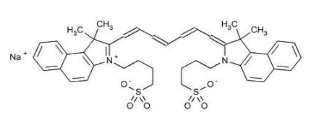
Physicochemical properties: Indocyanine green is a water soluble, tricarbocyanine dye with a peak spectral absorption at 800 nm. SPY AGENT Green has a pH of approximately 7.0 when reconstituted.
13 CLINICAL TRIALS
13.1 Trial Design and Study Demographics
The efficacy and safety of SPY AGENT™ Green (indocyanine green for injection, USP) was assessed in one open-label, controlled, multicenter, single arm, within patient, lymphatic mapping study in patients with uterine and cervical cancer (FILM study) and in four (4) systematic summary reviews, in the form of meta analyses, of published literature with use of indocyanine green as the imaging agent for Novadaq Fluorescence Imaging Systems and the da Vinci® Surgical Robotic System. The four (4) meta-analysis included one in lymphatic mapping in patients with uterine and cervical cancer (Study LNM-UC01), and three related systematic summary reviews, one for visualization of macrovascular blood flow, one for visualization of microvascular tissue perfusion and one for visualization of extrahepatic biliary anatomy (Studies Macro-IAM-01, Micro-IAM-01 and Biliary-IAM-01, respectively).
A total of 176 patients were treated with SPY AGENT™ Green in the FILM study and further clinical experience with lymphatic mapping using indocyanine green in 1,512 patients with uterine and cervical cancer was provided from Study LNM-UC01.
Data from 1,184 and 2,055 patients undergoing various surgical procedures requiring visualization of blood flow and tissue perfusion, are provided from Studies Macro-IAM-01 and Micro-IAM-01, respectively. Clinical experience with visualization of the main extrahepatic biliary ducts using indocyanine green and SPY Fluorescence Imaging Systems in 314 patients is provided from Study Biliary-IAM-01.
13.2 Study Results
13.2.1 Imaging Blood Flow and Tissue Perfusion
Study Macro-IAM-01
Study Macro-IAM-01 was a systematic review, in the form of a meta-analysis, of 13 studies examining the use of Novadaq fluorescence imaging systems, including the da Vinci® system, for visualization of macrovascular blood flow in vessels (i.e., arteries, veins and bypass grafts) during various procedures including, but not limited to, coronary bypass surgery, organ transplant procedures, plastic reconstructive surgery utilizing autologous flaps, renal cancer and vascular surgeries. A total of 1,184 patients were evaluated in this meta-analysis. The indocyanine green was administered as a single administration pre- or intraoperatively and may have included a repeat administration during surgery. Indocyanine green doses ranged from 0.0125 mg – 25 mg across all 13 studies. The primary endpoint was the success rate of intraoperative visualization of macrovascular blood flow in vessels.
The results showed an overall visualization success rate of 97.0% with 95% CI of 96.3% to 97.6%, meeting the success criteria of the study. There were a total of 2,854 visualizations attempted and 2,768 visualizations succeeded. Across the 13 studies reviewed, 12 studies exceeded the success criteria of a 90% visualization rate and 10/13 studies showed a 100% visualization rate.
Study Micro-IAM-01
Study Micro-IAM-01 was a systematic review, in the form of a meta-analysis, of 33 studies examining the use of Novadaq fluorescence imaging systems, including the da Vinci® system, for visualization of microvascular blood flow in tissues during various procedures including, but not limited to, myocardial perfusion in cardiac and cardiovascular surgeries, tissue flap perfusion in plastic reconstructive surgery, perfusion in vascular surgeries (such as wound, amputation and coronary vessels), GI tract perfusion during surgery of the colon, stomach or esophagus and parathyroid perfusion during endocrine surgery. A total of 2,055 patients were evaluated in this meta-analysis. The indocyanine green was administered as a single administration pre- or intraoperatively and may have included a repeat administration during surgery. Indocyanine green doses ranged from 2.5 mg – 17.5 mg across all 33 studies. The primary endpoint was the success rate of intraoperative visualization of microvascular blood flow in tissues.
The results showed an overall visualization success rate of visualization success rate of 99.9% with 95% CI of 99.7% to 100.0%. All of the studies showed a success rate greater than 90% and therefore, met the study success criteria. There were a total of 2,696 visualizations attempted and 2,693 succeeded. Across the 33 studies reviewed, all of the studies exceeded the success criteria of a 90% visualization rate and 31/33 studies showed a 100% visualization rate.
13.2.2 Imaging Extrahepatic Biliary Anatomy
Study Biliary-IAM-01
Study Biliary-IAM-01 was a systematic review, in the form of a meta-analysis, of 4 studies examining the use of Novadaq fluorescence imaging systems, including the da Vinci® system, for visualization of extrahepatic biliary anatomy. A total of 314 patients were evaluated in this meta-analysis. The indocyanine green was administered as a single administration pre operatively and may have included a repeat administration during surgery. Indocyanine green doses ranged from 1.4 mg – 17.5 mg across the 4 studies. The primary endpoint was the success rate of intraoperative visualization of at least one of the major extrahepatic bile ducts (cystic duct, common bile duct or common hepatic duct).
The results showed an overall visualization success rate of 99.3%, with 95% CI of 97.3% to 100.0%. There were a total of 286 visualizations attempted and 284 succeeded. All 4 studies showed a success rate greater than 90% and therefore, met the study success criteria.
13.2.3 Imaging of Lymph Nodes and Lymphatic Vessels During Lymphatic Mapping
FILM Study
The FILM study was a randomized, prospective, multi-center, open-label within patient comparative study in patients with early stage uterine or cervical cancers, with no known regional nodal or metastatic disease by standard clinical evaluation. The efficacy of SPY AGENT™ Green and the PINPOINT® Fluorescence Imaging System in the detection of lymphatic vessels and lymph nodes during lymphatic mapping procedures was determined by the number of histology-confirmed lymph nodes detected by SPY AGENT™ Green and/or the blue dye comparator. SPY AGENT™ Green and the blue dye comparator were injected into the cervix of patients at the beginning of the operative procedure. Lymphatic mapping was performed intraoperatively using the PINPOINT® Fluorescence Imaging System followed by excision of lymph nodes identified by SPY AGENT™ Green, blue dye, or the surgeon’s visual and palpation examination. The resected lymph nodes were evaluated by histopathology. SPY AGENT ™Green and the PINPOINT® Fluorescence Imaging System identification rate in pathology-positive lymph nodes was also determined.
A total of 176 patients received either SPY AGENT™ Green followed by blue dye or blue dye followed by SPY AGENT™ Green. A total of four (4) 1 mL injections of a 1.25 mg/mL solution of SPY AGENT™ Green was administered into the cervix at the 3 and 9 o’clock positions with a superficial (1-3 mm) and a deep (1-3 cm) injection at each position for a total dose of 5 mg per patient. During lymphatic mapping evaluable lymph nodes were resected. Table 4 shows the distribution of resected, confirmed lymph nodes by the presence or absence of SPY AGENT™ Green or blue dye. Most of the resected lymph nodes were identified by either SPY AGENT™ Green or blue dye or both. Significantly more resected lymph nodes were identified by SPY AGENT™ Green comparison to blue dye. Of the confirmed lymph nodes identified, 96.5% were identified using SPY AGENT™ Green, and 46.4% were identified using blue dye, a difference of 50.1% with 95% confidence interval 39.2% to 46.4%.
Table 4: Number of Resected Confirmed Lymph Nodes Detected by SPY AGENT™ Green (SPY) and/or Blue Dye (BD) in Patients with Uterine and Cervical Cancers
| Analysis population | Nodes (n) | % Detected with BD | % Detected with SPY | % Difference | 95% CI limits |
| mITT | 545 | 46.4% | 96.5% | 50.1% | 39.2%, 60.1% |
| PP | 517 | 46.6% | 97.1% | 50.5% | 39.3%, 61.7% |
mITT= Modified Intent-to-Treat population; PP=Per Protocol population
95% Confidence Intervals (CI) represent the spread in the difference between the individual treatments.
Tables 5 and 6 show the number of patients with greater than one resected confirmed lymph node and the number of patients with resected confirmed bilateral lymph nodes identified with SPY AGENT™ Green or blue dye.
Table 5: Number of Patients with Uterine or Cervical Cancer with Greater than 1 Resected Confirmed Lymph Node Detected by SPY AGENT™ Green (SPY) and/or Blue Dye (BD)
| Identification Method | Number of Patients with ≥ 1 Lymph Node Detected | Percentage of Patients with ≥ 1 Lymph Node Detected |
| SPY | 168 | 95.5% |
| BD | 131 | 74.4% |
| Total | 176 | 100.0% |
| Difference | Lower Bound 95% CI | Upper Bound 95% CI | P-Value |
| 21.0 | 14.2 | 27.8 | < 0.0001 |
McNemar’s test for difference in percent of patients with lymph nodes identified by SPY compared to BD and 95% confidence interval for the difference.
Table 6: Number of Patients with Uterine or Cervical Cancer with Confirmed Bilateral Resected Lymph Node Detected by SPY AGENT™ Green (SPY) and/or Blue Dye (BD)
| Identification Method | Number of Patients with Bilateral Lymph Nodes Detected | Percentage of Patients with Bilateral Lymph Nodes Detected |
| SPY | 130 | 73.9% |
| BD | 51 | 29.0% |
| Total | 176 | 100.0% |
| Difference | Lower Bound 95% CI | Upper Bound 95% CI | P-Value |
| 49.9 | 37.0 | 52.7 | <0.0001 |
McNemar’s test for difference in percent of patients with lymph nodes identified by SPY compared to BD and 95% confidence interval for the difference.
Study LNM-UC01
Study LNM-UC01 was a meta-analysis of 11 published studies that examined the use of the investigational lymphatic mapping agent indocyanine green among patients undergoing surgery for uterine or cervical cancer. The 11 studies provided clinical data on 1,512 patients with approximately half of the data coming from studies that compared indocyanine green to blue dyes and half from studies designed only to assess indocyanine green lymph node detection rates. The predominance (70%) of the comparative clinical data came from published reports that cited use of the same indocyanine green dose (5 mg) and injection technique as that used in the FILM Study.
The results found an overall indocyanine green lymph node detection rate of 81% compared to a blue dye lymph node detection rate of 52% with an odds ratio of 3.83 and 95% CI of 2.82 to 5.21 (p < 0.0001). Lymph node detection was shown to be superior with indocyanine green compared to blue dye, with statistical success reported by the publication authors in four of the five comparative studies.
14 NON-CLINICAL TOXICOLOGY
14.1 Carcinogenesis, Mutagenesis, Impairment of Fertility
No studies have been performed to evaluate carcinogenicity, mutagenicity, or impairment of fertility.