


Product Monograph - Optiray 320
Recent Major Label Changes
| 2025-08 |
|---|---|
| 2025-08 |
| 2025-08 |
| 2025-08 |
| 2025-08 |
| 2025-08 |
Certain sections or subsections that are not applicable at the time of the preparation of the most recent authorized product monograph are not listed.
Table of Contents
Recent Major Label Changes ................................................................................................. 3
Table of Contents ................................................................................................................. 3
Part 1: Healthcare Professional Information.......................................................................... 7
- 1.
Indications..................................................................................................................... 7
1.1. Pediatrics .................................................................................................................... 7
1.2. Geriatrics .................................................................................................................... 7
- 2. Contraindications .......................................................................................................... 7
- 3. Serious Warnings and Precautions Box ........................................................................... 8
- 4.
Dosage and Administration ............................................................................................ 8
4.1. Dosing Considerations ............................................................................................... 8
4.2. Recommended Dose and Dosage Adjustment .......................................................... 9
4.2.1. Intra-arterial Procedures in Adults .................................................................... 9
4.2.1.1. Cerebral Arteriography .................................................................................. 9
4.2.1.2. Peripheral Arteriography ............................................................................... 9
4.2.1.3. Selective Coronary Arteriography with or without Left Ventriculography ... 9
4.2.1.4. Aortography and Visceral Arteriography ....................................................... 9
4.2.2. Intra-arterial Procedures in Pediatrics ............................................................. 10
4.2.2.1. Intra-arterial Digital Subtraction Angiography (IA-DSA) .............................. 10
4.2.2.2. Selective Coronary Arteriography with or without Left Ventriculography . 10
4.2.3. Intravenous Procedures in Adults and Pediatrics ............................................ 10
4.2.3.1. Intravenous Contrast Enhancement in Computed Tomography (CT) ......... 10
4.2.3.2. Venography .................................................................................................. 11
4.2.3.3. Excretory Urography .................................................................................... 11
4.3. Reconstitution .......................................................................................................... 12
4.4. Administration ......................................................................................................... 12
4.5. Missed Dose ............................................................................................................. 13
- 5. Overdose ..................................................................................................................... 13
- 6. Dosage Forms, Strengths, Composition, and Packaging ................................................ 13
- 7.
Warnings and Precautions ........................................................................................... 14
General ................................................................................................................................. 14
Anaesthetised patient .......................................................................................................... 15
Cardiovascular diseases ........................................................................................................ 15
Central nervous system disorders ........................................................................................ 15
Coagulation disorders........................................................................................................... 15
Extravasation ........................................................................................................................ 16
Fertility.................................................................................................................................. 16
Homozygous sickle cell disease ............................................................................................ 16
Hypersensitivity .................................................................................................................... 16
Neurologic ............................................................................................................................ 16
Phaeochromocytoma ........................................................................................................... 16
Renal ..................................................................................................................................... 17
Severe cutaneous adverse reactions (SCAR) ........................................................................ 17
Thromboembolic disorders .................................................................................................. 17
Thyroid disorders .................................................................................................................. 17
Thyroid dysfunction .............................................................................................................. 18
Cerebral Angiography ........................................................................................................... 18
Peripheral Arteriography ...................................................................................................... 18
Coronary Arteriography and Left Ventriculography ............................................................ 18
Intra-Arterial Digital Subtraction Arteriography .................................................................. 18
Aortography and Visceral Arteriography ............................................................................. 19
Intravenous Contrast Enhancement in Computed Tomography ......................................... 19
Venography .......................................................................................................................... 19
Excretory Urography ............................................................................................................ 19
7.1. Special Populations .................................................................................................. 19
7.1.1. Pregnancy ......................................................................................................... 19
7.1.2. Breastfeeding ................................................................................................... 20
7.1.3. Pediatrics .......................................................................................................... 20
7.1.4. Geriatrics .......................................................................................................... 21
- 8.
Adverse Reactions ....................................................................................................... 21
8.1. Adverse Reaction Overview ..................................................................................... 21
8.2. Clinical Trial Adverse Reactions ............................................................................... 22
8.2.1. Clinical Trial Adverse Reactions – Pediatrics.................................................... 24
8.3. Less Common Clinical Trial Adverse Reactions ........................................................ 24
8.3.1. Less Common Clinical Trial Adverse Reactions – Pediatrics ............................ 24
8.4. Abnormal Laboratory Findings: Hematologic, Clinical Chemistry, and Other Quantitative Data ................................................................................................................. 24
8.5. Post-Market Adverse Reactions ............................................................................... 24
- 9.
Drug Interactions ......................................................................................................... 27
9.1. Serious Drug Interactions ........................................................................................ 27
9.2. Drug Interactions Overview ..................................................................................... 27
9.3. Drug-Behaviour Interactions .................................................................................... 28
9.4. Drug-Drug Interactions ............................................................................................ 28
9.5. Drug-Food Interactions ............................................................................................ 28
9.6. Drug-Herb Interactions ............................................................................................ 28
9.7. Drug-Laboratory Test Interactions........................................................................... 28
- 10.
Clinical Pharmacology .................................................................................................. 29
10.1. Mechanism of Action ............................................................................................... 29
10.2. Pharmacodynamics .................................................................................................. 29
10.3. Pharmacokinetics ..................................................................................................... 31
- 11. Storage, Stability, and Disposal .................................................................................... 31
Part 2: Scientific Information .............................................................................................. 32
- 13. Pharmaceutical Information ........................................................................................ 32
- 14.
Clinical Trials ............................................................................................................... 32
14.1. Clinical Trials by Indication ...................................................................................... 32
14.2. Comparative Bioavailability Studies ........................................................................ 33
- 16. Non-Clinical Toxicology ................................................................................................ 33
Patient Medication Information ......................................................................................... 38
Part 1: Healthcare Professional Information
1. Indications
Adults
OPTIRAY 350 administered intravascularly is recommended in adults for coronary arteriography and ventriculography, peripheral and visceral arteriography, intravenous contrast enhancement in computed tomography of the head and body, excretory urography, intravenous digital subtraction angiography and venography.
OPTIRAY 320 administered intravascularly is recommended for angiography throughout the cardiovascular system in adults. The uses include cerebral, coronary, peripheral, visceral and renal arteriography, aortography and left ventriculography. OPTIRAY 320 is also recommended for contrast enhanced computed tomographic imaging of the head and body and in excretory urography.
OPTIRAY 300 administered intravascularly is recommended for use in adults for cerebral angiography, aortography, peripheral and visceral arteriography, intravenous contrast enhancement of computed tomography of the brain and body, excretory urography, intravenous digital subtraction angiography and venography.
1.1. Pediatrics
Pediatrics (0-18 years old): OPTIRAY 350 administered intravascularly is indicated in children for angiocardiography.
OPTIRAY 320 administered intravascularly is recommended in children one year of age or over for angiocardiography, contrast enhanced computed tomography of the head and body and for excretory urography.
OPTIRAY 300 administered intravascularly is recommended in children one year of age or over for intravenous excretory urography and intra-arterial digital subtraction angiography.
1.2. Geriatrics
Geriatrics (> 65 years of age): Ioversol is nearly completely excreted as parent drug by the kidney, and the risk of adverse reactions to OPTIRAY may be greater in patients with impaired renal function. Because elderly patients are more likely to have decreased renal function, dose selection should be cautious usually starting at the low end of the dosing range, reflecting the greater frequency of decreased hepatic, renal or cardiac function, and of concomitant disease or other drug therapy.
2. Contraindications
- OPTIRAY is contraindicated in patients who are hypersensitive to iodine-containing contrast media, the active substance or to any ingredient in the formulation or component of the container. For a complete listing see 6 Dosage Forms, Strengths, Composition, and Packaging of the product monograph
- Symptomatic hyperthyroidism
- Serious Warnings and Precautions Box
Serious Warnings and Precautions
- FOR INTRA-ARTERIAL AND INTRAVENOUS USE ONLY
- Inadvertent subarachnoidal administration may cause death, convulsions, cerebral hemorrhage, coma, paralysis arachnoiditis, acute renal failure, cardiac arrest, seizures, rhabdomyolysis, hyperthermia and brain edema
- USE THE RECOMMENDED OPTIRAY CONCENTRATION FOR THE PARTICULAR PROCEDURE TO BE UNDERTAKEN (see 7 Warning and Precaution)
4. Dosage and Administration
4.1. Dosing Considerations
- Introduction of the catheter or needle is normally performed with local anaesthesia. General anaesthesia is rarely required.
- Use the lowest dose necessary to obtain adequate visualization.
- Use only the recommended concentration for the particular procedure to be undertaken.
- Patients should be well hydrated prior to and following administration of OPTIRAY (ioversol). Do not dehydrate patients for any procedures.
- The volume of each individual injection is a more important consideration than the total dose used. When large individual volumes are administered, sufficient time should be permitted to elapse between each injection to allow for subsidence of hemodynamic disturbances.
- In angiographic procedures, the possibility of dislodging plaque or damaging or perforating the vessel wall should be considered during catheter manipulation and contrast medium injection. Test injections to ensure proper catheter placement are recommended.
Patients with renal impairment:
- Patients with mild or moderate renal impairment do not require dose adjustment. OPTIRAY should not be used with subjects with severe (eGFR < 30 mL/min) renal impairment.
- Patients with a serum creatinine level above 3 mg/dL should not undergo excretory urography or other radiological procedures unless the benefits clearly outweigh the risks incurred.
- In patients with advanced renal disease, iodinated contrast media should be used with caution and only when the examination is essential since excretion of the medium is impaired. Use of OPTIRAY is not recommended in patients with anuria or severe oliguria.
Patients with hepatic impairment:
- Patients with mild or moderate (Child-Pugh A or B) hepatic impairment do not require dose adjustment. OPTIRAY should not be used with subjects with severe (Child-Pugh C) hepatic impairment.
- For patients undergoing excretory urography, a low residue diets the day preceding the examination, and a laxative the evening before the examination, may be given unless contraindicated.
4.2. Recommended Dose and Dosage Adjustment
4.2.1. Intra-arterial Procedures in Adults
4.2.1.1. Cerebral Arteriography
OPTIRAY 320 or 300 may be used to visualize the cerebral arteries. The recommended dose for visualization of cerebral arteries is shown below (may repeat as necessary).
| Diagnostic Area | Single Dose | Maximum Cumulative Dose |
|---|---|---|
| Carotid arteries | 5 mL to 10 mL (for OPTIRAY 300 or OPTIRAY 320) | 200 mL |
| Vertebral arteries | 4 mL to 8 mL (for OPTIRAY 300 or OPTIRAY 320) | 200 mL |
Aortic arch injection (four vessel studies) | 15 mL to 25 mL (For OPTIRAY 320 only) |
|
4.2.1.2. Peripheral Arteriography
OPTIRAY 350, 320 or 300 may be used for arteriograms of the lower extremities. The recommended dose for visualization of peripheral arteries is shown below (may repeat as necessary).
| Diagnostic Area | Single Dose | Maximum Cumulative Dose |
|---|---|---|
| Aorto-iliac run-off studies | 20 mL to 50 mL | 250 mL |
| Iliac and femoral arteries | 10 mL to 30 mL | 250 mL |
4.2.1.3. Selective Coronary Arteriography with or without Left Ventriculography
OPTIRAY 320 or 300 is recommended for selective coronary arteriography. The recommended dose for visualization of the coronary arteries and left ventricle is shown below. Doses may be repeated if indicated; however, several minutes should be allowed to elapse between injections to allow for subsidence of hemodynamic disturbance.
| Diagnostic Area | Single Dose | Maximum Cumulative Dose |
|---|---|---|
| Left coronary | 2 mL to 10 mL | 250 mL |
| Right coronary | 2 mL to 6 mL | 250 mL |
| Left ventricle | 30 mL to 40 mL |
|
4.2.1.4. Aortography and Visceral Arteriography
OPTIRAY 300, 320 or 350 are recommended for aortography and visceral arteriography. The recommended dose for aorta and visceral arteries is shown below (may repeat as necessary):
| Diagnostic Area | Single Dose | Maximum Cumulative Dose |
|---|---|---|
| Abdominal aorta | 20 mL to 50 mL | 250 mL |
| Superior mesenteric artery | 20 mL to 40 mL | 250 mL |
| Renal artery | 4mL to 10 mL | 250 mL |
4.2.2. Intra-arterial Procedures in Pediatrics
4.2.2.1. Intra-arterial Digital Subtraction Angiography (IA-DSA)
The usual pediatric dose of OPTIRAY 300 for IA-DSA is 1 to 3 mL/kg.
4.2.2.2. Selective Coronary Arteriography with or without Left Ventriculography
OPTIRAY 320 or 300 is recommended for left coronary arteriography in children 1 year of age and over. The usual single injection dose of OPTIRAY 320 or 350 is 1.25 mL/kg with a range of 1 mL/kg to 1.5 mL/kg. When multiple injections are given, the total administered dose should not exceed 5 mL/kg up to a total volume of 250 mL.
4.2.3. Intravenous Procedures in Adults and Pediatrics
4.2.3.1. Intravenous Contrast Enhancement in Computed Tomography (CT)
Computed Tomography of the Head
Neoplastic Conditions – OPTIRAY 300, 320 or 350 may be used to enhance the demonstration of the presence and extent of certain primary or metastatic malignancies.
Non-Neoplastic Conditions - The use of OPTIRAY 300, 320 or OPTIRAY 350 may be beneficial in the image enhancement of non-neoplastic lesions, such as cerebral infarctions of recent onset; however, some infarctions are obscured if contrast media are used.
Adult Dosage and Administration - The usual adult dosage of OPTIRAY 300, 320 or 350 are 50 to 100 mL. A maximum dose of 150 mL of OPTIRAY 320 or 350 should not be exceeded. Scanning is performed immediately after injection.
Pediatric Dosage and Administration - The recommended dose of OPTIRAY 320 for children one year of age and over is 1 mL/kg to 3 mL/kg.
Computed Tomography of the Body
OPTIRAY 300, 320 or 350 may be administered for contrast enhancement of the organs, tissues and larger blood vessels of the chest, abdomen and pelvis.
Adult Dosage and Administration – OPTIRAY 300, 320 or 350 may be administered by bolus injection, rapid infusion, or a combination of both. Depending on the area to be examined, the usual dose range for infusion is 30 to 100 mL. When prolonged enhancement is required, 25 to 50 mL may be given as a rapid bolus and the remainder as an infusion. The total dose should not exceed 150 mL of OPTIRAY 300, 320 or 350. Scanning is performed immediately after injection.
Pediatric Dosage and Administration - The recommended dose of OPTIRAY 320 for use in children one year of age and over is 2 mL/kg, with a range of 1 mL/kg to 3 mL/kg.
4.2.3.2. Venography
OPTIRAY 300 or OPTIRAY 350 may be used to visualize the peripheral venous circulation. Venograms are obtained by injection or infusion into an appropriate vein in the lower extremity.
Adult Dosage and Administration - The usual adult dose of OPTIRAY 300 or 350 will range from 20 to 100 mL for the lower extremity.
Following the procedure, the venous system should be flushed with normal or heparinized saline solution. Massage and elevation of the leg are also helpful for clearing the contrast medium from the extremity to prevent post-procedural thrombophlebitis. The maximum dose should not exceed 250 mL.
4.2.3.3. Excretory Urography
OPTIRAY 350, 320 or 300 may be used for excretory urography. Following intravenous injection in patients with normal renal function, OPTIRAY is excreted mostly by the kidneys. Maximum radiographic density in the calyces and pelves occurs in most instances within 5 to 15 minutes after injection.
Adult Dosage and Administration - The usual adult dose of OPTIRAY 300, 320 or 350 is 50 mL in the average normal adult. In these patients, high dose urography may be preferred using OPTIRAY 320 at a dose of 1.5 to 2 mL/kg. The dose is injected intravenously, usually within 1 to 3 minutes. Maximum doses of 150 mL of OPTIRAY 300 or 320 and 140 mL of OPTIRAY 350 should not be exceeded.
Pediatric Dosage and Administration - OPTIRAY 300 and OPTIRAY 320 are recommended at a dose of 1 mL/kg and may range from 0.5 mL/kg to 3 mL/kg. Dosage for children over 1 year of age should be administered in proportion to age and body weight. The total administered dose should not exceed 3 mL/kg.
Summary of Intravenous Dosage
Table 1 Adult Intravenous Dosage
| Procedure | Concentration of Solution (mgI/mL) | Usual Recommended Single Dose (mL) |
| Intravenous Contrast Enhanced CT | ||
| 300 | ||
| 320 | ||
| 350 | ||
| Head CT | 50 to 100 | |
| Body CT | 30 to 100 (infusion) | |
| 25 to 50 (bolus) | ||
| Venography | ||
| 300 | 20 to 100 | |
| 350 | ||
| Excretory urography | ||
| 300 | 50 | |
| 320 | 50 | |
| 350 | 50 |
Table 2 Pediatric Intravenous Dosage
| Procedure | Concentration of Solution (mgI/mL) | Usual Recommended Single Dose (mL) |
| Excretory urography | 300 320 | > 1 year old: 2 mL/kg > 1 year old: 1 to 1.5 mL/kg |
| Computed Tomography of the Head | 320 | 1 to 3 mL/kg |
| Computed Tomography of the Body | 320 | 1 to 3 mL/kg |
4.3. Reconstitution
Not applicable
4.4. Administration
- OPTIRAY should be inspected visually for particulate matter and discoloration prior to administration. If either is present, the vial should be discarded.
- OPTIRAY should not be transferred into other delivery systems except immediately before use and should be used immediately once the deal has been punctured.
- It is advisable that OPTIRAY be at, or close to, body temperature when injected.
- Under no circumstances should other drugs be administered concomitantly in the same syringe or I.V. administration set as OPTIRAY because of a potential for chemical incompatibility
- Patency of the vessel and other position of the catheter tip or needle should be checked with a small pilot dose of OPTIRAY before injecting the full dose. The catheter tip should be kept free of aspirated blood. Prolonged contact of OPTIRAY with blood must be avoided because of potential thromboembolic complications.
- Any unused portion of one container should be discarded.
4.5. Missed Dose
Not applicable
5. Overdose
The adverse events of overdosage are life-threatening and affect mainly the pulmonary, cardiovascular, and central nervous systems. Treatment of an overdosage is directed toward the support of all vital functions, and prompt institution of specific therapy. OPTIRAY does not bind to plasma or serum proteins and is, therefore, dialyzable.
For the most recent information in the management of a suspected drug overdose, contact your regional poison control centre or Health Canada’s toll-free number, 1-844 POISON-X (1-844-764-7669).
- 6. Dosage Forms, Strengths, Composition, and Packaging
Each millilitre of OPTIRAY 300 (ioversol injection 64% w/v) provides 636 mg of ioversol with 3.6 mg of tromethamine as buffer and 0.2 mg of edetate calcium disodium as a stabilizer. OPTIRAY 300 provides 30% (300 mg/mL) organically bound iodine.
Each millilitre of OPTIRAY 320 (ioversol injection 68% w/v) provides 678 mg of ioversol with 3.6 mg of tromethamine as a buffer and 0.2 mg of edetate calcium disodium as a stabilizer. OPTIRAY 320 provides 32% (320 mg/mL) organically bound iodine.
Each millilitre of OPTIRAY 350 (ioversol injection 74% w/v) provides 741 mg of ioversol with 3.6 mg of tromethamine as a buffer and 0.2 mg of edetate calcium disodium as a stabilizer. OPTIRAY 350 provides 35% (350 mg/mL) organically bound iodine.
OPTIRAY 300
30 mL vials, boxes of 10
50 mL vials, boxes of 10
100 mL fill/150 mL bottles, boxes of 10
150 mL bottles, boxes of 10
Ultraject hand-held prefilled syringes of 30 mL and 50 mL, boxes of 10 and 20
Ultraject power injector prefilled syringes of 50 mL fill/125 mL; 100 mL fill/125 mL; 125 mL, boxes of 10 and 20.
OPTIRAY 320
20 mL vials, boxes of 10
30 mL vials, boxes of 10
50 mL vials, boxes of 10
100 mL fill/150 mL bottles, boxes of 10
150 mL bottles, boxes of 10
200 mL fill/250 mL bottles, boxes of 10
Ultraject hand-held prefilled syringes of 30 mL and 50 mL , boxes of 10 and 20
Ultraject power injector prefilled syringes of 50 mL fill/125 mL , 75 mL fill/125 mL; 100 mL fill/125 mL and 125 mL, boxes of 10 and 20.
OPTIRAY 350
30 mL vials, boxes of 10
50 mL vials, boxes of 10
75 mL fill/150 mL bottles, boxes of 10
100 mL fill/150 mL bottles, boxes of 10
150 mL bottles, boxes of 10
200 mL fill/250 mL bottles, boxes of 10
Ultraject hand-held prefilled syringes of 30 and 50 mL, boxes of 10 and 20
Ultraject power injector prefilled syringes of 50 mL fill/125 mL; 75 mL fill/125 mL; 100 mL fill/125 mL and 125 mL, boxes of 10 and 20.
Not all formats and packaging configurations are applicable for all global markets
Pharmacy Bulk Vial for OPTIRAY 320 and 350 (500 mL Bottle); Boxes of 5 For Multiple Dispensing
This Bulk Pharmacy Vial is intended for multiple dispensing for intravenous use only, it must be spiked only once.
Directions for Use
Use proper aseptic techniques when handling injection device for maintenance of sterility during multiple dispensing contrast agent at room temperature.
The availability of the Bulk Pharmacy Vial is restricted to hospitals with a recognized intravenous admixture program for multiple dispensing.
Once punctured, use the contents of the Pharmacy Bulk Vial within four (4) hours and diluted solutions within 24 hours if kept at room temperature, and 72 hours if refrigerated from the time of initial puncture.
7. Warnings and Precautions
See 3 Serious Warnings and Precautions.
General
Serious or fatal reactions have been associated with the administration of iodinated X-ray contrast media. It is of utmost importance to be completely prepared to treat any contrast medium reaction. Diagnostic Procedures should be performed under the direction of personnel skilled and experienced in the particular procedure to be performed. A fully equipped emergency card, or equivalent supplies and equipment and personnel competent in recognising and treating adverse reactions of all types should always be available. Since severe delayed reactions have been known to occur, emergency facilities and competent personnel should be available for at least 30 to 60 minutes after administration.
Anaesthetised patient
General anaesthesia may be indicated in some procedures; however, one should be aware of possible increased incidence of adverse reactions in such circumstances.
Cardiovascular diseases
Patients with congestive heart failure should be observed for several hours following the procedure to detect delayed haemodynamic disturbances, which may be associated with a transitory increase in the circulating osmotic load.
Central nervous system disorders
Serious neurologic events have been observed following direct injection into cerebral arteries or vessels supplying the spinal cord or in angiocardiography, due to inadvertent filling of the carotids. A cause-effect relationship to the contrast medium has not been established, since the patient’s pre-existing condition and procedural techniques are causative factors in themselves.
Encephalopathy has been reported with the use of ioversol. Contrast-induced encephalopathy may manifest with symptoms and signs of neurological dysfunction such as headache, visual disturbance, cortical blindness, confusion, seizures, loss of coordination, hemiparesis, aphasia, unconsciousness, coma, and cerebral oedema. Symptoms usually occur within minutes to hours after administration of ioversol and generally resolve within days.
Factors which increase blood-brain barrier permeability facilitate the passage of the contrast medium into cerebral tissue, which can lead to central nervous system reactions, e.g. encephalopathy.
If contrast encephalopathy is suspected, appropriate medical management should be initiated, and administration of ioversol must not be repeated.
Coagulation disorders
Non-ionic iodinated contrast media, including OPTIRAY, inhibit blood coagulation less than ionic contrast media. Therefore, meticulous intravascular administration technique is necessary, particularly during angiographic procedures, to minimize thromboembolic events. Frequent flushing of standard angiographic catheters and avoiding prolonged contact of blood with contrast agent in syringes and catheters is recommended.
Extreme caution should be used in patients who are known to have multiple myeloma and other paraproteinemias, because of the risk of inducing transient to fatal renal failure. In these instances, anuria has developed resulting in progressive uremia, renal failure, and eventually death. A minimal diagnostic dose should be employed and renal function, as well as extent of urinary precipitation of the myelomatous protein, should be monitored for a few days subsequent to the procedure. The patients should be normally hydrated for the examination, since dehydration may predispose to precipitation of myeloma protein in the renal tubules. No form of therapy, including dialysis, has been successful in reversing the effect.
Extravasation
Ioversol should be injected with caution to avoid perivascular application. This is especially important in patients with severe arterial or venous disease. However, significant extravasation of ioversol may occur especially during the use of power injectors. Generally, it is tolerated without substantial tissue injury applying conservative treatment. However, serious tissue damage (e.g. ulceration) has been reported in isolated cases requiring surgical treatment.
Fertility
Animal studies did not indicate direct or indirect harmful effects with respect to fertility in humans. There are, however, no adequate and well controlled clinical studies on fertility.
Homozygous sickle cell disease
In patients with homozygous sickle cell disease, hyperosmolar agents such as ioversol may affect sickling of erythrocytes. Hence, there is a need for careful consideration before the intra-arterial administration of such agents to patients with homozygous sickle cell disease.
Hypersensitivity
OPTIRAY is contraindicated in patients with hypersensitivity to iodine-containing contrast media.
The patient should also be informed that allergic reactions may develop up to several days post administration; in such case, a physician should be consulted immediately. The occurrence of severe idiosyncratic reactions has prompted the use of several pre-testing methods. However, pre-testing cannot be relied upon to predict severe reactions and may itself be hazardous to the patient. T is suggested that a thorough medical history with emphasis on allergy and hypersensitivity, prior to the injection of any contrast medium, may be more accurate than pre-testing in predicting potential adverse reactions.
A positive history of allergies does not arbitrarily contraindicate the use of a contrast agent when a diagnostic procedure is thought essential, but caution should be exercised (see 2 Contraindications). Appropriate resuscitation measures should be immediately available.
Pre-medication with antihistamines and corticosteroids to avoid or minimise allergic reactions should be considered. Reports indicate that such pre-treatment does not prevent serious life-threatening reactions but may reduce both their incidence and severity.
Neurologic
OPTIRAY, as with other contrast medium, can cause serious neurologic sequelae, including permanent paralysis, following cerebral arteriography and injection into vessels supplying the spinal cord. The injection of a contrast medium should never be made following the administration of vasopressors, since they strongly potentiate neurologic effects.
Phaeochromocytoma
Administration of radiopaque materials to patients known or suspected to have pheochromocytoma should be performed with extreme caution if, in the opinion of the physician, the possible benefits of such procedures outweigh the considered risks. The amount of radiopaque medium injected should be kept to an absolute minimum. The blood pressure should be assessed throughout the procedure and measures for treatment of a hypertensive crisis should be available.
Renal
Combinations with nephrotoxic medicines should be avoided. If this cannot be avoided, laboratory monitoring of renal function must be intensified. Caution must be exercised in patients with severely impaired renal function, combined renal and hepatic disease, diabetes mellitus, homozygous sickle cell disease, multiple myeloma or other paraproteinaemia, anuria, particularly when large doses are administered. Serious renal effects, including acute renal failure, may occur in these patients. Although neither the contrast agent nor dehydration has been proved separately to be the cause of renal failure, it has been speculated that the combination of both may be causative. The risk in patients with impaired renal function is not a contraindication to the procedure: however, special precautions, including maintenance of normal hydration and close monitoring, are required.
An effective hydration prior to the administration of ioversol is essential and may decrease the risk of renal injury. Preparatory dehydration is dangerous and may contribute to acute renal failure.
Patients with a serum creatinine level above 3mg/dL should not undergo excretory urography or other radiological procedures unless the benefits clearly outweigh the risks incurred. In patients with advanced renal disease, iodinated contrast media should be used with caution and only when the examination is essential since excretion of the medium is impaired. Use of OPTIRAY is not recommended in patients with anuria or severe oliguria. Avid combinations with nephrotoxic medicines. If this cannot be avoided, laboratory monitoring of renal function must be intensified.
Severe cutaneous adverse reactions (SCAR)
Severe cutaneous adverse reactions (SCAR) may develop from 1 hour to several weeks after intravascular contrast agent administration. These reactions include Stevens-Johnson syndrome and toxic epidermal necrolysis (SJS/TEN), acute generalized exanthematous pustulosis (AGEP), and drug reaction with eosinophilia and systemic symptoms (DRESS). Reaction severity may increase and time to onset may decrease with repeat administration of a contrast agent; prophylactic medications may not prevent or mitigate severe cutaneous adverse reactions. Avoid administering OPTIRAY to patients with a history of a severe cutaneous adverse reaction to OPTIRAY.
Thromboembolic disorders
Angiography should be avoided whenever possible in patients with homocystinuria due to an increased risk of thrombosis and embolism.
In patients with advanced atherosclerosis, serious hypertension, cardiac decompensation, senility, preceding cerebral thrombosis or embolism, special caution should be exercised. Cardiovascular reactions as bradycardia, rising or falling of blood pressure may occur more often.
Thyroid disorders
OPTIRAY is contraindicated in patients who manifest hyperthyroidism. Reports of thyroid storm following intravascular use of iodinated radiopaque agents in patients with hyperthyroidism or with an autonomously functioning thyroid nodule, suggest that this additional risk be carefully evaluated in such patients before use of any contrast medium.
Thyroid dysfunction
Iodinated contrast media may induce changes in thyroid function in some patients. Transient hyperthyroidism or hypothyroidism has been reported following iodinated contrast media administration to adult and pediatric patients. Decreased levels of thyroxine (T4) and triiodothyronine (T3) and increased level of TSH were reported after exposure to ICM in infants, especially preterm infants, which remained for up to a few weeks or even more than a month (see 8 Adverse Reactions). Individualize thyroid function monitoring based on underlying risk factors, especially in term and pre-term neonates.
Cerebral Angiography
Cerebral angiography with OPTIRAY should be used with caution in elderly patients or patients in poor clinical condition. Additionally, caution is warranted in patients with advanced arteriosclerosis, severe hypertension, cardiac decompensation, senility, recent cerebral thrombosis, embolism or subarachnoid hemorrhage, recent migraine, and any condition compromising the integrity of the blood brain barrier. Cerebral angiography with OPTIRAY should only be used in these patients if the examination is considered to be necessary for the welfare of the patient. The patient should be watched carefully for possible adverse reactions.
Peripheral Arteriography
Moderate decreases in blood pressure occur frequently with intra-arterial injections. This change is usually transient; however, the blood pressure should be monitored for approximately 10 minutes following injection.
Injection of OPTIRAY in patients with severe arterial disease (e.g. thromboangitis obliterans, severe atherosclerosis, ischemia, thrombosis, significant obstruction) should be undertaken with extreme caution and only when absolutely necessary. Pulsation must be present in the artery to be injected.
Coronary Arteriography and Left Ventriculography
Since the risk in coronary arteriography is increased if the procedure is performed shortly after acute myocardial infarction, it is recommended that this procedure not be performed for approximately 4 weeks following the diagnosis of myocardial infarction. Patients should be monitored continuously by ECG and vital signs throughout the procedure. The injection of relatively large volumes of hypertonic solutions (e.g. contrast media) into the heart chambers can cause significant hemodynamic disturbances. Caution is advised especially in patients with incipient heart failure because of the possibility of aggravating the pre-existing condition. Hypotension should be corrected promptly since it may induce serious arrhythmias.
Intra-Arterial Digital Subtraction Arteriography
In addition to the general precautions already described, the risks and adverse reactions associated with IA-DSA are usually associated with the conventional procedure performed in the area of the specific vessel.
In IA-DSA of the distal aorta, great care is necessary to avoid entry of a large aortic bolus into an aortic branch, since this could cause deleterious effects on the organs supplied by the branch. Patient motion, including respiration and swallowing, can result in misregistration leading to image degradation and non diagnostic studies.
Aortography and Visceral Arteriography
Avoid entry of a large bolus into an aortic branch. Excessive dose injection of OPTIRAY into an aortic branch or arterial trucks (i.e., those supplying the spinal arteries), or prolonged contact time between the concentrated contrast medium and the CNS tissue has been associated with adverse events, including: mesenteric necrosis, acute pancreatitis, renal infarction, acute tubular necrosis, renal shutdown, and serious neurologic complications (e.g.paraplegia and quadriplegia). Conditions that can contribute to prolonged contact time include: decreased circulation, aortic stenosis or partial occlusions distal to the site of injection, abdominal compression, hypotension, general anesthesia, or the administration of vasopressors. When these conditions exist or occur, the necessity of performing or continuing the procedure should be carefully evaluated and the dose and number of repeat injections should be maintained at a minimum with appropriate intervals between injections.
Intravenous Contrast Enhancement in Computed Tomography
Patients with diabetes mellitus, impaired renal function, and congestive heart failure are considered to be at greater risk of developing acute renal failure following injection of the large doses of contrast media required for contrast enhancement in CT scanning.
Convulsions and other serious neurologic complications (e.g. stroke) have occurred in patients with primary or metastatic cerebral lesions, breached blood-brain barrier, or slowed cerebral circulation, following the administration of iodine-containing radiopaque media for enhancement of CT brain images.
Venography
In addition to the general precautions previously described, specific caution is advised when venography is required in patients with suspected thrombosis, phlebitis, severe ischemic disease, local infection or a significantly obstructed venous system.
Extreme caution is necessary to avoid extravasation and fluoroscopy is recommended. This is especially important in patients with severe venous disease. Significant extravasation of OPTIRAY may occur: Generally, it is tolerated without substantial tissue injury applying conservative treatment. However, serious tissue damage (e.g. necrosis) has been reported in isolated cases requiring surgical treatment.
Excretory Urography
Adequate renal function must be present. Dehydration will not improve contrast quality in patients with impaired renal function and will increase the risk of contrast induced renal damage. The examination should not be repeated for at least 72 hours because of the potential of additive renal damage.
7.1. Special Populations
7.1.1. Pregnancy
Animal studies do not indicate direct or indirect harmful effects with respect to pregnancy, embryonal/foetal development, parturition, or postnatal development (see 16 Non-Clinical Toxicology) . There are, however, no adequate and well controlled studies in pregnant women. Literature reports show that ioversol crosses the placenta, reaches foetal tissues in small amounts, and is visualized in the digestive tract of exposed infants after birth. Many injectable contrast agents cross the placental barrier in humans and appear to enter fetal tissue passively.
Because animal teratology studies are not always predictive of human response, this drug should be used during pregnancy only if clearly needed. X-ray procedures include a certain risk related to the exposure of the fetus.
The transient iodine overload following administration to the mother may induce foetal dysthyroidism if the examination takes place after more than 14 weeks of amenorrhoea. However, in view of the reversibility of the effect and expected benefit to the mother, the isolated administration of an iodinated contrast agent is justifiable if the indication for the radiological examination in a pregnant woman has been carefully evaluated.
Thyroid function of neonates should be closely monitored if iodinated contrast was administered to the mother during pregnancy
7.1.2. Breastfeeding
Because contrast media are secreted in human milk, if the administration of OPTIRAY is considered to be essential, breastfeeding should be discontinued for at least 48 hours following the procedure.
7.1.3. Pediatrics
Pediatrics (0-18 years of age): Some pediatric patients have a higher risk of adverse reactions to contrast media. Such patients may include those with sensitivity to allergens, including other drugs, those with asthma, congestive heart failure, a serum creatinine >1.5 mg/dL, or ages under 12 months.
Hypothyroidism or transient thyroid suppression may be observed after exposure to iodinated contrast media.
This adverse reaction has been more frequently observed in neonates and premature infants, or following diagnostic procedures associated with higher doses. It may also be observed in newborns whose mothers have received an iodinated contrast medium during pregnancy.
The incidence of hypothyroidism in patients younger than 3 years of age exposed to iodinated contrast media ranges between 1% and 15% depending on the age of the subjects and the dose of the iodinated contrast agent.
Younger age, very low birth weight, prematurity, and the presence of other conditions, such as, admission to neonatal or pediatric intensive care units, and cardiac conditions are associated with an increased risk.
Pediatric patients with cardiac conditions may be at the greatest risk given that they often require high doses of contrast during invasive cardiac procedures, such as catheterization, and computed tomography (CT).
Special attention should be paid to pediatric patients below 3 years of age because an incident underactive thyroid during early life may be harmful for motor, hearing, and cognitive development and may require transient thyroxin (T4) replacement therapy.
Thyroid function should be evaluated in all pediatric patients younger than 3 years of age, 7 -10 days and 1 month after exposure to iodinated contrast media, especially in premature infants and neonates. If hypothyroidism is detected, thyroid function should be monitored as appropriate even when replacement treatment is given.
Infants: Decreased levels of thyroxine (T4) and triiodothyronine (T3) and increased level of TSH were reported after exposure to ICM in infants, especially preterm infants, which remained for up to a few weeks or more than a month (see 8 Adverse Reactions). Hypothyroidism in infants may be harmful for growth and development, including mental development and may require treatment. Thyroid function in infants exposed to ICM should therefore be evaluated and monitored until thyroid function is normalized.
7.1.4. Geriatrics
(>65 years of age): The tolerance of elderly patients to drugs in general is diminished. These patients may have reduced renal reserve, impaired general health and may be taking medication (e.g. adrenergic B-blockers) that make them more susceptible to the potentially harmful effects of procedures involving the use of contrast media. The need for and the expected benefits of the procedure have to be carefully evaluated. Dosage should be very conservative.
- 8. Adverse Reactions
8.1. Adverse Reaction Overview
OPTIRAY is an iodinated contrast agent with an adverse reaction profile similar to other non-ionic contrast media. Adverse events associated with the use of any contrast agent may occur with OPTIRAY.
Most adverse reactions following the use of OPTIRAY are of mild or moderate intensity, however, serious, life-threatening and fatal adverse reactions, mostly of cardiovascular origin, have been reported.
Although most adverse reactions occur soon after the administration of the contrast medium, some adverse reactions can be delayed and can be of long-lasting nature.
The reported incidence of adverse reactions to contrast media in patients with a history of allergy is twice that of the general population. Patients with a history of previous reactions to a contrast medium are three times more susceptible to adverse reactions than other patients.
Injection of OPTIRAY, as with other contrast media, is commonly associated with sensations of warmth and pain.
Serious neurological reactions that have been associated with cerebral arteriography include, stroke, seizures, amnesia, hemiparesis, visual field loss, cortical blindness, aphasia, confusion, disorientation, hallucination, convulsions, coma and death. Cardiovascular reactions that may occur are: bradycardia, arrhythmia, change in systemic blood pressure and ECG changes.
Adverse reactions observed during peripheral arteriography and IA-DSA may be due to trauma during the procedure or to the injection of the contrast material. Adverse reactions reported with the use of iodinated contrast media include: hypotension, soreness in extremities, transient arterial spasm, contrast medium induced thrombosis, embolism, gangrene, perforation of vessels, extravasation, hemorrhage, hematoma formation with tamponade, injury to spinal cord and nerves and other structures in close proximity to the artery, transverse myelitis, thrombosis, dissecting aneurysm, arteriovenous fistula, dislodgment of atheromatous plaques, subintimal injection, leg pain, and renal damage including infarction and tubular necrosis due to accidental filling of the renal arteries.
The following adverse events have occurred in conjunction with the administration of iodinated intravascular contrast agents for coronary arteriography: hypotension, shock, anginal pain, coronary thrombosis, myocardial infarction, cardiac arrhythmias (bradycardia, ventricular tachycardia, heart block, ventricular fibrillation), cardiac arrest, and death.
Severe adverse reactions, especially arrhythmias, are likely to occur with greater frequency following right coronary artery injection. Fatalities have been reported. Complications to the procedures include: dissection of coronary arteries, dislodgement of atheromatous plaques, embolization from the catheter, perforation of heart chambers or coronary arteries with cardiac tamponade, hemorrhage, and thrombosis.
Aortic injection of contrast medium may also be associated with the following adverse events: injury to the aorta and neighbouring organs, pleural puncture, renal damage including infarction and acute tubular necrosis with oliguria and anuria due to accidental filling of the renal arteries, retroperitoneal hemorrhage from the translumbar approach, spinal cord injury, and pathology associated with the syndrome of transverse myelitis. Occasional serious neurological complications (e.g. paraplegia) have been reported in patients with aorto-iliac or femoral artery obstruction, abdominal compression, hypotension, hypertension, spinal anesthesia, and injection of vasopressor drugs to enhance contrast.
Complications of the venography procedure include bleeding, thrombosis, embolism, contrast medium-induced thrombophlebitis, gangrene, and major systemic adverse reactions.
All adverse reactions known to occur with the intravenous use of OPTIRAY can also occur with excretory urography.
8.2. Clinical Trial Adverse Reactions
Clinical trials are conducted under very specific conditions. Therefore, the frequencies of adverse reactions observed in the clinical trials may not reflect frequencies observed in clinical practice and should not be compared to frequencies reported in clinical trials of another drug. Adverse reaction information from clinical trials may be useful in identifying and approximating rates of adverse drug reactions in real-world use.
Table 3 lists reactions based upon phase II and III clinical trials with OPTIRAY formulations in 1506 adult patients to support the marketing authorization application. These adverse reactions are listed regardless of their direct attributability to the drug or the procedure and are coded according to the MedDRA (ver. 19.1) system organ classes.
Table 3 Clinical Trial Adverse Drug Reactions Supporting Marketing Authorization
| System | Adverse Reactions* | |
|---|---|---|
| System | Adverse Reactions* | |
| Cardiac disorders | Angina pectoris | |
Arrhythmia Atrioventricular block complete Bradycardia Vascular trauma** | ||
| Ear and labyrinth disorders | Vertigo | |
| Eye disorders | Periorbital oedema Vision blurred | |
| Gastrointestinal disorders | Nausea Vomiting | |
| General disorders and administration site conditions | Feeling hot Pain Chills Extravasation Face oedema Injection site haematoma Malaise | |
| Injury, poisoning and procedural complications | Vascular pseudoaneurysm | |
| Musculoskeletal and connective tissue disorders | Muscle spasms | |
| Nervous system disorders | Aphasia Cerebral infarction Dizziness Dysgeusia Headache Paraesthesia Presyncope | |
| Psychiatric disorders | Disorientation Visual hallucination | |
| Respiratory, thoracic and mediastinal disorders | Coughing Dyspnoea Hypoxia Laryngeal oedema Nasal congestion Pulmonary oedema sneezing | |
| Skin and subcutaneous tissue disorders | Pruritus Urticaria | |
| Vascular disorders | Arterial spasm Flushing Hypertension, hypotension | |
* Less Common Adverse Drug Reactions with a frequency of less than or equal to 1%.
** Adverse reaction with no equivalent preferred term in MedDRA version 19.1
Controlled clinical trials with OPTIRAY formulations supporting marketing authorization involved 128 patients for pediatric angiocardiography, contrast enhanced computed tomography of the head and body, and intravenous excretory urography. The adverse reactions reported were as follows: pyrexia (1.6%), nausea (0.8%), muscle spasm (0.8%), LV pressure change (0.8%).
8.2.1. Clinical Trial Adverse Reactions – Pediatrics
No data available.
8.3. Less Common Clinical Trial Adverse Reactions
Not applicable.
8.3.1. Less Common Clinical Trial Adverse Reactions – Pediatrics
Not applicable.
8.4. Abnormal Laboratory Findings: Hematologic, Clinical Chemistry, and Other Quantitative Data
Not applicable.
8.5. Post-Market Adverse Reactions
Table 4 lists additional reactions observed during the post-approval phase II and III clinical trials with OPTIRAY formulations in 2370 adult patients. These adverse reactions are listed regardless of their direct attributability to the drug or the procedure and are coded according to the MedDRA (ver. 19.1) system organ classes.
Table 4 Post-Market Adverse Drug Reactions From Clinical Trials
| System |
|---|
Adverse Reactions
| Cardiac disorders | Atrioventricular block Cardiac arrest Cardiovascular disorder Myocardial infarction Nodal rhythm Palpitations Tachycardia |
|---|---|
| Ear and labyrinth disorders | Tinnitus |
| Eye disorders | Conjunctivitis allergic (including eye irritation, ocular hyperaemia, lacrimation increased, conjunctival oedema) Eye swelling |
| Gastrointestinal disorders | Abdominal pain Dry mouth Dysphagia Salivary hypersecretion Tongue oedema |
General disorders and administration site conditions
Asthenia
Chest pain
Fatigue
Injection site reactions (including pain, erythema and haemorrhage up to necrosis especially after extravasation)
Pyrexia
Feeling abnormal
Oedema
Sluggishness
Infections and infestations
Rhinitis
Injury, poisoning and procedural complications
Heart injury
Investigations
Blood pressure decreased blood pressure increased
ECG ST segment depression
Electrocardiogram abnormal
Metabolism and nutrition disorders
Acidosis
Musculoskeletal and connective tissue disorders
Back pain
Muscular weakness
Nervous system disorders
Brain mass
syncope
Somnolence
Tremor
Loss of consciousness
Hypoaesthesia
Psychiatric disorders
Anxiety
Agitation
Confusional state
Hallucination
Renal and urinary disorders
Micturition urgency
Acute kidney injury
Polyuria
Renal pain
Urinary retention
Respiratory, thoracic and mediastinal disorders
Hyperventilation
Pulmonary embolism
Laryngospasm
Laryngeal obstruction (incl. throat tightness, stridor)
Throat irritation
Skin and subcutaneous tissue disorders
Angioedema
Hyperhidrosis (incl. Cold sweat)
Erythema
Hyperhidrosis
Rash
Vascular disorders
phlebitis
Vasodilatation
Vasospasm
Immune system disorders
Anaphylactoid (hypersensitivity) reaction
Cumulative frequency from all clinical trials was 1.3% for nausea. Controlled post-approval clinical trials with OPTIRAY formulations involved 183 pediatric patients. Pyrexia and nausea remained the most commonly reported adverse events following angiocardiography, contrast enhanced computed tomographic imaging of the head and body, and intravenous excretory urography.
In addition to the above reported reactions, the following adverse reactions have been identified during post-market use of OPTIRAY:
Endocrine disorders: Thyroid dysfunction and tests indicative of hypothyroidism or transient thyroid suppression have been uncommonly reported following iodinated contrast media administration to adult and pediatric patients, including infants. Some patients were treated for hypothyroidism.
Thyroid dysfunction has been observed in younger children following the administration of iodinated radiopaque agents.
Eye disorders: transient cortical blindness
General disorders and administrative site conditions: Pyrexia injection site reactions (rash, erythema, swelling, induration, bruising, nerve injury)
Immune system disorders: anaphylactic/hypersensitivity reaction and anaphylactoid reaction (mild to moderate manifestations such as but not limited to rash, erythema, asthenia, pallor, angioedema and peripheral edema); anaphylactic shock and anaphylactoid shock (with multi-organ failure and cardio-respiratory arrest which may be fatal); Type I hypersensitivity, Type IV hypersensitivity
Renal and urinary disorders: toxic nephropathy (such as contrast-induced nephropathy), anuria, dysuria
Respiratory, thoracic and mediastinal disorders: bronchospasm, laryngospasm, pharyngeal edema, respiratory arrest, asthma, dysphonia.
Skin and subcutaneous tissue disorders: acute generalized exanthematous pustulosis (AGEP), erythema multiforme (EM), Drug Reaction with Eosinophilia and Systemic Symptoms (DRESS syndrome), Stevens Johnson Syndrome (SJS) / Toxic Epidermal Necrolysis (TEN)
The following may occur with any contrast agent, including OPTIRAY:
Allergic Type Reaction: apnea, bullous or pleomorphic rashes, cyanosis, edema of glottis, erythematous, lacrimation, purpura
Blood and lymphatic system disorders: disseminated intravascular coagulation, neutropenia
Cardiac disorders: atrioventricular block, bundle branch block, cardiogenic shock, cardiovascular insufficiency, coronary artery thrombosis, cyanosis
Eye disorders: blindness, lacrimation increased
Gastrointestinal disorders: diarrhea
General disorders and administration site conditions: death
Immune system disorders: allergic type reactions, anaphylactic reaction
Musculoskeletal and connective tissue disorders: musculoskeletal stiffness
Nervous system disorders: coma, convulsion, dysgeusia, hemiparesis, hemiplegia, motor dysfunction, nystagmus, paralysis
Psychiatric disorders: confusional state, photomas, psychotic disorder, restlessness
Renal and urinary disorders: anuria, hematuria, oliguria, proteinuria, renal failure
Respiratory, thoracic and mediastinal disorders: apnea, laryngeal oedema
Skin and subcutaneous tissue disorders: purpura, rash erythematous
Vascular disorders: circulatory collapse, hypertensive crisis, hypotensive shock, thrombophlebitis, vasodilatation, venous and arterial thrombosis
Related to procedure: arterial spasm, brachial plexus palsy following axillary artery injections, dislodgment of atheromatous plaques, dissection of blood vessels, ecchymosis and tissue necrosis, hematoma, hemorrhage, injury to nerves and neighbouring organs, perforation, rupture, thrombophlebitis, thrombosis embolization
Pediatric population
Thyroid dysfunction was observed in paediatric patients 0 to 3 years of age following the administration of iodinated radiopaque agents.
9. Drug Interactions
9.1. Serious Drug Interactions
- Drugs which lower seizure threshold, especially phenothiazine derivatives, including those used for their antihistaminic or antinauseant properties, should not be used with OPTIRAY.
9.2. Drug Interactions Overview
No information available.
9.3. Drug-Behaviour Interactions
The interaction of OPTIRAY with individual behavioural risks (e.g. cigarette smoking, cannabis use, and/or alcohol consumption) has not been submitted.
9.4. Drug-Drug Interactions
Metformin: Acute renal failure has been associated with lactic acidosis in patients receiving Metformin at the time of an x-ray examination involving parenteral administration of iodinated contrast media.
Therefore, in diabetic patients taking Metformin, the examination should be performed, and intake of Metformin stopped from the time of the examination. The use of Metformin should not be resumed for 48 hours and should only be restarted if renal function/serum creatinine remains within the normal range.
Interleukin: The literature reports that patients who had been treated with Interleukin may develop a higher rate of adverse reactions: cutaneous eruption, hypotension, oliguria, and renal failure.
Diuretics: In case of diuretic-induced dehydration, patients are at increased risk of acute renal failure, especially when using important doses of iodinated contrast media.
Vasopressors: The arterial injection of a contrast medium should not be made following the administration of vasopressors since they strongly potentiate neurologic effects.
Oral cholecystographic agents: Renal toxicity has been reported in patients with liver dysfunction who were given oral cholecystographic agentsssss followed by intravascular contrast agents. Therefore, administration of a contrast agent should be postponed by at least 48 hours following use of an oral cholecystographic agent.
No drugs should be mixed with OPTIRAY (ioversol).
9.5. Drug-Food Interactions
Interactions with food have not been established.
9.6. Drug-Herb Interactions
Interactions with herbal products have not been established.
9.7. Drug-Laboratory Test Interactions
Protein-Bound Iodine, Radioactive Iodine Determinations
The results of protein-bound iodine and radioactive iodine uptake studies, which depend on iodine estimation, will not accurately reflect thyroid function for up to 16 days following administration of iodinated contrast media. However, thyroid function tests that do not depend on iodine estimations, e.g., T3 resin uptake and total or free thyroxine (T4) assays are not affected.
10. Clinical Pharmacology
10.1. Mechanism of Action
Intravascular injection of OPTIRAY opacifies those vessels in the path of flow of the contrast bolus, permitting their radiographic visualization.
Following intravenous contrast medium administration, the increase in density in non-neural tissue is dependent on the presence of iodine in the vascular and extravascular (extra cellular) compartments. This is related to the rate and amount of contrast material administered, blood flow, vascularity, capillary permeability, extravascular effusion, and renal filtration.
10.2. Pharmacodynamics
- A. Intravascular
Peak iodine blood levels occur immediately following rapid intravenous administration, then fall rapidly as the contrast medium is diluted in the plasma volume and diffuses from the vascular into the extravascular spaces. Equilibration between plasma and extravascular iodine concentration occurs within a few minutes.
Contrast enhancement (increase in the difference in density between adjacent tissues) is the result of differential vascular and extravascular iodine concentration between normal and abnormal tissues, which may accentuate inherent differences in pre-existent tissue density. With contrast enhancement, a pathological lesion may demonstrate increased or decreased density compared to the surrounding normal tissue. Some lesions, however, will remain or become isodense and thus undetectable by attempted contrast enhancement. Contrast enhancement in most cases is greatest immediately after bolus injection.
OPTIRAY may be visualized in the renal parenchyma within 30 to 60 seconds following rapid intravenous injection. Opacification of the calyces and pelves in patients with normal renal function becomes apparent within 1 to 3 minutes, with optimum contrast occurring within 5 to 15 minutes.
In nephropathic conditions, particularly when excretory capacity has been altered, the rate of excretion varies unpredictably, and opacification may be delayed for up to several hours after injection. Severe renal impairment may result in a lack of diagnostic opacification of the urinary tract, and depending on the degree of renal impairment, prolonged plasma ioversol levels may be anticipated in these patients as well as in infants with immature kidneys.
OPTIRAY (32%I) was compared in intra-carotid studies in 45 anesthetized rats to iopamidol (33%I), iohexol (32%I) and diatrizoate (30%I). There was no detectable damage to the blood-brain barrier with any of these substances.
Generally, less warmth and pain are associated with the injection of OPTIRAY than with conventional ionic media. Comparative studies using diatrizoate and iothalamate showed significantly less heat sensation and pain with OPTIRAY. Other non-ionic agents, iohexol and iopamidol, gave results similar to OPTIRAY.
OPTIRAY had significantly less effect on cardiovascular and ECG parameters than did diatrizoate. For example, it produced significantly less bradycardia, tachycardia, T-wave changes, ST depression, ST elevation and hypotension than were seen with diatrizoate.
- B. Computerized Tomography
CT Scanning of the Head
In brain scanning, the contrast medium does not accumulate in normal brain tissue due to the presence of the blood-brain barrier. The increase in X-ray absorption in the normal brain is due to the presence of the contrast agent within the blood pool. A break in the blood-brain barrier, such as occurs in malignant tumors of the brain allows accumulation of the contrast medium within the interstitial tumor tissue; adjacent normal brain tissue does not retain the contrast medium.
Rapid infusion of the dose yields peak blood iodine concentrations immediately following infusion (within 15 to 120 seconds), which fall rapidly over the next 5 to 10 minutes.
Diagnostic contrast enhancement images of the brain have been obtained up to 1 hour after intravenous bolus administration.
CT Scanning of the Body
During CT of the body, OPTIRAY diffuses rapidly from the vascular to the extra-vascular space. Increase in X-ray absorption is related to blood flow, concentration of the contrast medium, and extraction of the contrast medium by interstitial tissue. Contrast enhancement is thus due to the relative differences in extra-vascular diffusion between normal and abnormal tissue - a situation quite different from that in the brain.
Contrast enhancement appears to be greatest immediately after bolus infusion (15 to 120 seconds).
Utilization of a continuous scanning technique (dynamic CT scanning) may improve enhancement of tumor and other lesions, such as an abscess.
- C. General
OPTIRAY like other non-ionic contrast media, has an insignificant effect on blood coagulation (as shown by slightly increased prothrombin time and partial thromboplastin time, and delayed platelet aggregation) and does not possess the anti-coagulant properties of ionic media.
OPTIRAY causes concentration-dependent hemolysis, aggregation and crenation of red blood cells.
Elevations of several laboratory parameters (AST, ALT, LDH, bilirubin, creatinine and BUN) following intravascular administration have been reported in several patients, which were not considered clinically significant.
- D. Effect on Cardiac Repolarization
No specific thorough QT study was conducted.
10.3. Pharmacokinetics
The pharmacokinetics of OPTIRAY in normal subjects conform to an open two compartment model with first order elimination (a rapid alpha phase of 6.8 minutes for drug distribution and a slower beta phase of 92 minutes, for drug elimination). Based on the blood clearance curves for 12 healthy volunteers (6 receiving 50 mL and 6 receiving 150 mL of OPTIRAY 320), the biological half-life was 1.5 hours for both dose levels and there was no evidence of any dose related difference in the rate of elimination. The mean half-life for urinary excretion following a 50 mL dose was 118 minutes (105 to 156) and following a 150 mL dose was 105 minutes.
Distribution
OPTIRAY does not notably bind to serum or plasma proteins to any marked extent.
Metabolism
No significant metabolism, deiodination, or biotransformation occurs with OPTIRAY.
Elimination
OPTIRAY is excreted mainly through the kidneys following intravascular administration. Fecal elimination is 3% to 9%. Approximately 50% of the injected dose is excreted at 1.5 hours and 86% at 48 hours; about 1.5% is retained, mostly by the thyroid and liver. In patients with impaired renal function and in infants with immature kidneys, the elimination half-life is prolonged. In patients with severe renal disease, excretion does not occur.
- 11. Storage, Stability, and Disposal
Store at controlled room temperature between 15° and 30° C. Discard unused portion. Submersion of syringes in water is not recommended. Do not re-autoclave plastic container because of possible damage to syringe. Protect from light and freezing.
Part 2: Scientific Information
13. Pharmaceutical Information
Drug Substance
Non-proprietary name of the drug substance(s): Ioversol
Chemical name: N, N’ – Bis(2,3-dihydroxypropyl)-5-[N-(2-hydroxyethyl)-glycolamido]-2,4,6-triiodoisophthalamide
Molecular formula and molecular mass: C18H24I3N3O9, 807.12
Structure (for biologics)/Structural formula:
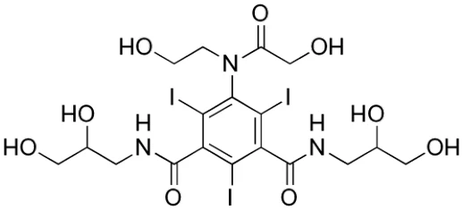
Physicochemical properties: OPTIRAY formulations are clear, colourless to pale yellow, sterile, non-pyrogenic aqueous solutions. Crystallization does not occur at room temperature.
The pH of the OPTIRAY formulation is adjusted between 6.0 and 7.4 with hydrochloric acid or sodium hydroxide.
Table 5 OPTIRAY physicochemical properties
| OPTIRAY | OPTIRAY | OPTIRAY | |
| 300 | 320 | 350 | |
| Ioversol content (mg/mL) | 636 | 678 | 741 |
| Iodine content (mg/mL) | 300 | 320 | 350 |
| Osmolality (mOsm/kg) | 651 | 702 | 792 |
| Viscosity (cps) | |||
| 25°C | 8.2 | 9.9 | 14.3 |
| 37°C | 5.5 | 5.8 | 9.0 |
14. Clinical Trials
14.1. Clinical Trials by Indication
Please refer to sections 8 Adverse Reactions and 10 Clinical Pharmacology.
14.2. Comparative Bioavailability Studies
No information available
- 16. Non-Clinical Toxicology
General toxicology
Non-clinical pharmacology
The biodistribution, metabolism and excretion of OPTIRAY were evaluated in rats and dogs following intravenous injection of 0.2 and 2,0 g I/kg, for these studies, OPTIRAY was exchange-labelled with 125I and radioactivity was assayed in biological samples.
Following intravenous injection of two lots of 125I ioversol into rats at dose levels of 0.2 and 1.0 g I/kg, 6 male and 6 female rats being used at each dose level, radioactivity was rapidly excreted, primarily via the urine. Two male and 2 female rats were killed at 2, 24 and 48 h for assay of radioactivity in organs, urine and feces. Seventy-three to 90% of the radioactivity had been excreted by 2 hours. By 24 hours, 91-99% of the injected dose was recovered in urine and feces, and approximately 1 % of the dose was retained by all assayed organs and tissues (thyroid 1.1 %, liver 0.5%). The experiment was repeated with 24 additional rats with similar results.
Following a single intravenous injection of 125I ioversol in 12 conscious beagle dogs, 6 male and 6 female, the disappearance of radioactivity from blood was typically bi-exponential with an initial distribution half-life ranging from approximately 1 to 4 minutes followed by a terminal elimination phase with a half-life ranging from approximately 40-55 minutes. No significant organ retention of radioactivity was apparent after 48 hours and 86-88% of the radioactivity was excreted, primarily in the urine, within 48 hours. Fecal excretion amounted to about 3-9%. Upon reverse phase HPLC analysis, ioversol accounted for essentially all of the radioactivity excreted in the urine, suggesting that ioversol is excreted in an unchanged form in dogs.
Following intracisternal injection of 125I-ioversol (240 mgI/kg) in 5 beagle dogs, 1 male and 4 female, activity was measured in the blood at 5 minutes and peaked at 3.9 hours post-injection. The elimination t1/2 in the blood was 1.04 hours. The main route of excretion was in the urine, where 74% of the dose was recovered within 24 hours. By 72 hours post-injection, approximately 94% of the injected dose was eliminated in the urine and feces. Thus, elimination of ioversol injected into the cisterna magna was found to be fairly rapid and comparable to that reported for iopamidol in similar studies.
Four female beagle dogs received ioversol and 4 iopamidol. Plasma concentrations were assayed by HPLC and CSF and urine concentrations by UV spectrophotometry, Both contrast media appeared in the plasma 15 min. after injection. Peak concentrations were attained within 56 min. The contrast media then rapidly disappeared from the plasma, the elimination t½ for ioverosl being 1.37 hr. and for iopamidol1.23 hr. Urinary excretion for ioversol and iopamidol respectively was 37% and 39% after 2 hours and 69% and 74% after 6 hours. The results show that intrathecal administration of the two-contrast media is followed by rapid absorption into the plasma and prompt urinary excretion. The absorption and elimination patterns of the two media via the route of administration were identical.
Upon incubation with dog red blood cells, ioversol caused hemolysis ranging from 0% at 2.5%I to 61% at 18.5%I w/v. Crenation (70-100%) and aggregation of red blood cells occurred only at high levels of ioversol (10% and 18.5%I). The results of this study indicate that ioversol is compatible with dog red blood cells at clinically relevant intravascular doses. It is likely also to be compatible with human red blood cells.
Ioversol does not notably bind to serum or plasma proteins to any marked extent and no significant metabolism, deiodination or biotransformation occurs. Using gel exclusion chromatography, ioversol exhibited a low order of human plasma protein binding (9 to 13%), which is of no consequence from a clinical standpoint.
The effects on blood coagulation of OPTIRAY were compared with those of iopamidol, iohexol, diatrizoate and ioxaglate at concentration up to 32 mgI/mL when mixed with plasma (1:10 v/v) from human volunteers. Coagulation parameters were measured by effects on activated partial thromboplastin time, prothrombin time and thrombin time, and platelet aggregation. Coagulation times were increased in a concentration-related manner with all of the contrast media.
Where significant differences existed, the prolongation of coagulation times was less with ioversol, iopamidol and iohexol than with the ionic agent diatrizoate. OPTIRAY was not significantly different from iopamidol or iohexol in the coagulation parameters measured. Ioversol, iopamidol and iohexol were found to inhibit platelet aggregation less than diatrizoate and ioxaglate. These findings are in line with the known greater anticoagulant effect of ionic agents such as diatrizoate when compared to non-ionic agents.
Histamine release from rat peritoneal mast cells was found to be significantly less (p<0.01) with ioversol, iohexol and iopamidol than with amidotrizoate. Transient elevations of ASAT, ALAT, LDH, bilirubin, creatinine and BUN were seen in several human subjects, but these were not clinically significant.
Single does intracarotid administration of 2 mL ioversol (28%I) in anesthetized rats, 6 rats per treatment group (total 18), showed a 26% drop in mean arterial pressure, compared with a 3% increase after saline, and a 46% drop after diatrizoate (28%I). Heart rate was reduced by 12%-a value which did not differ significantly from the saline control (-0.42%) but was significantly different from the drop with diatrizoate (-26%). A 7% increase in respiratory rate did not differ significantly from the saline control but was significantly different from diatrizoate (-93%). The incidence of arrhythmias with ioversol was the same as with the saline control (2/6 rats). Damage to the blood brain barrier was evaluated in 18 rats (6 each with ioversol 28%I, diatrizoate 28%I or saline) by intracarotid injection of sodium Tc-99m) pertechnetate one minute after test substance administration and determining the Tc-99m brain/blood ratio one minute. Later, the Tc-99m retention index was three times greater for the sodium diatrizoate than for the OPTIRAY and saline groups, indicating that OPTIRAY did not alter the blood-brain barrier, whereas sodium diatrizoate caused disruption of the barrier integrity.
Ioversol and iopamidol were each injected intracisternally into 16 female rats at dose levels of 60, 120, 240 and 480 mgI/kg (4 rats for each concentration, 32 in all). Sedation and hyper-activity were alternating events which were seen during the first hour after injection. The animals recovered uneventfully overnight. Two animals at the 480 mgI/kg dose level of iopamidol displayed mild convulsions during the first hour after dosing, but none were seen in the ioversol group. Mild teeth gnashing was seen with 3 animals: 2 with ioversol at 480 mgI/kg and 1 with iopamidol at 240 mgI/kg. Drug-related weight losses were seen during a 48 hour post-injection period in 4/4 rats given ioversol and in 2/4 given iopamidol - a significant difference. Such weight losses are not uncommon in animals who are given contrast media while under general anesthesia. Thus, by the intracisternal route in rats, ioversol is not more toxic than iopamidol.
Ioversol 30%I was relatively well tolerated by 3 cynomolgus monkeys given a single dose of 0.2 mL/kg intrathecally. Ioversol caused muscular fasciculations and/or tremor in 2/3 animals during the first hour after injection. No convulsions or pre-convulsive activity were seen. There were no other adverse events, no deaths and no signs of adhesive arachnoiditis at necropsy 12 weeks after injection. OPTIRAY, therefore, does not appear to be toxic at the above dose by this route.
Cardiovascular Effects
Ioversol (37%I) caused a transient positive inotrophy and bradycardia in the isolated perfused rabbit heart following a single bolus injection. At the highest dose level intracoronary injection of 4.0 mL caused fibrillation in 5/6 preparations with ioversol, 3/5 with iohexol and 4/6 with iopamidol. The coronary cardiotoxicity of ioversol in the isolated perfused rabbit heart was qualitatively and quantitatively similar to that of the other non-ionic X-ray contrast media, iohexol (35%I) and iopamidol (37%I). The non-ionic agents differed from diatrizoate (37%I) in that they were more liable to cause fibrillation at the 4.0 mL dose and to cause increases in contractile force, while the diatrizoate caused marked dose-related decreases.
The hemodynamic effects of intravenously injected 37%I ioversol and iopamidol, 35%I iohexol and 37%I sodium meglumine diatrizoate were compared in 16 pentobarbital anesthetized dogs (4 per compound). Changes were grades as minimal (1-10%), slight (11-20%), moderate (21-30%) or marked (>30%). The non-ionics, at doses of 1.2 and 4 mL/kg, caused minimal to slight perturbations of left ventricular pressure, myocardial contractility, heart rate and blood pressure. Diatrizoate, in contrast, caused markedly reduced contractility and lowering of left ventricular, systolic and diastolic pressures. Therefore, while the non-ionic caused only minimal to slight changes in cardiovascular parameters, those produced by diatrizoate were distinguishable because of their greater magnitude. It may therefore be concluded that non-ionic agents cause less cardiovascular perturbations than ionic agents.
The cardiotoxicity of 37%I ioversol and 37%I sodium meglumine diatrizoate were compared using left intra ventricular doses of 1 and 3 mL and selective coronary injection (left: 5 and 10 mL, right: 5 and 8 mL) in 16 pentobarbital-anesthetized, closed chest dogs, 4 per test substance. Each dog received doses of 1, 2 and 4 mL/kg. As before, changes were graded as minimal (1-10%), slight (11-20%), moderate (21-30%) or marked (30%). Thirty seconds after intra ventricular injection of 3 mL/kg, diatrizoate induced a fall in arterial pressure of 46 mmHg (32%) compared to a fall after ioversol of 23 mmHg (16%). The mean fall of arterial pressure 30 seconds after intra-coronary injections was: for the left coronary: ioverol-5.0 mmHg, diatrizoate -22mmHg; for the right coronary: ioversol-11.5 mmHg, diatrizoate -3mmHg. At 15 seconds following injection the means were: for the left coronary: ioversol-7.5 mmHg, diatrizoate -22 mmHg; for the right coronary: ioversol-l1.5 mmHg, diatrizoate -24 mmHg. These effects were therefore minimal or slight with ioversol versus moderate with diatrizoate. Arrhythmias occurred with ioversol in 1/3 dogs with left coronary injections of 10 mL and in 3/3 with diatrizoate. With right coronary injections of 5 mL, 3/3 dogs had arrhythmias with both agents and one of the dogs fibrillated and dies following diatrizoate. After 8 mL, two out of 3 dogs died with fibrillation following right coronary artery injection with ioversol and 1/3 after diatrizoate injection. The incidence of arrhythmias, consisting mainly of premature ventricular contractions and fibrillation, was similar for each compound. In clinical studies with ioverol and in 2 to 3 years' use in patients worldwide, no corresponding effects have been seen in humans.
Toxicology
Acute Toxicity
In a mouse study, with 150m, 50f, 20 saline controls and 20 untreated controls, dosed intravenously 16-22 gl/kg, LD50 was 18.4 gl/kg. Mice showed hypoactivity and respiratory depression. Livers showed chronic granulomatous inflammation, with necrosis at two highest dose groups.
In a rat study, with 50m, 50f and 20 saline controls, dosed intravenously 14-18 gl/kg, LD50 was 15 gl/kg. Rats showed hypoactivity and respiratory depression (former resolved by 4h and the latter by ½ h), vacuolation of renal tubules, and convulsions occurred in 4/20 animals.
In a dog (purebred beagle) study, with 6m, 6f, 4 untreated controls and 4 saline controls, dosed intravenously 3-12 gl/kg, at highest dose trembling, licking, urination, retching, vomiting and hematuria were seen (resolved by 30 mins.).
In a mouse study, with 48m, 48f and 64 mice in iohexol control group, dosed intravenously 3.0, 6.0 and 12.0 gl/kg (same doses for iohexol controls), 8 mice per timepoint (3, 7, 14 and 29 days after dosing) were killed and autopsied for evidence of hepatotoxicity. Significant degenerative changes were seen microscopically in the livers of some mice of all groups treated with both ioversol and iohexol. Subcapsular, granulomatous inflammation occurred in 4.7% of ioversol mice and 6.5% of iohexol mice and was most marked at the 12 gI/kg dose. Vacuolative hepatocellular degeneration occurred only at this dose and was seen at day 3 only with incidences of 5/8 and 4/8 for ioversol and iohexol respectively. The changes were not seen at 29 days post-injection. It was concluded that both contrast agents showed a similar incidence of reversible hepatic toxicity in 50 to 60% of mice.
Acute EEG effects of intracisteral metrizamide, ioversol, iohexol and iopamidol in rats.
Subacute toxicity
In a rat study, with 72m, 72f, 40 untreated controls and 40 saline controls, dosed intravenously daily at 0.2, 0.8 and 3.2 gl/kg for 28 days (with recovery observations to 56 days), microscopic changes consisting of minimal to moderate dose-related renal vacuolation at the 0.8 and 3.2 g/I/kg/day dose levels were observed. Renal vacuolation was not seen at lower dose levels or in any rats after 28 and 56 day recovery periods. It was concluded that ioversol showed a low order of toxicity.
In a dog (purebread beagle), with 30m, 30f, 12 untreated controls and 12 saline controls, dosed intravenously daily at 0.2, 0.8 and 3.2 gl/kg for 28 days, dose-related emesis was seen during the dosing period of the study. Mild hepato-cellular vacuolation was noted at the 3.2 g/I/kg/day dose level in 4/8 dogs. They were not seen in dogs at the end of an 8 week recovery period.
Genotoxicity
Two in vitro mutagenicity and chromosomal aberration studies indicated that ioversol 32%I w/v solution, in doses 0.1 to 150 mL per plate, did not possess mutagenic activity. Incubation with ioversol 32%I did not transform Salmonella typhimurium, Escheriscia coli or mouse lymphoma cells under non-metabolic, or metabolic activation conditions (using Arochlor® 1254-induced rat liver metabolic activation system). In addition, ioversol did not induce chromosomal aberrations in Chinese hamster ovary cells in vitro under both non-metabolic and metabolic activation conditions.
Carcinogenicity
No information available
Reproductive and developmental toxicology
Ioversol was given intravenously to 3 groups of 30 female rats at dose levels of 0.2, 0.8 and 3.2 gI/kg once daily during days 7 through 17 of pregnancy. Higher values for litter size in test groups (not statistically significant) were related to higher implantation rates. There was a dose-related tendency to reduced fetal weights which was significant compared to the controls but not between dosage groups. There was also a dose-related reduction in litter size and implantation rate and a higher pup mortality rate. There was a non-significant, dose-related increase in fetuses with skeletal abnormalities. Group mean incidences of malformations and visceral anomalies were unrelated to dosage.
Intravenous administration of ioversol to 3 groups of 20 male Sprague-Dawley rats daily from 9 weeks prior to mating and throughout the mating period did not affect mating performance.
Intravenous administration of ioversol to 3 groups of 30 female Sprague-Dawley rats at dose levels of 0.2,0.8 and 3.2 gI/kg daily from 2 weeks prior to mating and throughout mating, pregnancy and lactation did not affect duration of gestation, pre- and post-implantation loss, litter size or mean litter weight. No treatment-related adverse events were noted in the Fl or F2 offspring. Although 3.2 gI/kg ioversol was a no-effect level in this study, at 4.8 gI/kg tremors, languid behaviour and polynea were noted. At 3.2 gI/kg/day some reduction in food intake was observed, with differences attaining statistical significance on days 1 to 6 post-partum; also a slight retardation of body weight gain during late gestation occurred. Also at this highest dosage level there was a reduction in litter size. Mean pup weight was also lower, which resulted in a reduction of litter weight from day 4 to day 21 post-partum. It was not considered that treatment with ioversol had any significant adverse events on the dam or resulting litter parameters at 0.2 or 0.8 gI/kg/day; neither were there any adverse events on the maturation of the Fl generation.
Ioversol was given intravenously at doses of 0.2, 0.8 and 3.2 gI/kg/day to three groups of pregnant rabbits (total 54) once daily during days 6 through 18 of pregnancy, Ioversol treatment was well tolerated in the pregnant rabbits and no treatment-related visceral or skeletal abnormalities were noted in fetuses derived from those killed on day 29 of pregnancy.
Juvenile toxicity
No information available.
Special toxicology
No information available.