


Product Monograph - Cellcept
1. Indications
CellCept® (mycophenolate mofetil) is indicated for: Adults
- The prophylaxis of organ rejection in patients receiving allogeneic renal, cardiac or hepatic transplants. CellCept should be used concomitantly with cyclosporine and corticosteroids.
CellCept i.v. (mycophenolate mofetil hydrochloride for intravenous infusion) is an alternative dosage form to CellCept capsules, tablets and oral suspension. CellCept i.v. should be administered within 24 hours following transplantation. CellCept i.v. can be administered for up to 14 days; patients should be switched to oral CellCept as soon as they can tolerate oral medication.
1.1. Pediatrics
Pediatrics (2-18 years of age): CellCept is indicated for the prophylaxis of organ rejection in pediatric patients (2 to 18 years) receiving allogeneic renal transplants. CellCept should be used concomitantly with cyclosporine and corticosteroids.
1.2. Geriatrics
- No data are available to Health Canada; therefore, Health Canada has not authorized an indication for geriatric use.
2. Contraindications
- CellCept (mycophenolate mofetil) is contraindicated in patients with a history of hypersensitivity, including anaphylaxis, to mycophenolate mofetil, mycophenolic acid or any component of the drug product (see 7. Dosage Forms, Strengths, Composition, and Packaging).
- CellCept i.v. (mycophenolate mofetil hydrochloride for injection) is contraindicated in patients who are allergic to Polysorbate 80 (TWEEN) (see 7. Dosage Forms, Strengths, Composition, and Packaging).
- CellCept is contraindicated during pregnancy due to its mutagenic and teratogenic potential (see 7. Warnings and Precautions).
- CellCept is contraindicated in women of childbearing potential not using highly effective contraceptive methods and without providing a pregnancy test result (see 7. Warnings and Precautions).
- CellCept is contraindicated in women who are breastfeeding (see 7. Warnings and Precautions).).
3. Serious Warnings and Precautions Box
|
|---|
|
4. Dosage and Administration
4.1. Dosing Considerations
- CellCept (mycophenolate mofetil) should be used concomitantly with standard cyclosporine and corticosteroid therapy
CellCept Capsules, Tablets and Powder for Oral Suspension
- The initial oral dose of CellCept should be given as soon as possible following renal, cardiac or hepatic transplantation. Food had no effect on MPA AUC, but has been shown to decrease MPA Cmax by 40%. It is recommended that CellCept be administered on an empty stomach.
- Note: If required CellCept Oral Suspension can be administered via a nasogastric tube with a minimum size of 8 French.
CellCept i.v.
- CellCept i.v. is an alternative dosage form to CellCept capsules and tablets recommended for patients unable to take CellCept capsules or tablets. CellCept i.v. should be administered within 24 hours following transplantation. CellCept i.v. can be administered for up to 14 days; patients should be switched to oral CellCept as soon as they can tolerate oral medication.
Caution: CellCept i.v. solution should never be administered by rapid or bolus intravenous injection.
4.2. Recommended Dose and Dosage Adjustment
Adults
Renal Transplantation
A dose of 1 g administered orally or intravenously (over 2 hours) twice a day (daily dose of 2 g) is recommended for use in renal transplant patients. Although a dose of 1.5 g administered twice daily (daily dose of 3 g) was used in clinical trials and was shown to be safe and effective, no efficacy advantage could be established for renal transplant patients. Patients receiving 2 g per day of CellCept in these trials demonstrated an overall better safety profile than did patients receiving 3 g per day of CellCept.
Cardiac Transplantation
A dose of 1.5 g twice daily administered intravenously (over no less than 2 hours) or 1.5 g twice daily oral (daily dose of 3 g) is recommended for use in adult cardiac transplant patients.
Hepatic Transplantation
A dose of 1 g twice daily administered intravenously (over no less than 2 hours or 1.5 g twice daily oral (daily dose of 3 g) is recommended for use in adult hepatic transplant patients.
Pediatrics (2 to 18 years)
The recommended dose of CellCept oral suspension for renal transplant patients is
600 mg/m2 body surface area twice daily (up to a maximum of 2 g daily).
Patients with a body surface area of 1.25 to 1.5 m2 may be dosed with CellCept capsules at a dose of 750 mg twice daily (1.5 g daily dose). Patients with a body surface area >1.5 m2 may be dosed with CellCept capsules or tablets at a dose of 1 g twice daily (2 g daily dose).
Dosage Adjustment
Renal Impairment
In renal transplant patients with severe chronic renal impairment (GFR <25mL/min/1.73m2) outside the immediate post-transplant period, doses of CellCept greater than 1 g administered twice a day should be avoided. These patients should also be carefully observed (see 10.3. Pharmacokinetics, Special Populations and Conditions, Renal Insufficiency).
No data are available for cardiac or hepatic transplant patients with severe chronic renal impairment. CellCept should be used for cardiac or hepatic transplant patients with severe chronic renal impairment if the potential benefits outweigh the potential risks.
If neutropenia develops (ANC <1.3 x 10³/µL), dosing with CellCept should be interrupted or the dose reduced, appropriate diagnostic tests performed, and the patient managed appropriately. (See 7. Warnings and Precautions, Immune, Monitoring and Laboratory Tests and 8. Adverse Reactions)
Delayed Renal Graft Function Post Transplant
No dose adjustment is recommended for these patients, however, they should be carefully observed (see 10.3. Pharmacokinetics, Special Populations and Conditions, Renal Insufficiency).
4.3. Reconstitution
Oral Solutions:
Preparation of Oral Suspension:
- 1. Tap the closed bottle several times to loosen the powder.
- 2. Measure 94 mL of purified water in a graduated cylinder.
- 3. Add approximately half of the total amount of the purified water for reconstitution to the bottle and shake the closed bottle well for about 1 minute.
- 4. Add the remainder of water and shake the closed bottle well for about 1 minute.
- 5. Remove the child-resistant cap and push bottle adapter into neck of bottle.
- 6. Close bottle with child-resistant cap tightly. This will assure the proper seating of the bottle adapter in the bottle and child-resistant status of the cap.
Dispense with patient information leaflet and oral dispensers. Oral dispensers are for use with CellCept oral suspension only. It is recommended to write the date of expiration of the reconstituted suspension on the bottle label in the space provided. (The shelf life of the reconstituted suspension is 60 days.)
Net contents after reconstitution of the oral suspension is 175 mL, containing 200 mg/mL mycophenolate mofetil. Store reconstituted suspension at 15-30ºC or in the refrigerator between 2-8ºC.. Do not freeze. Discard any unused portion 60 days after reconstitution.
Parenteral Products:
Preparation of Infusion Solution (6 mg/mL)
CellCept i.v. does not contain an antibacterial preservative; therefore reconstitution and dilution of the product must be performed under asceptic conditions.
CellCept i.v. infusion solution must be prepared in two steps: the first step is a reconstitution step with 5% Dextrose Injection, USP and the second step is a dilution step with 5% Dextrose Injection, USP. A detailed description of the preparation is given below:
Step 1
a. Two (2) vials of CellCept i.v. are used for preparing each 1 g dose, whereas three (3) vials are needed for each 1.5 g dose. Reconstitute the contents of each vial by injecting 14 mL of 5% Dextrose Injection, USP.
b. Gently shake the vial to dissolve the drug.
c. Inspect the resulting slightly yellow solution for particulate matter and discoloration prior to further dilution. Discard the vial if particulate matter or discoloration is observed.
Step 2
a. To prepare a 1 g dose, further dilute the contents of the two reconstituted vials (approx. 2 x 15 mL) into 140 mL of 5% Dextrose Injection USP. To prepare a 1.5 g dose, further dilute the contents of the three reconstituted vials (approx. 3 x 15 mL) into 210 mL of 5% Dextrose Injection USP. The final concentration of both solutions is 6 mg mycophenolate mofetil per mL.
b. As with all parenteral drug products, diluted solution should be inspected visually for clarity, particulate matter, precipitate, discoloration and leakage prior to administration whenever solution and container permit. Solutions showing haziness, particulate matter, precipitate, discoloration or leakage should not be used. Discard unused portion.
If the infusion solution is not prepared immediately prior to administration, the commencement of administration of the infusion solution should be within 4 hours from reconstitution and dilution of the drug product. Keep solutions at 15-30°C.
CellCept i.v. should not be mixed or administered concurrently via the same infusion catheter with other intravenous drugs or infusion admixtures.
4.4. Administration
CellCept (tablets, capsules) should be administered orally, and should be taken on an empty stomach (see 10.3. Pharmacokinetics, Absorption).
It is recommended that CellCept Powder for Oral Suspension be reconstituted by the pharmacist prior to dispensing to the patient (see 4.3. Reconstitution,, Preparation of Oral Suspension).
CellCept i.v. must be reconstituted and diluted to a concentration of 6 mg/mL using 5% Dextrose Injection USP (see 4.3. Reconstitution, Preparation of Infusion Solutions). CellCept i.v. is incompatible with other intravenous infusion solutions. Following reconstitution, CellCept i.v. must be administered by slow intravenous infusion over a period of no less than 2 hours by either peripheral or central vein.
5. Overdose
Reports of overdoses with mycophenolate mofetil have been received from clinical trials and during post-marketing experience. In many of these cases no adverse events were reported. In those overdose cases in which adverse events were reported, the events fall within the known safety profile of the drug.
It is expected that an overdose of mycophenolate mofetil could possibly result in oversuppression of the immune system and increase susceptibility to infections and bone marrow suppression (see 7. Warnings and Precautions, Immune). If neutropenia develops, dosing with CellCept should be interrupted or the dose reduced (see 7. Warnings and Precautions, Immune)
The highest dose administered to renal transplant patients in clinical trials has been 4 g per day. In limited experience with cardiac and hepatic transplant patients in clinical trials, the highest doses used were 4 g or 5 g per day. At doses of 4 g or 5 g per day, there appears to be a higher rate, compared to the use of 3 g per day or less, of gastrointestinal intolerance (nausea, vomiting, and/or diarrhea), and occasional hematologic abnormalities, principally neutropenia, leading to a need to reduce or discontinue dosing.
At clinically encountered concentrations, MPA and MPAG are not removed by hemodialysis. However, at high MPAG plasma concentrations (>100 µg/mL), small amounts of MPAG are removed. By interfering with enterohepatic recirculation of the drug, bile acid sequestrants, such as cholestyramine reduce the MPA AUC.
| For the most recent information in the management of a suspected drug overdose, contact your regional poison control centre or Health Canada’s toll-free number, 1-844 POISON-X (1-844-764-7669). |
|---|
- 6. Dosage Forms, Strengths, Composition, and Packaging
Table 1 Dosage Forms, Strengths, Composition and Packaging
| Route of Administration | Dosage Form / Strength/Composition | Non-medicinal Ingredients |
| Oral | Capsules/250 mg | None |
| Oral | Tablets/500 mg | None |
| Oral | Powder/200 mg/mL (reconstituted) | Aspartame (See 7. Warnings and Precautions, Endocrine and Metabolism, Phenylketonurics) |
| Intravenous | Lyophilized powder /500 mg/vial (as hydrochloride) | Polysorbate 80 (TWEEN) (See 2. Contraindications) |
Description
Capsules
Composition: CellCept (mycophenolate mofetil) is available for oral administration as capsules containing 250 mg of MMF. Inactive ingredients include croscarmellose sodium, magnesium stearate, povidone (K-90) and pregelatinized starch. The capsule shells contain black iron oxide, indigotine (FD&C blue #2), gelatin, potassium hydroxide, red iron oxide, shellac, titanium dioxide, and yellow iron oxide.
Availability: CellCept capsules are oblong, blue/brown, two-piece hard gelatin capsules, printed in black with "CellCept 250" on the blue cap and "Roche" on the brown body. They are provided in unit dose of 10 capsules in blister packs, 10 packs per box.
Film-Coated Tablets
Composition: CellCept is also available for oral administration as tablets containing 500 mg of MMF. Inactive ingredients include croscarmellose sodium, hydroxypropyl cellulose, hydroxypropyl methylcellulose, indigotine (FD&C blue #2) aluminum lake, iron oxide, magnesium stearate, microcrystalline cellulose, polyethylene glycol 400, povidone (K-90), titanium dioxide
Availability: CellCept tablets are lavender coloured, caplet-shaped, film coated tablets, engraved with “CellCept 500" on one side and “Roche” on the other. They are provided in unit dose of 10 tablets in blister packs, 5 packs per box.
Powder for Oral Suspension
Composition: CellCept is also available for oral administration as a powder for oral suspension, which when reconstituted contains 200 mg/mL of MMF. Each bottle of CellCept powder for oral suspension contains 35 g of MMF in a 110 g powder for oral suspension. Inactive ingredients in CellCept Powder for Oral Suspension are aspartame, citric acid, colloidal silicon dioxide, fruit flavour, methylparaben, sodium citrate, sorbitol, soybean lecithin, and xanthan gum.
Availability: CellCept powder for oral suspension is supplied as a white to off-white powder blend for reconstitution to a white to off-white fruit flavour suspension. It is provided in a 225 mL bottle with bottle adapter and 2 oral dispensers*. The deliverable volume after reconstitution is 165 mL.
* Oral dosing dispenser manufactured by B. Braun Melsungen AG. 34209 Melsungen Germany
Lyophilized Powder for Solution for Intravenous Infusion
Composition: CellCept i.v. is available as a sterile white to off-white lyophilized powder in vials containing MMF hydrochloride for administration by intravenous infusion only. Each vial of CellCept i.v. contains the equivalent of 500 mg MMF as the hydrochloride salt. The inactive ingredients are citric acid, 5 mg, polysorbate 80, 25 mg, and sodium hydroxide and/or hydrochloric acid to adjust pH.
Availability: CellCept i.v. is supplied in a 20 mL, sterile vial containing the equivalent of 500 mg MMF, as the hydrochloride salt in cartons of 4 vials. Each vial is intended for single use only.
7. Warnings and Precautions
Please see 3. Serious Warnings and Precautions Box.
General
Caution: CellCept i.v. solution should never be administered by rapid or bolus intravenous injection.
Caution should be exercised when switching combination therapy from regimens containing immunosuppressants, which interfere with mycophenolic acid (MPA) enterohepatic recirculation e.g. cyclosporine to others devoid of this effect e.g. tacrolimus, sirolimus, belatacept, or vice versa, as this might result in changes of MPA exposure (See 9. Drug Interactions). Drugs which interfere with MPA’s enterohepatic cycle (e.g. cholestyramine, sevelamer, antibiotics) should be used with caution due to their potential to reduce the plasma levels and efficacy of CellCept (see 9.4. Drug-Drug Interactions). Therapeutic drug monitoring of MPA may be appropriate when switching combination therapy (e.g. from ciclosporin to tacrolimus or vice versa) or to ensure adequate immunosuppression in patients with high immunological risk (e.g. risk of rejection, treatment with antibiotics, addition or removal of an interacting medication).
It is recommended that CellCept (mycophenolate mofetil) should not be administered concomitantly with azathioprine because both have the potential to cause bone marrow suppression and such concomitant administration has not been studied clinically.
Hypersensitivity Reactions
Post-marketing cases of serious hypersensitivity reactions, including angioedema and anaphylaxis, have been reported with MMF with or without concomitant therapies, premedication, and/or a previous history of hypersensitivity reaction. These reactions generally occur within minutes to a day after initiating MMF. It is recommended that if signs or symptoms of anaphylactic reaction occur, MMF should be discontinued permanently. The patients should be provided appropriate medical treatment immediately and monitored until the symptoms resolve (see 2. Contraindications).
Blood Donation
Patients should not donate blood during therapy and for at least 6 weeks following discontinuation of CellCept.
Carcinogenesis and Genotoxicity
Neoplasms
Patients receiving CellCept, as part of an immunosuppressive regimen are at increased risk of developing lymphomas and other malignancies, particularly of the skin. The risk appears to be related to the intensity and duration of immunosuppression rather than to the use of any specific agent. As with all patients at an increased risk for skin cancer, exposure to sunlight and UV light should be limited by wearing protective clothing and using a sunscreen with a high protection factor (see 8. Adverse
Reactions).
Driving and Operating Machinery
CellCept may have a moderate influence on the ability to drive and use machines.
Patients should be advised to use caution when driving or using machines if they experience adverse drug reactions such as somnolence, confusion, dizziness, tremor or hypotension during treatment with CellCept.
Endocrine and Metabolism
CellCept is an inosine monophosphate dehydrogenase (IMPDH) inhibitor, therefore it should be avoided in patients with rare hereditary deficiency of hypoxanthine-guanine phosphoribosyl-transferase (HGPRT) such as Lesch-Nyhan and Kelley-Seegmiller syndrome.
Gastrointestinal
CellCept should be administered with caution in patients with active serious digestive system disease. Gastrointestinal bleeding (requiring hospitalization) has been observed in approximately 3% of renal, in 1.7% of cardiac and in 5.4% of hepatic transplant patients treated with CellCept 3g daily. CellCept has been associated with an increased incidence of digestive system adverse events, including infrequent cases of gastrointestinal tract ulceration, and rarely perforation (colon, gall bladder). Most patients receiving CellCept were also receiving other drugs that are known to be associated with these complications. Patients with active peptic ulcer disease were excluded from enrollment in studies with mycophenolate mofetil.
Hematologic
Cases of pure red cell aplasia (PRCA) have been reported in patients treated with CellCept in combination with other immunosuppressive agents. The mechanism for mycophenolate mofetil induced PRCA is unknown. In some cases PRCA was found to be reversible with dose reduction or cessation of CellCept therapy. In transplant patients however reduced immunosuppression may place the graft at risk.
Patients receiving CellCept should be instructed to report immediately any evidence of infection, unexpected bruising, bleeding or any other manifestation of bone marrow depression.
Patients receiving CellCept should be monitored for neutropenia. Complete blood counts should be performed weekly during the first month, twice monthly for the second and third months of treatment, then monthly through the first year (see 7. Warnings and Precautions, Monitoring and Laboratory Tests and 4.2. Recommended Dose and Dosage Adjustment). The development of neutropenia may be related to CellCept itself, concomitant medications, viral infections, or some combination of these causes. If neutropenia develops (absolute neutrophil count [ANC] <1.3 x 10³/µL), dosing with CellCept should be interrupted or the dose should be reduced, appropriate diagnostic tests performed, and the patient managed appropriately. Neutropenia has been observed most frequently in the period from 31 to 180 days post-transplant for patients treated for prevention of renal, cardiac and hepatic rejection.
Severe neutropenia (ANC < 0.5 x 10³/µL) developed in up to 2.0% of renal, up to 2.8% of cardiac and up to 3.6% hepatic transplant patients receiving CellCept 3g daily (see 8. Adverse Reactions).
Immune
CellCept has been administered in combination with the following agents in clinical trials: anti-thymocyte globulin [equine] (Atgam®) induction, muromonab-CD3 (Orthoclone OKT®3), cyclosporine (Sandimmune®, Neoral®), and corticosteroids. The efficacy and safety of the use of CellCept in combination with other immunosuppressive agents has not been determined.
Oversuppression of the immune system can also increase susceptibility to infection, including opportunistic infections, fatal infections and sepsis. Such infections include latent viral reactivation, such as hepatitis B or hepatitis C reactivation or infections caused by polyomaviruses. Cases of hepatitis due to reactivation of hepatitis B or hepatitis C have been reported in carrier patients treated with immunosuppresants.
Cases of progressive multifocal leukoencephalopathy (PML) associated with the JC virus, sometimes fatal, have been reported in CellCept treated patients. The reported cases had risk factors for PML, including immunosuppressant therapies and impairment of immune function. In immunosuppressed patients, physicians should consider PML in the differential diagnosis in patients reporting neurological symptoms and consultation with a neurologist should be considered as clinically indicated.
BK virus-associated nephropathy has been observed during the use of CellCept in patients post renal transplant. This infection can be associated with serious outcomes, sometimes leading to renal graft loss. Patient monitoring may help detect patients at risk for BK virus-associated nephropathy. Due to the cytostatic effect of CellCept on B- and T-lymphocytes, increased severity of COVID-19 may occur. Dose reduction or discontinuation of CellCept in immunosuppression should be considered for patients who develop evidence of BK virus-associated nephropathy, or in cases of clinically significant COVID-19.
In patients receiving CellCept (2 g or 3 g) in controlled studies for prevention of renal, cardiac or hepatic rejection, fatal infection/sepsis occurred in approximately 2% of renal and cardiac patients and in 5% of hepatic patients (see 8. Adverse Reactions).
Monitoring and Laboratory Tests
Complete blood counts should be performed weekly during the first month, twice monthly for the second and third months of treatment, then monthly through the first year (see 7. Warnings and Precautions, Immune and 4. Dosage and Administration).
Information for Patients
Patients should be informed of the need for repeated appropriate laboratory tests while they are receiving CellCept (see 7. Warnings and Precautions, Immune). Patients should be given complete dosage instructions and informed of the increased risk of lymphoproliferative disease and certain other malignancies.
Renal
Administration of doses of CellCept greater than 1 g administered twice a day to renal transplant patients with severe chronic renal impairment (GFR < 25 mL/min/1.73m2) should be avoided and patients should be carefully observed (see 10.3. Pharmacokinetics, Special Populations and Conditions, Renal Insufficiency and 4.2. Recommended Dose and Dosage Adjustment, Dosage Adjustment, Renal Impairment).
No data are available for cardiac or hepatic transplant patients with severe chronic, renal impairment. CellCept should be used for cardiac or hepatic transplant patients with severe, chronic, renal impairment if the potential benefits outweigh the potential risks.
Reproductive Health
- Fertility
Cellcept is contraindicated in women of childbearing potential not using highly effective contraceptive methods (see 2. Contraindications).
Before the start of treatment, female and male patients of reproductive potential must be made aware of the increased risk of pregnancy loss and congenital malformations and must be counseled regarding pregnancy prevention, and planning. Women of child bearing potential should use two reliable forms of contraception simultaneously, at least one of which must be highly effective, before beginning CellCept therapy, during therapy, and for six weeks following discontinuation of therapy, unless abstinence is the chosen method of contraception.
Prior to starting therapy with CellCept, female patients of childbearing potential must have two negative serum or urine pregnancy tests with a sensitivity of at least 25 mIU/mL; the second test should be performed 8-10 days later. Repeat pregnancy tests should be performed during routine follow-up visits. Results of all pregnancy tests should be discussed with the patient. Patients should be instructed to consult their physician immediately should pregnancy occur.
Limited clinical evidence is currently available on paternal exposure to CellCept. Based on the animal data, the risk of genotoxic effects on sperm cells cannot completely be excluded. In absence of sufficient data to exclude a risk of harm to the fetus conceived during or directly after the treatment of the father, the following precautionary measure is recommended: sexually active male patients and/or their female partners are recommended to use effective contraception during treatment of the male patient and for at least 90 days after cessation of treatment. If pregnancy does occur during treatment, the physician and patient should discuss the desirability of continuing the pregnancy.
Men should not donate semen during therapy and for 90 days following discontinuation of CellCept.
7.1. Special Populations
7.1.1. Pregnancy
CellCept is contraindicated during pregnancy and in women of childbearing potential not using highly effective contraceptive methods and without providing a pregnancy test result (see 2. Contraindications and 8.5. Post-Market Adverse Reactions). Cellcept is a powerful teratogen and mutagen. Spontaneous abortion (rate of 45-49% compared to a reported rate between 12 and 33% in solid organ transplant patients treated with other immunosuppressants) and congenital malformations (estimated rate of 23-27%) have been reported following MMF exposure during pregnancy (see 8.5. Post-Market Adverse Reactions). For comparison the risk of malformations is estimated at approximately 2% of live births in the overall population and at approximately 4 to 5 % in solid organ transplant patients treated with immunosupressants other than mycophenolate mofetil.
Studies in animals have shown reproductive toxicity (see 16. Non-Clinical Toxicology, Reproductive and Developmental Toxicity).
Labor and delivery
The safe use of CellCept during labor and delivery has not been established.
7.1.2. Breastfeeding
CellCept is contraindicated during breastfeeding due to the potential for serious adverse reactions in nursing infants (see 2. Contraindications). Studies in rats have shown mycophenolate mofetil is excreted in milk. Limited data shows that mycophenolic acid may be excreted in human breast milk.
7.1.3. Pediatrics
Safety and efficacy in children receiving allogeneic cardiac or hepatic transplants have not been established.
For pediatric patients receiving renal transplants also see 10.3. Pharmacokinetics, Special Populations and Conditions, Pediatrics; 14. Clinical Trials; 8.2.1. Clinical Trial Adverse Reactions - Pediatrics; and 4.2. Recommended Dose and Dosage Adjustment, Pediatrics
7.1.4. Geriatrics
Geriatric patients may be at an increased risk of adverse events such as certain infections (including cytomegalovirus tissue invasive disease) and possibly gastrointestinal haemorrhage and pulmonary oedema, compared with younger individuals.
8. Adverse Reactions
8.1. Adverse Reaction Overview
The adverse event profile associated with the use of immunosuppressive drugs is often difficult to establish owing to the presence of underlying disease and the concurrent use of many other medications. The principal adverse reactions associated with the administration of CellCept (mycophenolate mofetil) include diarrhea, leukopenia, sepsis and vomiting, and there is evidence of a higher frequency of certain types of infections.
The adverse event profile associated with the administration of CellCept i.v. (mycophenolate mofetil hydrochloride for injection) has been shown to be similar to that observed after administration of oral dosage forms of CellCept.
8.2. Clinical Trial Adverse Reactions
Clinical trials are conducted under very specific conditions. The adverse reaction rates observed in the clinical trials; therefore, may not reflect the rates observed in practice and should not be compared to the rates in the clinical trials of another drug. Adverse reaction information from clinical trials may be useful in identifying and approximating rates of adverse drug reactions in real-world use.
CellCept (oral)
The incidence of adverse events for CellCept was determined in randomized comparative double-blind trials in prevention of rejection in renal (2 active, 1 placebo controlled trials), cardiac (1 active controlled trial) and hepatic (1 active controlled trial) transplant patients.
Safety data are summarized below for all active controlled trials in renal (2 trials), cardiac
(1 trial) and hepatic (1 trial) transplant patients. Approximately 53% of renal patients,
65% of the cardiac patients and 45% of the hepatic patients have been treated for more than one year.
Adverse events, whether or not deemed to be causally associated with the study medication, reported in ≥ 10% of patients in treatment groups are presented below.
Table 2 Adverse Events in Controlled Studies in Prevention of Renal, Cardiac or Hepatic Allograft Rejection (Reported in ≥10% of Adult Patients Randomized to CellCept)
| Renal Studies | Cardiac Study | Hepatic Study | ||||||||||||||||||||||||
| Renal Studies | Cardiac Study | Hepatic Study | ||||||||||||||||||||||||
| Renal Studies | Cardiac Study | Hepatic Study | ||||||||||||||||||||||||
| Renal Studies | Cardiac Study | Hepatic Study | ||||||||||||||||||||||||
| CellCept 2 g/day | CellCept 3 g/day | Azathioprine 1-2 mg/kg/day or 100-150 mg/day | CellCept 3 g/day | Azathioprine 1.5-3 mg/kg/day | CellCept 3 g/day | Azathioprine 1-2 mg/kg/day | ||||||||||||||||||||
| CellCept 2 g/day | CellCept 3 g/day | Azathioprine 1-2 mg/kg/day or 100-150 mg/day | CellCept 3 g/day | Azathioprine 1.5-3 mg/kg/day | CellCept 3 g/day | Azathioprine 1-2 mg/kg/day | ||||||||||||||||||||
| CellCept 2 g/day | CellCept 3 g/day | Azathioprine 1-2 mg/kg/day or 100-150 mg/day | CellCept 3 g/day | Azathioprine 1.5-3 mg/kg/day | CellCept 3 g/day | Azathioprine 1-2 mg/kg/day | ||||||||||||||||||||
| CellCept 2 g/day | CellCept 3 g/day | Azathioprine 1-2 mg/kg/day or 100-150 mg/day | CellCept 3 g/day | Azathioprine 1.5-3 mg/kg/day | CellCept 3 g/day | Azathioprine 1-2 mg/kg/day | ||||||||||||||||||||
| (N=336) | (N=330) | (N=326) | (N=289) | (N=289) | (N=277) | (N=287) | ||||||||||||||||||||
| % | % | % | % | % | % | % | ||||||||||||||||||||
| Body as a Whole | ||||||||||||||||||||||||||
| Pain | 33.0 | 31.2 | 32.2 | 75.8 | 74.7 | 74.0 | 77.7 | |||||||||||||||||||
| Abdominal pain | 24.7 | 27.6 | 23.0 | 33.9 | 33.2 | 62.5 | 51.2 | |||||||||||||||||||
| Fever | 21.4 | 23.3 | 23.3 | 47.4 | 46.4 | 52.3 | 56.1 | |||||||||||||||||||
| Headache | 21.1 | 16.1 | 21.2 | 54.3 | 51.9 | 53.8 | 49.1 | |||||||||||||||||||
| Infection | 18.2 | 20.9 | 19.9 | 25.6 | 19.4 | 27.1 | 25.1 | |||||||||||||||||||
| Sepsis | 17.6 | 19.7 | 15.6 | 18.7 | 18.7 | 27.4 | 26.5 | |||||||||||||||||||
| Asthenia | 13.7 | 16.1 | 19.9 | 43.3 | 36.3 | 35.4 | 33.8 | |||||||||||||||||||
| Chest pain | 13.4 | 13.3 | 14.7 | 26.3 | 26.0 | 15.9 | 13.2 | |||||||||||||||||||
| Back pain | 11.6 | 12.1 | 14.1 | 34.6 | 28.4 | 46.6 | 47.4 | |||||||||||||||||||
| Accidental injury | - | - | - | 19.0 | 14.9 | 11.2 | 15.0 | |||||||||||||||||||
| Chills | - | - | - | 11.4 | 11.4 | 10.8 | 10.1 | |||||||||||||||||||
| Ascites | - | - | - | - | - | 24.2 | 22.6 | |||||||||||||||||||
| Abdomen enlarged | - | - | - | - | - | 18.8 | 17.8 | |||||||||||||||||||
| Hernia | - | - | - | - | - | 11.6 | 8.7 | |||||||||||||||||||
| Peritonitis | - | - | - | - | - | 10.1 | 12.5 | |||||||||||||||||||
| Cardiovascular | ||||||||||||||||||||||||||
| Hypertension | 32.4 | 28.2 | 32.2 | 77.5 | 72.3 | 62.1 | 59.6 | |||||||||||||||||||
| Hypotension | - | - | - | 32.5 | 36.0 | 18.4 | 20.9 | |||||||||||||||||||
| Cardiovascular disorder | - | - | - | 25.6 | 24.2 | - | - | |||||||||||||||||||
| Tachycardia | - | - | - | 20.1 | 18.0 | 22.0 | 15.7 | |||||||||||||||||||
| Arrhythmia | - | - | - | 19.0 | 18.7 | - | - | |||||||||||||||||||
| Bradycardia | - | - | - | 17.3 | 17.3 | - | - | |||||||||||||||||||
| Pericardial effusion | - | - | - | 15.9 | 13.5 | - | - | |||||||||||||||||||
| Heart failure | - | - | - | 11.8 | 8.7 | - | - | |||||||||||||||||||
| Digestive | ||||||||||||||||||||||||||
| Diarrhea | 31.0 | 36.1 | 20.9 | 45.3 | 34.3 | 51.3 | 49.8 | |||||||||||||||||||
| Constipation | 22.9 | 18.5 | 22.4 | 41.2 | 37.7 | 37.9 | 38.3 | |||||||||||||||||||
| Nausea | 19.9 | 23.6 | 24.5 | 54.0 | 54.3 | 54.5 | 51.2 | |||||||||||||||||||
| Dyspepsia | 17.6 | 13.6 | 13.8 | 18.7 | 19.4 | 22.4 | 20.9 | |||||||||||||||||||
| Vomiting | 12.5 | 13.6 | 9.2 | 33.9 | 28.4 | 32.9 | 33.4 | |||||||||||||||||||
| Nausea and vomiting | 10.4 | 9.7 | 10.7 | 11.1 | 7.6 | - | - | |||||||||||||||||||
| Oral moniliasis | 10.1 | 12.1 | 11.3 | 11.4 | 11.8 | 10.1 | 10.1 | |||||||||||||||||||
| Flatulence | - | - | - | 13.8 | 15.6 | 12.6 | 9.8 | |||||||||||||||||||
| Anorexia | - | - | - | - | - | 25.3 | 17.1 | |||||||||||||||||||
| Liver function tests abnormal | - | - | - | - | - | 24.9 | 19.2 | |||||||||||||||||||
| Cholangitis | - | - | - | - | - | 14.1 | 13.6 | |||||||||||||||||||
| Hepatitis | - | - | - | - | - | 13.0 | 16.0 | |||||||||||||||||||
| Cholestatic jaundice | - | - | - | - | - | 11.9 | 10.8 | |||||||||||||||||||
| Hemic and Lymphatic | ||||||||||||||||||||||||||
| Anemia | 25.6 | 25.8 | 23.6 | 42.9 | 43.9 | 43.0 | 53.0 | |||||||||||||||||||
| Leukopenia | 23.2 | 34.5 | 24.8 | 30.4 | 39.1 | 45.8 | 39.0 | |||||||||||||||||||
| Thrombocytopenia | 10.1 | 8.2 | 13.2 | 23.5 | 27.0 | 38.3 | 42.2 | |||||||||||||||||||
| Hypochromic anemia | 7.4 | 11.5 | 9.2 | 24.6 | 23.5 | 13.7 | 10.8 | |||||||||||||||||||
| Leukocytosis | 7.1 | 10.9 | 7.4 | 40.5 | 35.6 | 22.4 | 21.3 | |||||||||||||||||||
| Ecchymosis | - | - | - | 16.6 | 8.0 | - | - | |||||||||||||||||||
| Metabolic and Nutritional | ||||||||||||||||||||||||||
| Peripheral edema | 28.6 | 27.0 | 28.2 | 64.0 | 53.3 | 48.4 | 47.7 | |||||||||||||||||||
| Hypercholesteremia | 12.8 | 8.5 | 11.3 | 41.2 | 38.4 | - | - | |||||||||||||||||||
| Hypophosphatemia | 12.5 | 15.8 | 11.7 | - | - | 14.4 | 9.1 | |||||||||||||||||||
| Edema | 12.2 | 11.8 | 13.5 | 26.6 | 25.6 | 28.2 | 28.2 | |||||||||||||||||||
| Hypokalemia | 10.1 | 10.0 | 8.3 | 31.8 | 25.6 | 37.2 | 41.1 | |||||||||||||||||||
| Hyperkalemia | 8.9 | 10.3 | 16.9 | 14.5 | 19.7 | 22 | 23.7 | |||||||||||||||||||
| Hyperglycemia | 8.6 | 12.4 | 15.0 | 46.7 | 52.6 | 43.7 | 48.8 | |||||||||||||||||||
| Creatinine increased | - | - | - | 39.4 | 36.0 | 19.9 | 21.6 | |||||||||||||||||||
| BUN increased | - | - | - | 34.6 | 32.5 | 10.1 | 12.9 | |||||||||||||||||||
| Lactic dehydrogenase increased | - | - | - | 23.2 | 17 | - | - | |||||||||||||||||||
| Bilirubinemia | - | - | - | 18 | 21.8 | 14.4 | 18.8 | |||||||||||||||||||
| Hypervolemia | - | - | - | 16.6 | 22.8 | - | - | |||||||||||||||||||
| Generalized edema | - | - | - | 18.0 | 20.1 | 14.8 | 16 | |||||||||||||||||||
| Hyperuricemia | - | - | - | 16.3 | 17.6 | - | - | |||||||||||||||||||
| SGOT increased | - | - | - | 17.3 | 15.6 | - | - | |||||||||||||||||||
| Hypomagnesemia | - | - | - | 18.3 | 12.8 | 39 | 37.6 | |||||||||||||||||||
| Acidosis | - | - | - | 14.2 | 16.6 | - | - | |||||||||||||||||||
| Weight gain | - | - | - | 15.6 | 15.2 | - | - | |||||||||||||||||||
| SGPT increased | - | - | - | 15.6 | 12.5 | - | - | |||||||||||||||||||
| Hyponatremia | - | - | - | 11.4 | 11.8 | - | - | |||||||||||||||||||
| Hyperlipemia | - | - | - | 10.7 | 9.3 | - | - | |||||||||||||||||||
| Hypocalcemia | - | - | - | - | - | 30 | 30 | |||||||||||||||||||
| Hypoproteinemia | - | - | - | - | - | 13.4 | 13.9 | |||||||||||||||||||
| Hypoglycemia | - | - | - | - | - | 10.5 | 9.1 | |||||||||||||||||||
| Healing abnormal | - | - | - | - | - | 10.5 | 8.7 | |||||||||||||||||||
| Musculoskeletal System | ||||||||||||||||||||||||||
| Leg cramps | - | - | - | 16.6 | 15.6 | - | - | |||||||||||||||||||
| Myasthenia | - | - | - | 12.5 | 9.7 | - | - | |||||||||||||||||||
| Myalgia | - | - | - | 12.5 | 9.3 | - | - | |||||||||||||||||||
| Nervous System | ||||||||||||||||||||||||||
| Tremor | 11.0 | 11.8 | 12.3 | 24.2 | 23.9 | 33.9 | 35.5 | |||||||||||||||||||
| Insomnia | 8.9 | 11.8 | 10.4 | 40.8 | 37.7 | 52.3 | 47.0 | |||||||||||||||||||
| Dizziness | 5.7 | 11.2 | 11.0 | 28.7 | 27.7 | 16.2 | 14.3 | |||||||||||||||||||
| Anxiety | - | - | - | 28.4 | 23.9 | 19.5 | 17.8 | |||||||||||||||||||
| Paresthesia | - | - | - | 20.8 | 18.0 | 15.2 | 15.3 | |||||||||||||||||||
| Hypertonia | - | - | - | 15.6 | 14.5 | - | - | |||||||||||||||||||
| Depression | - | - | - | 15.6 | 12.5 | 17.3 | 16.7 | |||||||||||||||||||
| Agitation | - | - | - | 13.1 | 12.8 | - | - | |||||||||||||||||||
| Somnolence | - | - | - | 11.1 | 10.4 | - | - | |||||||||||||||||||
| Confusion | - | - | - | 13.5 | 7.6 | 17.3 | 18.8 | |||||||||||||||||||
| Nervousness | - | - | - | 11.4 | 9.0 | 10.1 | 10.5 | |||||||||||||||||||
| Respiratory | ||||||||||||||||||||||||||
| Infection | 22.0 | 23.9 | 19.6 | 37.0 | 35.3 | 15.9 | 19.9 | |||||||||||||||||||
| Dyspnea | 15.5 | 17.3 | 16.6 | 36.7 | 36.3 | 31 | 30.3 | |||||||||||||||||||
| Cough increased | 15.5 | 13.3 | 15.0 | 31.1 | 25.6 | 15.9 | 12.5 | |||||||||||||||||||
| Pharyngitis | 9.5 | 11.2 | 8.0 | 18.3 | 13.5 | 14.1 | 12.5 | |||||||||||||||||||
| Lung disorder | - | - | - | 30.1 | 29.1 | 22 | 18.8 | |||||||||||||||||||
| Sinusitis | - | - | - | 26.0 | 19.0 | 11.2 | 9.8 | |||||||||||||||||||
| Rhinitis | - | - | - | 19.0 | 15.6 | - | - | |||||||||||||||||||
| Pleural effusion | - | - | - | 17.0 | 13.8 | 34.3 | 35.9 | |||||||||||||||||||
| Asthma | - | - | - | 11.1 | 11.4 | - | - | |||||||||||||||||||
| Pneumonia | - | - | - | 10.7 | 10.4 | 13.7 | 11.5 | |||||||||||||||||||
| Atelectasis | - | - | - | - | - | 13 | 12.9 | |||||||||||||||||||
| Skin and Appendages | ||||||||||||||||||||||||||
| Acne | 10.1 | 9.7 | 6.4 | 12.1 | 9.3 | - | - | |||||||||||||||||||
| Rash | - | - | - | 22.1 | 18.0 | 17.7 | 18.5 | |||||||||||||||||||
| Skin disorder | - | - | - | 12.5 | 8.7 | - | - | |||||||||||||||||||
| Pruritus | - | - | - | - | - | 14.1 | 10.5 | |||||||||||||||||||
| Sweating | - | - | - | - | - | 10.8 | 10.1 | |||||||||||||||||||
| Special Senses | ||||||||||||||||||||||||||
| Amblyopia | - | - | - | 14.9 | 6.6 | - | - | |||||||||||||||||||
| Urogenital | ||||||||||||||||||||||||||
| Urinary tract infection | 37.2 | 37.0 | 33.7 | 13.1 | 11.8 | 18.1 | 17.8 | |||||||||||||||||||
| Hematuria | 14 | 12.1 | 11.3 | - | - | - | - | |||||||||||||||||||
| Kidney tubular necrosis | 6.3 | 10.0 | 5.8 | - | - | - | - | |||||||||||||||||||
| Kidney function abnormal | - | - | - | 21.8 | 26.3 | 25.6 | 28.9 | |||||||||||||||||||
| Oliguria | - | - | - | 14.2 | 12.8 | 17 | 20.6 | |||||||||||||||||||
The placebo-controlled renal transplant study generally showed fewer adverse events occurring in ≥10% of patients. In addition, those that occurred were not only qualitatively similar to the azathioprine-controlled renal transplant studies, but also occurred at lower rates, particularly for infection, leukopenia, hypertension, diarrhea and respiratory infection. However, the following adverse events were reported in the placebo-controlled renal transplant study but not reported in the azathioprine-controlled renal transplant studies with an incidence of ≥10%: urinary tract disorder, bronchitis and pneumonia.
The above data demonstrate that in three pivotal trials for prevention of renal rejection, patients receiving 2 g per day of CellCept had an overall better safety profile than did patients receiving 3 g per day of CellCept.
The above data demonstrate that the types of adverse events observed in multicentre controlled trials in renal, cardiac and hepatic transplant patients are qualitatively similar except for those that are unique to the specific organ involved.
Sepsis, which was generally CMV viremia, was slightly more common in renal transplant patients treated with CellCept compared to patients treated with azathioprine. The incidence of sepsis was comparable in patients treated with CellCept or azathioprine in cardiac and hepatic studies.
In the digestive system, diarrhea was increased in renal and cardiac transplant patients receiving CellCept compared to patients receiving azathioprine, but was comparable in hepatic transplant patients treated with CellCept or azathioprine.
The incidence of malignancies among the 1,483 patients treated in controlled trials for the prevention of renal allograft rejection who were followed for ≥1 year was similar to the incidence reported in the literature for renal allograft recipients.
Lymphoproliferative disease or lymphoma developed in 0.4%-1% of patients receiving CellCept (2g or 3g daily) with other immunosuppressive agents in controlled clinical trials of renal, cardiac and hepatic transplant patients followed for at least 1 year (see 7. 7. Warnings and Precautions, Carcinogenesis and Genotoxicity). Non-melanoma skin carcinomas occurred in 1.6%-4.2% of patients, other types of malignancy in 0.7%-2.1% of patients. Three-year safety data in renal and cardiac transplant patients did not reveal any unexpected changes in incidence of malignancy compared to the 1-year data.
Severe neutropenia (ANC < 0.5 x 10³/µL) developed in up to 2.0% of renal transplant patients, up to 2.8% of cardiac transplant patients and up to 3.6% of hepatic transplant patients receiving CellCept 3g daily (see 7. Warnings and Precautions, Immune and Monitoring and Laboratory Tests and 4.2. Recommended Dose and Dosage Adjustment)
The following table shows the incidence of opportunistic infections that occurred in the renal, cardiac and hepatic transplant populations in the azathioprine-controlled prevention trials:
Table 3 Viral and Fungal Infections in Controlled Studies in Prevention of Renal, Cardiac or Hepatic Transplant Rejection
| Renal Studies | Cardiac Study | Hepatic Study | |||||||||||||||||||||
| CellCept 2 g/day | CellCept 3 g/day | Azathioprine 1-2 mg/kg/day or 100-150 mg/day | CellCept 3 g/day | Azathioprine 1.5-3 mg/kg/day | CellCept 3 g/day | Azathioprine 1-2 mg/kg/day | |||||||||||||||||
| % | % | % | % | % | % | % | |||||||||||||||||
| Herpes simplex | 16.7 | 20.0 | 19.0 | 20.8 | 14.5 | 10.1 | 5.9 | ||||||||||||||||
| CMV | |||||||||||||||||||||||
| 13.4 | 12.4 | 13.8 | 12.1 | 10.0 | 14.1 | 12.2 | ||||||||||||||||
| -Tissue invasive disease | 8.3 | 11.5 | 6.1 | 11.4 | 8.7 | 5.8 | 8.0 | ||||||||||||||||
| Herpes zoster | 6.0 | 7.6 | 5.8 | 10.7 | 5.9 | 4.3 | 4.9 | ||||||||||||||||
| -Cutaneous disease | 6.0 | 7.3 | 5.5 | 10.0 | 5.5 | 4.3 | 4.9 | ||||||||||||||||
| Candida | 17.0 | 17.3 | 18.1 | 18.7 | 17.6 | 22.4 | 24.4 | ||||||||||||||||
| -Muco-cutaneous | 15.5 | 16.4 | 15.3 | 18.0 | 17.3 | 18.4 | 17.4 | ||||||||||||||||
The following other opportunistic infections occurred with an incidence of less than 4% in patients treated with CellCept in the above azathioprine-controlled studies: Herpes zoster, visceral disease; Candida, urinary tract infection, fungemia/disseminated disease, tissue invasive disease; Cryptococcosis; Aspergillus/Mucor; Pneumocystis jiroveci.
In the placebo-controlled renal transplant study, the same pattern of opportunistic infection was observed compared to the azathioprine-controlled renal studies, with a notably lower incidence of the following: Herpes simplex and CMV tissue-invasive disease.
In patients receiving CellCept (2 g or 3 g) in controlled studies for prevention of renal, cardiac or hepatic rejection, fatal infection/sepsis occurred in approximately 2% of renal and cardiac patients and in 5% of hepatic patients (see 7. Warnings and Precautions, Immune).
In cardiac transplant patients, the overall incidence of opportunistic infections was approximately 10% higher in patients treated with CellCept than in those receiving azathioprine, but this difference was not associated with excess mortality due to infection/sepsis among patients treated with CellCept.
The following adverse events were reported with ≥ 3% - < 10% incidence in renal, cardiac and hepatic transplant patients treated with CellCept, in combination with cyclosporine and corticosteroids.
CellCept i.v.
The adverse event profile of CellCept i.v. was determined from a single, double-blind, controlled comparative study of the safety of 2 g/day of intravenous and oral CellCept in renal transplant patients in the immediate post-transplant period (administered for the first 5 days). The potential venous irriration of CellCept i.v. was evaluated by comparing the adverse events attributable to peripheral venous infusion of CellCept i.v. with those observed in the IV placebo group; patients in this group received active medication by the oral route.
Adverse events attributable to peripheral venous infusion were phlebitis and thrombosis, both observed at 4% in patients treated with CellCept i.v.
In the active controlled study in hepatic transplant patients, 2 g/day of CellCept i.v. was administered in the immediate posttransplant period (up to 14 days). The safety profile of intravenous CellCept was similar to that of intravenous azathioprine.
8.2.1. Clinical Trial Adverse Reactions – Pediatrics
The type and frequency of adverse events in a clinical study in 100 pediatric patients 3 months to 18 years of age dosed with CellCept oral suspension 600 mg/m2 twice daily (up to 1 g twice daily) were generally similar to those observed in adult patients dosed with CellCept capsules at a dose of 1 g twice daily. However, the following treatment-related adverse events occurred with a frequency of ≥10% in children and were more frequent in the pediatric population, particularly in children under 6 years of age, when the frequency of treatment-related adverse events were compared to adults: diarrhea, anemia, leucopenia, sepsis, and infection.
8.3. Less Common Clinical Trial Adverse Reactions
Table 4 Adverse Events Reported in ≥ 3% - <10% of Adult Patients Treated with
CellCept in Combination with Cyclosporine and Corticosteroids
| Body System | Renal | Cardiac | Hepatic |
| Body System | Renal | Cardiac | Hepatic |
| Body as a whole | abdomen enlarged, accidental injury, chills occurring with fever, cyst, face edema, flu syndrome, hemorrhage, hernia, malaise, pelvic pain | abdomen enlarged, cellulitis, chills occurring with fever, cyst, face edema, flu syndrome, hemorrhage, hernia, malaise, neck pain, pelvic pain | abscess, cellulitis, chills occurring with fever, cyst, flu syndrome, hemorrhage, lab test abnormal, malaise, neck pain |
| Cardiovascular | angina pectoris, atrial fibrillation, cardiovascular disorder, hypotension, palpitation, peripheral vascular disorder, postural hypotension, tachycardia, thrombosis, vasodilatation | angina pectoris, atrial fibrillation, atrial flutter, congestive heart failure, extrasystole, heart arrest, palpitation, pallor, peripheral vascular disorder, postural hypotension, pulmonary hypertension, supraventricular tachycardia, supraventricular extrasystoles, syncope, vasospasm, ventricular extrasystole, ventricular tachycardia, venous pressure increased | arrhythmia, arterial thrombosis, atrial fibrillation, bradycardia, palpitation, syncope, vasodilatation |
| Digestive | anorexia, esophagitis, flatulence, gastritis, gastroenteritis, gastrointestinal hemorrhage, gastrointestinal moniliasis, gingivitis, gum hyperplasia, hepatitis, ileus, infection, liver function tests abnormal, mouth ulceration, rectal disorder | anorexia, dysphagia, esophagitis, gastritis, gastroenteritis, gastrointestinal disorder, gingivitis, gum hyperplasia, infection, jaundice, liver damage, liver function tests abnormal, melena, rectal disorder, stomatitis | dysphagia, esophagitis, gastritis, gastrointestinal disorder, gastrointestinal hemorrhage, ileus, infection, jaundice, melena, mouth ulceration, nausea and vomiting, rectal disorder, stomach ulcer |
| Endocrine | diabetes mellitus, parathyroid disorder | Cushing’s syndrome, diabetes mellitus, hypothyroidism | diabetes mellitus |
| Hemic and Lymphatic | ecchymosis, polycythemia | petechia, prothrombin time increased, thromboplastin time increased | coagulation disorder, ecchymosis, pancytopenia, prothrombin time increased |
| Metabolic and Nutritional | acidosis, alkaline phosphatase increased, creatinine increased, dehydration, gamma glutamyl transpeptidase increased, hypercalcemia, hyperlipemia, hyperuricemia, hypervolemia, hypocalcemia, hypoglycemia, hypoproteinemia, lactic dehydrogenase increased, SGOT increased, SGPT increased, weight gain | abnormal healing, alkaline phosphatase increased, alkalosis, dehydration, gout, hypocalcemia, hypochloremia, hypoglycemia, hypoproteinemia, hypophosphatemia, hypovolemia, hypoxia, respiratory acidosis, thirst, weight loss | acidosis, alkaline phosphatase increased, dehydration, hypercholesteremia, hyperlipemia, hyperphosphatemia, hypervolemia, hyponatremia, hypoxia, hypovolemia, SGOT increased, SGPT increased, weight gain, weight loss |
| Muskoskeletal | arthralgia, joint disorder, leg cramps, myalgia, myasthenia | arthralgia, joint disorder | arthralgia, leg cramps, myalgia, myasthenia, osteoporosis |
| Nervous | anxiety, depression, hypertonia, paresthesia, somnolence | convulsion, emotional lability, hallucinations, neuropathy, thinking abnormal, vertigo | agitation, convulsion, delirium, dry mouth, hypertonia, hypesthesia, neuropathy, psychosis, thinking abnormal, somnolence |
| Respiratory | asthma, bronchitis, lung edema, lung disorder, pleural effusion, pneumonia, rhinitis, sinusitis | apnea, atelectasis, bronchitis, epistaxis, hemoptysis, hiccup, lung edema, neoplasm, pain, pneumothorax, respiratory | asthma, bronchitis, epistaxis, hyperventilation, lung edema, pneumothorax, respiratory |
| disorder, sputum increased, voice alteration | disorder, respiratory moniliasis, rhinitis | ||
| Skin and Appendages | alopecia, fungal dermatitis, hirsutism, pruritus, rash, skin benign neoplasm, skin carcinoma, skin disorder, skin hypertrophy, skin ulcer, sweating | fungal dermatitis, hemorrhage, pruritus, skin benign neoplasm, skin carcinoma, skin hypertrophy, skin ulcer, sweating | acne, fungal dermatitis, hemorrhage, hirsutism, skin benign neoplasm, skin disorder, skin ulcer, vesiculobullous rash |
| Special Senses | amblyopia, cataract (not specified), conjunctivitis | abnormal vision, conjunctivitis, deafness, ear disorder, ear pain, eye hemorrhage, tinnitus, lacrimation disorder | abnormal vision, amblyopia, conjunctivitis, deafness |
| Urogenital | albuminuria, dysuria, hydronephrosis, impotence, pain, pyelonephritis, urinary frequency, urinary tract disorder | dysuria, hematuria, impotence, kidney failure, nocturia, prostatic disorder, urine abnormality, urinary frequency, urinary incontinence, urinary retention | acute kidney failure, dysuria, hematuria, kidney failure, scrotal edema, urinary frequency, urinary incontinence |
8.5. Post-Market Adverse Reactions
The following adverse reactions have been reported from marketing experience with CellCept. Because these reactions are reported voluntarily from a population of uncertain size, it is not always possible to reliably estimate their frequency or establish a causal relationship to drug exposure.
Infections and infestations: protozoal infections
Neoplasms benign, malignant and unspecified (including cysts and polyps): lymphoma, lymphoproliferative disorder
Blood and lymphatic system disorders: aplasia pure red cell, bone marrow failure
Gastrointestinal disorders: pancreatitis
Immune system disorders: hypersensitivity, anaphylactic reaction, hypogammaglobulinemia
Respiratory, thoracic and mediastinal disorders: bronchiectasis, interstitial lung disease, pulmonary fibrosis
Vascular disorders: lymphocele
The following adverse events, not mentioned above, were reported in clinical trials and in postmarketing experience in patients treated with CellCept:
Congenital disorders: Congenital malformations, including multiple malformations, have been reported post marketing in children of female patients exposed to mycophenolate mofetil in combination with other immunosuppressants during pregnancy (See 7.1. Special Populations). The following malformations were most frequently reported:
- Facial malformations such as cleft lip, cleft palate, micrognathia and hypertelorism of the orbits;
- Abnormalities of the ear (e.g. abnormally formed or absent external/middle ear) and eye (e.g. coloboma, microphthalmos);
- Malformations of the fingers (e.g. polydactyly, syndactyly, brachydactyly);
- Cardiac abnormalities such as atrial and ventricular septal defects;
- Oesophageal malformations (e.g. oesophageal atresia);
- Nervous system malformations (such as spina bifida).
Pregnancy, puerperium and perinatal conditions: Spontaneous abortions have been reported in patients exposed to mycophenolate mofetil (45-49%), mainly in the first trimester (See 7.1. Special Populations).
Digestive: colitis (sometimes caused by cytomegalovirus), pancreatitis, isolated cases of intestinal villous atrophy.
Hemic and Lymphatic: Cases of pure red cell aplasia (PRCA) and hypogammaglobulinemia have been reported in patients treated with CellCept in combination with other immunosuppressive agents. Consideration should be given, in patients developing recurrent infections, to have their serum immunoglobulins measured and monitored as needed.
Resistance Mechanism Disorders: Serious life-threatening infections such as meningitis and infectious endocarditis have been reported occasionally and there is evidence of a higher frequency of certain types of infection such as tuberculosis and atypical mycobacterial infection.
Cases of progressive multifocal leukoencephalopathy (PML), sometimes fatal, have been reported in CellCept treated patients. The reported cases had risk factors for PML, including immunosuppressant therapies and impairment of immune function.
BK virus-associated nephropathy has been observed in patients treated with CellCept. This infection can be associated with serious outcomes, sometimes leading to renal graft loss.
Respiratory Disorders: There have been isolated reports of interstitial lung disease and pulmonary fibrosis in patients treated with CellCept in combination with other immunosuppressants, some of which have been fatal.
Bronchiectasis: In adult and pediatric transplant patients treated with CellCept in combination with other immunosuppressants, cases of bronchiectasis have been reported in the published literature. Considerations should be given, in patients developing persistent pulmonary symptoms such as coughing, dyspnea or recurring respiratory infections, to investigate further to determine definitively if they present bronchiectasis. In some literature reported cases, bronchiectasis was reported concurrently with hypogammaglobulinemia.
General disorders and administration site conditions: De novo purine synthesis inhibitors-associated acute inflammatory syndrome (AIS) has been observed and it is characterized by fever, arthralgia, arthritis, muscle pain and elevated inflammatory markers.
9. Drug Interactions
9.2. Drug Interactions Overview
It is recommended that CellCept (mycophenolate mofetil) should not be administered concomitantly with azathioprine because both have the potential to cause bone marrow suppression and such concomitant administration has not been studied clinically.
In view of the significant reduction in the AUC of mycophenolic acid (MPA) by cholestyramine, caution should be used in the concomitant administration of CellCept with drugs that interfere with enterohepatic recirculation because of the potential to reduce the efficacy of CellCept.
See 7. Warnings and Precautions.
Patients should be advised that during treatment with CellCept vaccinations may be less effective and the use of live attenuated vaccines should be avoided. Prescribers should refer to the Canadian Immunization Guideline for further guidance.
Drug interaction studies with mycophenolate mofetil have been conducted with acyclovir, antacids, cholestyramine, cyclosporine A, ganciclovir, tacrolimus, oral contraceptives, and trimethoprim/sulfamethoxazole. Drug interaction studies have not been conducted with other drugs that may be commonly administered to renal, cardiac or hepatic transplant patients. CellCept has not been administered concomitantly with azathioprine.
9.3. Drug-Behaviour Interactions
No formal drug-behavioural interaction studies have been conducted.
9.4. Drug-Drug Interactions
The drugs listed below are based on either drug interaction case reports or studies, or potential interactions due to the expected magnitude and seriousness of the interaction.
Acyclovir: Coadministration of mycophenolate mofetil (1 g) and acyclovir (800 mg) to twelve healthy volunteers resulted in no significant change in MPA AUC and Cmax. However, the phenolic glucuronide of MPA (MPAG) and acyclovir plasma AUCs were increased 10.6% and 21.9%, respectively. Because MPAG plasma concentrations are increased in the presence of renal impairment, as are acyclovir concentrations, the potential exists for mycophenolate and acyclovir or its prodrug e.g., valacyclovir to compete for tubular secretion, further increasing the concentrations of both drugs.
Antacids with magnesium and aluminum hydroxides and proton pump inhibitors (PPIs): Absorption of a single dose of mycophenolate mofetil (2 g) was decreased when administered to rheumatoid arthritis patients also taking Maalox® TC (10 mL four times daily). The Cmax and AUC values for MPA were 38% and 17% lower, respectively, than when mycophenolate mofetil was administered alone under fasting conditions. CellCept may be administered to patients who are also taking antacids containing magnesium and aluminum hydroxides; however, it is recommended that CellCept and the antacid not be administered simultaneously. Decreased mycophenolic acid (MPA) exposure has also been observed when PPIs, including lansoprazole and pantoprazole, were administered with CellCept. This information from pharmacokinetic studies needs to be interpreted with caution as potential effects of decreased MPA exposure (when CellCept is given with PPIs or antacid medication) on efficacy endpoints, such as transplant rejection rates or graft loss, have not been studied.
Cholestyramine: Following single dose administration of 1.5g mycophenolate mofetil to normal healthy subjects pretreated with 4g three times daily of cholestyramine for 4 days, there was a mean 40% reduction in the AUC of MPA. This decrease is consistent with interruption of enterohepatic recirculation by irreversible binding, in the intestine, of recirculating MPAG with cholestyramine. Some degree of enterohepatic recirculation is also anticipated following IV administration of CellCept. Therefore, CellCept is not recommended to be given with cholestyramine or other agents that may interfere with enterohepatic recirculation.
Cyclosporine: CellCept has been investigated with Sandimmune® but not with the Neoral® formulation. Cyclosporine (Sandimmune®) pharmacokinetics (at doses of 275 mg/day to
415 mg/day) were unaffected by single and multiple doses of 1.5 g twice daily of mycophenolate mofetil in ten stable renal transplant patients. The mean (±SD) AUC0-12 and Cmax of cyclosporine after 14 days of multiple doses of mycophenolate mofetil were 3290 (±822) ng•h/mL and 753 (±161) ng/mL, respectively, compared to 3245 (±1088) ng•h/mL and 700 (±246) ng/mL, respectively, 1 week before administration of mycophenolate mofetil. The effect of cyclosporine on mycophenolate mofetil pharmacokinetics could not be evaluated in this study; however, plasma concentrations of MPA were similar to that for healthy volunteers. Cyclosporine A (CsA) interferes with MPA enterohepatic recycling, resulting in reduced MPA exposures by 30-50% in renal transplant patients treated with CellCept and CsA compared with patients receiving sirolimus or belatacept and similar doses of Cellcept. Conversely, changes of MPA exposure should be expected when switching patients from CsA to one of the immunosuppressants which do not interfere with MPA’s enterohepatic cycle (see 7. Warnings and Precautions).
Drugs affecting glucuronidation
Concomitant administration of drugs inhibiting glucuronidation of MPA may increase MPA exposure (e.g., increase of MPA AUC0-∞ by 35% was observed with concomitant administration of isavuconazole). Caution is therefore recommended when administering these drugs concomitantly with CellCept.
Ganciclovir: Following single-dose administration to twelve stable renal transplant patients, no pharmacokinetic interaction was observed between mycophenolate mofetil (1.5 g) and IV ganciclovir (5 mg/kg). Mean (±SD) ganciclovir AUC and Cmax (n=10) were 54.3 (±19.0) µg•h/mL and 11.5 (±1.8) µg/mL, respectively after coadministration of the two drugs, compared to 51.0 (±17.0) µg•h/mL and 10.6 (±2.0) µg/mL, respectively after administration of IV ganciclovir alone. The mean (±SD) AUC and Cmax of MPA (n=12) after coadministration were 80.9 (±21.6) µg•h/mL and 27.8 (±13.9) µg/mL, respectively compared to values of 80.3 (±16.4) µg•h/mL and 30.9 (±11.2) µg/mL, respectively after administration of mycophenolate mofetil alone. Therefore, no substantial alteration of MPA pharmacokinetics is anticipated and mycophenolate mofetil dose adjustment is not required. However, because MPAG plasma concentrations are increased in the presence of renal impairment, as are ganciclovir concentrations, the potential exists for the two drugs to compete for tubular secretion and thus further increases in concentrations of both drugs may occur. In patients with renal impairment in which mycophenolate mofetil and ganciclovir or its prodrug e.g., valganciclovir are coadministrated, the dose recommendations for ganciclovir, or its prodrug e.g., valganciclovir should be observed and patients monitored carefully.
Rifampicin: After correction for dose a 70% decrease in MPA exposure (AUC0-12h) has been observed with concomitant rifampicin administration in a single heart-lung transplant patient. It is therefore recommended to monitor MPA exposure levels and to adjust CellCept doses accordingly to maintain clinical efficacy when the drugs are administered concomitantly.
Tacrolimus: Exposure to tacrolimus concomitantly administered with CellCept had no effect on the AUC or Cmax of MPA in hepatic transplant recipients. A similar finding was observed in a recent study in kidney transplant recipients.
In renal transplant patients it was shown that the tacrolimus concentration did not appear to be altered by CellCept.
However, in hepatic transplant patients, there was a 20% increase in tacrolimus AUC when multiple doses of CellCept (1.5 g twice daily) were administered to patients on tacrolimus.
Telmisartan: Concomitant administration of telmisartan and CellCept resulted in an approximately 30% decrease of mycophenolic acid (MPA) concentrations. Telmisartan changes MPA’s elimination by enhancing PPAR gamma (peroxisome proliferator-activated receptor gamma) expression which in turn results in an enhanced UGT1A9 expression and glucuronidation. Experience with CellCept and telmisartan co-administration is limited. Caution should be exercised when CellCept is co-administered with telmisartan and monitoring of CellCept levels may be considered.
Oral contraceptives: Following single dose administration to healthy women, no pharmacokinetic interaction was observed between mycophenolate mofetil (1 g) and two tablets of Ortho-Novum® 7/7/7 (1 mg norethindrone [NET] and 35 µg ethinyl estradiol [EE]).
Similarly, a study of coadministration of CellCept (1 g twice daily) and combined oral contraceptives containing ethinylestradiol (0.02 mg - 0.04 mg) and levonorgestrel (0.05 mg - 0.20 mg), desogestrel (0.15 mg) or gestodene (0.05 mg - 0.10 mg), conducted in 18 women with psoriasis over 3 menstrual cycles and showed no clinically relevant influence of CellCept on serum levels of progesterone, LH and FSH, thus indicating no influence of CellCept on the ovulation-suppressing action of the oral contraceptives. The pharmacokinetics of oral contraceptives were not affected to a clinically relevant degree by coadministration of CellCept.
Although these studies demonstrate the lack of a gross pharmacokinetic interaction, one cannot exclude the possibility of changes in the pharmacokinetics of the oral contraceptive under long term dosing conditions with CellCept which might adversely affect the efficacy of the oral contraceptive.
Antibiotics: anibiotics eliminating β-glucuronidase-producing bacteria in the intestine (e.g. aminoglycoside, cephalosporin, fluoroquinolone, and penicillin classes of antibiotics) may interfere with MPAG/MPA enterohepatic recirculation thus leading to reduced systemic MPA exposure (see 7. Warnings and Precautions).
Trimethoprim/sulfamethoxazole, norfloxacin and metronidazole: Following single dose administration of mycophenolate mofetil (1.5 g) to twelve healthy male volunteers on day 8 of a 10 day course of Bactrim® DS (trimethoprim 160 mg/sulfamethoxazole 800 mg) administered twice daily, no effect on the bioavailability of MPA was observed. The mean (±SD) AUC and Cmax of MPA after concomitant administration were 75.2 (±19.8) µg•h/mL and 34.0 (±6.6) µg/mL, respectively compared to 79.2 (±27.9) µg•h/mL and 34.2 (±10.7) µg/mL, respectively after administration of mycophenolate mofetil alone.
No effect on the systemic exposure of MPA was observed when CellCept was concomitantly administered with any antibiotic separately. In contrast, the systemic exposure (AUC) of MPA was reduced by 10%, 19%, and 33% when CellCept was concomitantly administered with norfloxacin, metronidazole, and norfloxacin plus metronidazole, respectively, following a single dose of CellCept (statistically significant only for the differences seen in norfloxacin plus metronidazole when compared to baseline (P=.01)).
Ciprofloxacin and amoxicillin plus clavulanic acid: Reductions in pre-dose (trough) MPA concentrations of 54% have been reported in renal transplant recipients in the days immediately following commencement of oral ciprofloxacin or amoxicillin plus clavulanic acid. Effects tended to diminish with continued antibiotic use and cease after discontinuation. The change in pre-dose level may not accurately represent changes in overall MPA exposure therefore clinical relevance of these observations is unclear.
Live vaccines: Live vaccines should not be given to patients with an impaired immune response. The antibody response to other vaccines may be diminished.
Other interactions: The measured value for renal clearance of MPAG indicates removal occurs by renal tubular secretion as well as glomerular filtration. Consistent with this, co-administration of probenecid, a known inhibitor of tubular secretion, with mycophenolate mofetil in monkeys raises plasma AUC of MPAG by 3-fold. Thus, other drugs known to undergo renal tubular secretion may compete with MPAG and thereby raise plasma concentrations of MPAG or the other drug undergoing tubular secretion.
Drugs that alter the gastrointestinal flora may interact with mycophenolate mofetil by disrupting enterohepatic recirculation. Interference of MPAG hydrolysis may lead to less MPA available for absorption.
Concomitant administration of sevelamer and CellCept in adults and pediatric patients decreased the MPA Cmax and AUC0-12 by 30% and 25 %, respectively. This data suggests that sevelamer or other calcium free phosphate binders should not be administered simultaneously with CellCept to minimize the impact on the absorption of MPA.
9.5. Drug-Food Interactions
Interactions with food have not been established.
9.6. Drug-Herb Interactions
Interactions with herbal products have not been established.
9.7. Drug-Laboratory Test Interactions
Interactions with laboratory tests have not been established.
10. Clinical Pharmacology
10.1. Mechanism of Action
Mycophenolate mofetil (MMF) has been demonstrated in experimental animal models to prolong the survival of allogeneic transplants (kidney, heart, liver, intestine, limb, small bowel, pancreatic islets, and bone marrow). MMF has also been shown to reverse ongoing acute rejection in the canine renal and rat cardiac allograft models. MMF also inhibited proliferative arteriopathy in experimental models of aortic and heart allografts in rats, as well as in primate cardiac xenografts. MMF was used alone or in combination with other immunosuppressive agents in these studies. MMF has been demonstrated to inhibit immunologically-mediated inflammatory responses in animal models and to inhibit tumor development and prolong survival in murine tumor transplant models.
MMF is rapidly absorbed following oral administration and hydrolyzed to form MPA, which is the active metabolite. MPA is a potent, selective, uncompetitive and reversible inhibitor of inosine monophosphate dehydrogenase (IMPDH), and therefore inhibits the de novo pathway of guanosine nucleotide synthesis without incorporation into DNA. Because T- and B-lymphocytes are critically dependent for their proliferation on de novo synthesis of purines whereas other cell types can utilize salvage pathways, MPA has potent cytostatic effects on lymphocytes. MPA inhibits proliferative responses of T- and B-lymphocytes to both mitogenic and allospecific stimulation. Addition of guanosine or deoxyguanosine reverses the cytostatic effects of MPA on lymphocytes. MPA also suppresses antibody formation by B-lymphocytes. MPA prevents the glycosylation of lymphocyte and monocyte glycoproteins that are involved in intercellular adhesion to endothelial cells and may inhibit recruitment of leukocytes into sites of inflammation and graft rejection. MMF did not inhibit early events in the activation of human peripheral blood mononuclear cells, such as the production of interleukin-1 (IL-1) and interleukin-2 (IL-2), but did block the coupling of these events to DNA synthesis and proliferation.
10.2. Pharmacodynamics
Refer to Section 10.1 Mechanism of Action. Additional information in the Product Monograph not included at the time of authorization.
10.3. Pharmacokinetics
Following oral and IV administration, MMF undergoes rapid and complete metabolism to MPA, the active metabolite. Oral absorption of the drug is rapid and essentially complete. The parent drug MMF can be measured systemically during the intravenous infusion; however, shortly (about 5 minutes) after the infusion is stopped or after oral administration, MMF concentration is below the limit of quantitation (0.4 µg/mL).
Absorption
In 12 healthy volunteers, the mean absolute bioavailability of oral MMF relative to IV MMF (based on MPA AUC) was 94%. The area under the plasma-concentration time curve (AUC) for MPA appears to increase in a dose-proportional fashion in renal transplant patients receiving multiple doses of MMF up to a daily dose of 3 g (see Table 5 below for pharmacokinetic parameters).
Effect of Food: Food (27 g fat, 650 calories) had no effect on the extent of absorption (MPA AUC) of MMF when administered at doses of 1.5 g twice daily to renal transplant patients. However, MPA Cmax was decreased by 40% in the presence of food (see 4. Dosage and Administration).
Distribution:
The mean (±SD) apparent volume of distribution of MPA in twelve healthy volunteers is approximately 3.6 (±1.5) and 4.0 (±1.2) L/kg following IV and oral administration, respectively. MPA, at clinically relevant concentrations, is 97% bound to plasma albumin. MPAG is 82% bound to plasma albumin at MPAG concentration ranges that are normally seen in stable renal transplant patients; however, at higher MPAG concentrations (observed in patients with renal impairment or delayed graft function), the binding of MPA may be reduced as a result of competition between MPAG and MPA for protein binding. Mean blood to plasma ratio of radioactivity concentrations was approximately 0.6 indicating that MPA and MPAG do not extensively distribute into the cellular fractions of blood.
In vitro studies to evaluate the effect of other agents on the binding of MPA to human serum albumin (HSA) or plasma proteins showed that salicylate (at 25 mg/dL with HSA) and MPAG (at ≥460 µg/mL with plasma proteins) increased the free fraction of MPA. At concentrations exceeding those encountered clinically, cyclosporine, digoxin, naproxen, prednisone, propranolol, tacrolimus, theophylline, tolbutamide, and warfarin did not increase the free fraction of MPA. MPA at concentrations as high as 100 µg/mL had little effect on the binding of warfarin, digoxin or propranolol, but decreased the binding of theophylline from 53% to 45% and phenytoin from 90% to 87%.
Metabolism:
Following oral and intravenous dosing, MMF undergoes complete metabolism to MPA, the active metabolite. Metabolism to MPA occurs presystemically after oral dosing. MPA is metabolized principally by glucuronyl transferase (isoform UGT1A9) to form the phenolic glucuronide of MPA (MPAG).
In vivo, MPAG is converted back to free MPA via enterohepatic recirculation. A minor acylglucuronide (AcMPAG) is also formed. AcMPAG is pharmacologically active and is suspected to be responsible for some of MMF’s side effects (diarrhoea, leucopenia). The following metabolites of the 2-hydroxyethyl-morpholino moiety are also recovered in the urine following oral administration of MMF to healthy subjects: N-(2-carboxymethyl)-morpholine, N-(2-hydroxyethyl)-morpholine, and the N-oxide of N-(2-hydroxyethyl)-morpholine.
Secondary peaks in the plasma MPA concentration-time profile are usually observed 6-12 hours post-dose. The coadministration of cholestyramine (4 g three times daily) resulted in approximately a 40% decrease in the MPA AUC (largely as a consequence of lower concentrations in the terminal portion of the profile). These observations suggest that enterohepatic recirculation contributes to MPA plasma concentrations.
Renal insufficiency has no consistent effect on MPA pharmacokinetics. Mean MPA AUC was increased by 50% in severe renal impairment (GFR <25 mL/min/1.73 m2), however, there was considerable variation about the mean. For MPAG, there is an increase (3 - 6 fold) in mean AUC (see 10.3. Pharmacokinetics, Special Populations and Conditions, Renal Insufficiency).
Elimination
Negligible amount of drug is excreted as MPA (<1% of dose) in the urine. Orally administered radiolabeled MMF resulted in complete recovery of the administered dose; with 93% of the administered dose recovered in the urine and 6% recovered in feces. Most (about 87%) of the administered dose is excreted in the urine as MPAG. At clinically encountered concentrations MPA is not removed by hemodialysis. Similarly, MPAG concentrations normally encountered are unaffected by hemodialysis, however, at high MPAG plasma concentrations (>100 µg/mL), small amounts of this metabolite are removed.
Elimination parameters vary based on type of co-treatment with other immunosuppressants, time post-transplantation, plasma albumin concentration and renal function. In healthy volunteers and patients with autoimmune disease approximate clearance values of 10.6 L/h and 8.27 L/h respectively and half-life values of 17 h were observed. In transplant patients mean clearance values were higher (range 11.9-34.9 L/h) and mean half-life values shorter (5-11 h) with little difference between renal, hepatic or cardiac transplant patients. Plasma concentrations tend to increase over time compared to what is observed immediately after transplantation.
MPA’s disposition depends on several transporters. Organic anion transporting polypeptides (OATPs) and multidrug resistance-associated protein 2 (MRP2) are involved in MPA’s disposition; OATP isoforms, MRP2 and breast cancer resistance protein (BCRP) are transporters associated with the glucuronides’ biliary excretion. Multidrug resistance protein 1 (MDR1) is also able to transport MPA, but its contribution seems to be confined to the absorption process. In the kidney MPA and its metabolites potently interact with renal organic anion transporters.
Special Populations and Conditions
- Pediatrics
The pharmacokinetic parameters of MPA and MPAG have been evaluated in 55 pediatric patients (ranging from 1 year to 18 years of age) receiving CellCept oral suspension at a dose of 600 mg/m2 twice daily (up to a maximum of 1 g twice daily) after allogeneic renal transplantation. This dose achieved MPA AUC values in pediatric patients similar to those seen in adult renal transplant patients receiving CellCept capsules at a dose of 1g twice daily in the early post-transplant period. As observed in adults, early post-transplant MPA AUC values were approximately 45%-53% lower than those observed in the later post-transplant period (>3 months). MPA AUC values were similar in the early and late post-transplant period across the 1-18 year age range.
- Geriatrics
Pharmacokinetics in the elderly has not been formally evaluated.
- Sex
Data obtained from several studies were pooled to examine any gender-related differences in the pharmacokinetics of MPA (data were adjusted to 1 g oral dose). Mean (±SD) MPA AUC 0-12 for males (n=79) was 32.0 (±14.5) and for females (n=41) was 36.5 (±18.8) µg•h/mL while mean (±SD) MPA Cmax was 9.96 (±6.19) in the males and 10.6 (±5.64) µg/mL in the females. These differences are not of clinical significance.
- Pharmacokinetics in Healthy Volunteers, Renal, Cardiac and Hepatic Transplant Patients
Shown below are the mean (±SD) pharmacokinetic parameters for MPA following the administration of oral MMF given as single doses to healthy volunteers and multiple doses to renal, cardiac and hepatic transplant patients. In the early post-transplant period (< 40 days post-transplant), renal, cardiac and hepatic transplant patients had mean MPA AUCs approximately 30% lower and Cmax approximately 40% lower compared to the late transplant period (3-6 months post-transplant). This is reffered to as non-stationarity of MPA pharmacokinetics.
MPA AUC values obtained following administration of 1 g twice daily intravenous CellCept to renal transplant patients in the early post-transplant phase are comparable to those observed following 1 g twice daily oral CellCept. In hepatic transplant patients, administration of 1 g twice daily intravenous CellCept followed by 1.5 g twice daily oral CellCept resulted in MPA AUC values similar to those found in renal transplant patients administered 1 g CellCept twice daily.
Table 5 Pharmacokinetic Parameters For MPA [mean (±SD)] Following Administration Of MMF To Healthy Volunteers (Single Dose), Renal, Cardiac And Hepatic Transplant Patients (Multiple Doses)
| Dose/Route | Tmax (h) | Cmax (µg/mL) | Total AUC (µg•h/mL) | |
| Dose/Route | Tmax (h) | Cmax (µg/mL) | Total AUC (µg•h/mL) | |
| Healthy Volunteers (single dose) | 1 g/oral | 0.80 (±0.36) (N=129) | 24.5 (±9.5) (N=129) | 63.9 (±16.2) (N=117) |
| Renal Transplant Patients ( twice daily dosing) Time After Transplantation | Dose/Route | Tmax (h) | Cmax (µg/mL) | Interdosing Interval AUC0-12 (µg•h/mL) |
| 5 days | 1 g /iv | 1.58 (±0.46) (N=31) | 12.0 (±3.82) (N=31) | 40.8 (±11.4) (N=31) |
| 6 days | 1 g /oral | 1.33 (±1.05) (N=31) | 10.7 (±4.83) (N=31) | 32.9 (±15.0) (N=31) |
| Early (<40 days) | 1 g /oral | 1.31 (±0.76) (N=25) | 8.16 (±4.50) (N=25) | 27.3 (±10.9) (N=25) |
| Early (<40 days) | 1.5 g /oral | 1.21 (±0.81) (N=27) | 13.5 (±8.18) (N=27) | 38.4 (±15.4) (N=27) |
| Late (>3 months) | 1.5 g /oral | 0.90 (±0.24) (N=23) | 24.1 (±12.1) (N=23) | 65.3 (±35.4) (N=23) |
| Cardiac Transplant Patients (twice daily dosing) Time after Transplantation | Dose/Route | Tmax (h) | Cmax (µg/mL) | Interdosing Interval AUC0-12 (µg•h/mL) |
| Early (Day before discharge) | 1.5 g /oral | 1.8 (±1.3) (N=11) | 11.5 (±6.8) (N=11) | 43.3 (±20.8) (N=9) |
| Late (> 6 months) | 1.5 g /oral | 1.1 (±0.7) (N=52) | 20.0 (±9.4) (N=52) | 54.1* (±20.4) (N=49) |
| Hepatic Transplant Patients ( twice daily dosing) Time after Transplantation | Dose/Route | Tmax (h) | Cmax (µg/mL) | Interdosing Interval AUC0-12 (µg•h/mL) |
| 4 - 9 days | 1.0 g/iv | 1.50 (±0.517) (N=22) | 17.0 (±12.7) (N=22) | 34.0 (±17.4) (N=22) |
| Early (5 - 8 days) | 1.5 g/oral | 1.15 (±0.432) (N=20) | 13.1 (±6.76) (N=20) | 29.2 (±11.9) (N=20) |
| Late (3 months) | 1.5 g/oral |
- 1.44
(±1.03) (N=9) | 16.3 (±11.9) (N=9) | 38.6 (±10.8) (N=9) | ||
| Late (> 6 months) | 1.5 g/oral | 1.37 (±0.477) (N=9) | 19.6 (±9.86) (N=9) | 52.5 (±14.4) (N=9) |
* AUC0-12 values quoted are extrapolated from data from samples collected over 4 hours.
- Delayed Renal Graft Function Post-Transplant
In patients with delayed renal graft function post-transplant, mean MPA AUC0-12 was comparable, but MPAG AUC0-12 was 2-3 fold higher, compared to that seen in post-transplant patients without delayed renal graft function. In the three pivotal studies of prevention of rejection, 298 of 1,483 patients (20%) experienced delayed graft function. Although patients with delayed graft function have a higher incidence of certain adverse events (anemia, thrombocytopenia, hyperkalemia) than patients without delayed graft function, these events were not more frequent in patients receiving CellCept than azathioprine or placebo. No dose adjustment is recommended for these patients, however, they should be carefully observed (see 4.2. Recommended Dose and Dosage Adjustment, Delayed Renal Graft Function Post-Transplant).
- Hemodialysis
At clinically encountered concentrations, MPA is not removed by hemodialysis. Similarly, MPAG concentrations normally encountered are unaffected by hemodialysis, however, at high MPAG concentrations (>100 µg/mL), hemodialysis removes only small amounts of MPAG.
- Renal and Hepatic Insufficiency
Shown below are the mean (±SD) pharmacokinetic parameters for MPA following the administration of oral MMF given as single doses to non-transplant subjects with renal and hepatic impairment.
Table 6 Pharmacokinetic Parameters for MPA [mean (±SD)] Following Single Doses of MMF Capsules in Chronic Renal and Hepatic Impairment
| Renal Impairment (no. of patients) | Dose | Tmax (h) | Cmax (µg/mL) | AUC0-96 (µg•h/mL) |
| Healthy Volunteers GFR >80 mL/min/1.73m2 (N=6) | 1 g | 0.75 (±0.27) | 25.3 (±7.99) | 45.0 (±22.6) |
| Mild Renal Impairment GFR 50-80 mL/min/1.73m2 (N=6) | 1 g | 0.75 (±0.27) | 26.0 (±3.82) | 59.9 (±12.9) |
| Moderate Renal Impairment GFR 25-49 mL/min/1.73m2 (n=6) | 1 g | 0.75 (±0.27) | 19.0 (±13.2) | 52.9 (±25.5) |
| Severe Renal Impairment GFR <25 mL/min/1.73m2 (N=7) | 1 g | 1.00 (±0.41) | 16.3 (±10.8) | 78.6 (±46.4) |
| Hepatic Impairment (no. of patients) | Dose | Tmax (h) | Cmax (µg/mL) | AUC0-48 (µg•h/mL) |
| Healthy Volunteers (N=6) | 1 g | 0.63 (±0.14) | 24.3 (±5.73) | 29.0 (±5.78) |
| Alcoholic cirrhosis (N=18) | 1 g | 0.85 (±0.58) | 22.4 (±10.1) | 29.8 (±10.7) |
- Hepatic Insufficiency
In a single dose (1 g, oral) study of 18 volunteers with alcoholic cirrhosis and 6 healthy volunteers, hepatic MPA glucuronidation processes appeared to be relatively unaffected by hepatic parenchymal disease when pharmacokinetic parameters of healthy volunteers and alcoholic cirrhosis patients within this study were compared. However, it should be noted that for unexplained reasons, the healthy volunteers in this study had about a 50% lower AUC as compared to healthy volunteers in other studies, thus making comparisons between volunteers with alcoholic cirrhosis and healthy volunteers difficult. Effects of hepatic disease on this process probably depend on the particular disease. Hepatic disease with other etiologies may show a different effect. In a single-dose (1 g) intravenous study of 6 volunteers with alcoholic cirrhosis, MPA AUC was 44.1 µg•h/mL (±15.5).
- Renal Insufficiency
In a single-dose study, MMF was administered as capsule or intravenous infusion over 40 minutes. The mean plasma MPA AUC observed after oral dosing to volunteers with severe chronic renal impairment (glomerular filtration rate [GFR] <25 mL/min/1.73 m2) was about 75% higher relative to the mean observed in healthy volunteers (GFR >80 L/min/1.73 m2). However, the mean single dose plasma MPAG AUC was 3-6 fold higher in volunteers with severe renal impairment than in volunteers with mild renal impairment or healthy volunteers, consistent with the known renal elimination of MPAG. No data are available on the safety of long-term exposure to this level of MPAG.
Plasma MPA AUC observed after single dose (1 g) intravenous dosing to volunteers (n=4) with severe chronic renal impairment (GFR <25 mL/min/1.73 m2) was 62.4 µg•h/mL (±19.3). Multiple dosing of MMF in patients with severe chronic renal impairment has not been studied (see 4.2. Recommended Dose and Dosage Adjustment, Renal Impairment).
Subjects with severe chronic renal impairment who have received single doses of MMF showed higher mean plasma MPA and MPAG AUCs relative to subjects with lesser degrees of renal impairment or normal healthy subjects. No data are available on the safety of long-term exposure to these levels of MPAG.
- 11. Storage, Stability, and Disposal
CellCept Capsules 250 mg: Store at room temperature (15-25ºC). Store in original package.
CellCept Tablets 500 mg: Store at room temperature (15-25ºC). Protect from moisture. Store in original package.
CellCept Oral Suspension: Store dry powder at 15-30°C. Store reconstituted suspension at 15-30ºC or in the refrigerator between 2-8ºC, both for up to 60 days.
CellCept Intravenous: Store powder at 15-30°C. Reconstituted/Infusion solutions: Store at 15-30°C.
Disposal of unused/expired medicines
The release of pharmaceuticals in the environment should be minimized. Medicines should not be disposed of via wastewater and disposal through household waste should be avoided. Use established “collection systems”, if available in your location.
12. Special Handling Instructions
Mycophenolate mofetil has demonstrated teratogenic effects (see 7. Warnings and Precautions) therefore CellCept tablets and capsules should not be crushed or opened. Patients should also avoid inhalation or contact of the skin or mucous membranes with the powder contained in CellCept capsules and oral suspension (before reconstitution).Caution should be exercised in the handling and preparation of solutions of CellCept i.v. Avoid skin contact of the solution. If such contact occurs, wash thoroughly with soap and water; rinse eyes with plain water. Should a spill occur, wipe up using paper towels wetted with water to remove spilled powder or suspension. Wearing disposable gloves is recommended during reconstitution and when wiping the outer surface of the bottle/cap and the table after reconstitution.
Part 2: Scientific Information
13. Pharmaceutical Information
Drug Substance
Proper Name
Mycophenolate Mofetil
Chemical Name
2-morpholinoethyl (E)-6-(1,3-dihydro-4-hydroxy-6-methoxy-7-methyl-3-oxo-5-isobenzofuranyl)-4-methyl-4-hexenoate.
Molecular formula and molecular mass:
C23H31NO7, 433.50
Structural Formula
Physiochemical properties
Mycophenolate mofetil is a white to off-white crystalline powder.
Product Characteristics:
Solubility
Slightly soluble in water (43 µg/mL at pH 7.4); the solubility increases in acidic medium (4.27 mg/mL at pH 3.6). It is freely soluble in dimethyl sulfoxide, tetrahydrofuran, acetone, acetonitrile, dichloromethane, and ethyl acetate; soluble in methanol and propylene carbonate; sparingly soluble in anhydrous ethanol; slightly soluble in isopropanol and diethyl ether; and very slightly soluble in hexane.
PKa and PH Values
pKa1 = 5.6 for morpholino functional group pKa2 = 8.5 for phenolic functional group
Partition Co-efficient
The apparent partition co-efficient in 1-octanol/water buffer solution
(pH 7.4) is 238.
Melting point
96 ± 3°C
The chemical name for the hydrochloride salt of mycophenolate mofetil is 2-morpholinoethyl (E) 6-(1, 3-dihydro-4-hydroxy-6-methoxy-7-methyl-3-oxo-5-isobenzofuranyl)-4-methyl-4-hexenoate hydrochloride. It has an empirical formula of C23H31NO7 HCl and a molecular weight of 469.96. Mycophenolate mofetil hydrochloride salt is formed in-situ and has a solubility of 65.8 mg/mL in 5% Dextrose Injection USP (D5W).
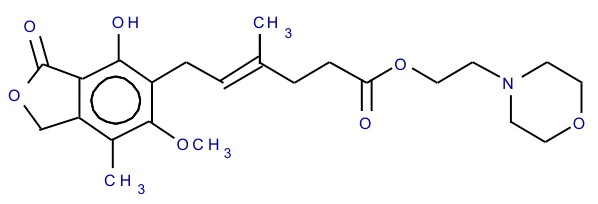
14. Clinical Trials
14.1. Clinical Trials by Indication
The safety and efficacy of CellCept (mycophenolate mofetil) as adjunctive therapy for the prevention of organ rejection were assessed in randomized, double-blind, multicentre trials in renal (3 trials), in cardiac (1 trial) and in hepatic (1 trial) transplant patients.
Renal Transplant
Adults
The three renal studies compared two dose levels of oral CellCept (1 g twice daily and 1.5 g twice daily) with azathioprine (2 studies) or placebo (1 study) when administered in combination with cyclosporine and corticosteroids to prevent acute rejection episodes. One study also included anti-thymocyte globulin [equine] (Atgam®) induction therapy. These studies are described by geographic location of the investigational sites. One study was conducted in the US at 14 sites, one study was conducted in Europe at 20 sites, and one study was conducted in Europe, Canada, and Australia at a total of 21 sites.
The primary efficacy endpoint was the proportion of patients in each treatment group who experienced biopsy-proven acute rejection or treatment failure (defined as early termination from the study for any reason without prior biopsy-proven rejection) within the first six months after transplantation. CellCept, when administered with anti-thymocyte globulin [equine] (Atgam®) induction (one study) and with cyclosporine and corticosteroids (all three studies), was shown to significantly reduce the incidence of treatment failure compared to the following three therapeutic regimens: (1) anti-thymocyte globulin [equine] (Atgam®) induction/ azathioprine/ cyclosporine/corticosteroids, (2) azathioprine/ cyclosporine /corticosteroids, and (3) cyclosporine/corticosteroids.
CellCept, in combination with corticosteroids and cyclosporine reduced (statistically significant at the <0.05 level) the incidence of treatment failure within the first 6 months following transplantation. The following tables summarize the results of these studies. These tables show (1) the proportion of patients experiencing treatment failure, (2) the proportion of patients who experienced biopsy-proven acute rejection on treatment, and (3) early termination, for any reason other than graft loss or death, without a prior biopsy-proven acute rejection episode. Patients who prematurely discontinued treatment were followed for the occurrence of death or graft loss, and the cumulative incidence of graft loss and patient death are summarized separately. Patients who prematurely discontinued treatment were not followed for the occurrence of acute rejection after termination. More patients discontinued receiving CellCept (without prior biopsy-proven rejection, death or graft loss) than discontinued in the control groups, with the highest rate in the CellCept 3 g/day group. Therefore, the acute rejection rates may be underestimated, particularly in the CellCept 3 g/day group.
Table 7 Renal Transplant Studies
Incidence of Treatment Failure
(Biopsy-Proven Rejection or Early Termination for Any Reason)
| USA Study (N=499 patients) | CellCept 2 g/day (N=167 patients) | CellCept 3 g/day (N=166 patients) | Azathioprine 1-2 mg/kg/day (N=166 patients) |
| All treatment failures | 31.1% | 31.3% | 47.6% |
| Early termination without prior acute rejection* | 9.6% | 12.7% | 6.0% |
| Biopsy-proven rejection episode on treatment | 19.8% | 17.5% | 38.0% |
| Europe/Canada/Australia Study (N=503 patients) | CellCept 2 g/day (N=173 patients) | CellCept 3 g/day (N=164 patients) | Azathioprine 100-150 mg/day (N=166 patients) |
| All treatment failures | 38.2% | 34.8% | 50.0% |
| Early termination without prior acute rejection* | 13.9% | 15.2% | 10.2% |
| Biopsy-proven rejection episode on treatment | 19.7% | 15.9% | 35.5% |
| Europe Study (N=491 patients) | CellCept 2 g/day (N=165 patients) | CellCept 3 g/day (N=160 patients) | Placebo (N=166 patients) |
| All treatment failures | 30.3% | 38.8% | 56.0% |
| Early termination without prior acute rejection* | 11.5% | 22.5% | 7.2% |
| Biopsy-proven rejection episode on treatment | 17.0% | 13.8% | 46.4% |
* Does not include death and graft loss as reason for early termination.
Cumulative incidence of 12-month graft loss and patient death are presented below. No advantage of CellCept with respect to graft loss and patient death was established. Numerically, patients receiving CellCept 2 g/day and 3 g/day experienced a better outcome than controls in all three studies; patients receiving CellCept 2 g/day experienced a better outcome than CellCept 3 g/day in two of the three studies. Patients in all treatment groups who terminated treatment early were found to have a poor outcome with respect to graft loss and patient death at 1 year.
Table 8 Renal Transplant Studies
Cumulative Incidence of Combined Graft Loss and Patient Death at 12 Months
| Study | CellCept 2 g/day | CellCept 3 g/day | Control (Azathioprine or Placebo) |
| USA | 8.5% | 11.5% | 12.2% |
| Europe/Canada/ Australia | 11.7% | 11.0% | 13.6% |
| Europe | 8.5% | 10.0% | 11.5% |
Pediatrics
One open-label, safety and pharmacokinetic study of CellCept oral suspension 600 mg/m2 twice daily (up to 1g twice daily) in combination with cyclosporine and corticosteroids was performed at centers in the US (9), Europe (5) and Australia (1) in 100 pediatric patients (3 months to 18 years of age) for the prevention of renal allograft rejection.
CellCept was well tolerated in pediatric patients (see 8. Adverse Reactions), and the pharmacokinetics profile was similar to that seen in adult patients dosed with 1g twice daily CellCept capsules (see 10.3.
Pharmacokinetics, Special Populations and Conditions, Pediatrics). The rate of biopsy-proven rejection was similar across the age groups (3 months to <6 years, 6 to <12 years, 12 to 18 years). The overall biopsy-proven rejection rate at 6 months and the combined incidence of graft loss and patient death at 12 months post-transplant were similar to the rates observed in adult renal transplant patients.
Cardiac Transplant
A double-blind, randomized, comparative, parallel-group, multicentre study in primary cardiac transplant recipients was performed at 20 centers in the United States, one in Canada, five in Europe and two in Australia. The total number of patients enrolled was 650; 72 patients did not receive study drug and 578 patients received study drug. Patients received CellCept 1.5g twice daily (n=289) or azathioprine 1.5-3 mg/kg/day (n=289), in combination with cyclosporine (Sandimmune® or Neoral®) and corticosteroids as maintenance immunosuppressive therapy.
The two primary efficacy endpoints were: (1) the proportion of patients who, after transplantation, had at least one endomyocardial biopsy-proven rejection with hemodynamic compromise, or were retransplanted or died, within the first six months, and (2) the proportion of patients who died or were transplanted during the first twelve months following transplantation. Patients who prematurely discontinued treatment were followed for the occurrence of allograft rejection for up to 6 months and for the occurrence of death for 1 year.
(1) Rejection: No difference was established between CellCept and azathioprine (AZA) with respect to biopsy-proven rejection with hemodynamic compromise.
(2) Survival: CellCept was shown to be at least as effective as AZA in preventing death or retransplantation at 1 year (see table below).
Table 9 Cardiac Transplant Study
Rejection at 6 Months Death or Retransplantation at 1 Year
| All Patients | Treated Patients | |||||||
| AZA N = 323 | CellCept N = 327 | AZA N = 289 | CellCept N = 289 | |||||
| Biopsy-proven rejection with hemodynamic compromise at 6 months* | 121 (38%) | 120 (37%) | 100 (35%) | 92 (32%) | ||||
| Death or Retransplantation at 1 year | 49(15.2%) | 42(12.8%) | 33 (11.4%) | 18 (6.2%) | ||||
*Hemodynamic compromise occurred if any of the following criteria were met: pulmonary capillary wedge pressure ≥ 20 mm or a 25% increase; cardiac index < 2.0 L/min/m2 or a 25% decrease; ejection fraction ≤ 30%; pulmonary artery oxygen saturation <60% or a 25% decrease; presence of new S3 gallop; fractional shortening was ≤ 20% or a 25% decrease; inotropic support required to manage the clinical condition.
Hepatic Transplant
A double-blind, randomized, comparative, parallel-group, multicentre study in primary hepatic transplant recipients was performed at 16 centers in the United States, 2 in Canada, 4 in Europe and 1 in Australia. The total number of patients enrolled was 565. Patients received CellCept 1g twice daily intravenously for up to 14 days followed by CellCept 1.5 g twice daily orally or azathioprine 1-2 mg/kg/day intravenously followed by azathioprine 1-2 mg/kg/day orally, in combination with cyclosporine (Neoral®) and corticosteroids as maintenance immunosuppressive therapy.
The two primary endpoints were: (1) the proportion of patients who experienced, in the first 6 months post-transplantation, one or more episodes of biopsy-proven and treated rejection or death/retransplantation, and (2) the proportion of patients who experienced graft loss (death/retransplantation) during the first 12 months post-transplantation. Patients who prematurely discontinued treatment were followed for the occurrence of allograft rejection and for the occurrence of graft loss (death/retransplantation) for 1 year. Results: In the primary (intent-to-treat) analyses CellCept in combination with corticosteroids and cyclosporine was statistically significant (p < 0.02) compared to azathioprine for prevention of acute rejection and equivalent to azathioprine for survival.
Table 10 Hepatic Transplant Study
Rejection at 6 Months / Death or Retransplantation at 1 Year
| AZA N = 287 | CellCept N = 278 | |
| Biopsy-proven and treated rejection at 6 months | 137 (47.7%) | 106 (38.1%) |
| Death or retransplantation at 1 year | 42 (14.6%) | 39 (14.0%) |
14.2. Comparative Bioavailability Studies
Comparative Bioavailability Studies
It has been demonstrated that the 500 mg tablet (x2) is bioequivalent to the 250 mg capsule (x4) with respect to the extent of absorption (AUC), and with respect to the rate of absorption (Tmax). Shown in Table 11 below are the pharmacokinetic parameters for MPA following the administration of oral MMF in renal transplant patients.
Table 11 Summary Table of Measured Comparative Bioavailability Data for Mycophenolic Acid Following Administration of Mycophenolate Mofetil 2 x 500 mg Tablet vs 4 x 250 mg Capsule in Renal Transplant Patients
| Parameter | Geometric Mean and Arithmetic Mean (CV%) | Ratio of Geometric Means (%) | ||||||
| Test (500 mg Tablet) | Reference (250 mg Capsule) | |||||||
| AUC0 – 12 (µg·h/mL) | 31.7 33.5 (33.8) | 31.2 33.4 (38.2) | 102.1 | |||||
| Cmax (µg/mL) | 10.3 11.5 (46.8) | 10.6 12.2 (48.1) | 99.5 | |||||
| CMIN (µg/mL) | 1.40 (75) | 1.35 (81.5) | 110.3 | |||||
| Tmax* (h) | 1.09 (75.8) | 1.15 (70.5) | -- | |||||
| FL* (%) | 378 (51.3) | 404 (52.5) | -- | |||||
* Expressed as arithmetic mean (CV%) only
It has been demonstrated that 5 mL of the 200 mg/mL reconstituted suspension is bioequivalent to four 250 mg capsules. Shown below are the pharmacokinetic parameters for MPA following the administration of oral MMF to healthy volunteers.
Table 12 Summary Table of Measured Comparative Bioavailability Data for Mycophenolic Acid Following Administration of Mycophenolate Mofetil
1000 mg/5 mL Oral Suspension vs 4x250 mg Capsule in Healthy Volunteers
| Parameter | Geometric Mean and Arithmetic Mean (CV%) | Ratio of Geometric Means (%) | ||||||
| Test (200 mg/mL Suspension) | Reference (250 mg Capsule) | |||||||
| AUCT (µg•h/mL) | 64.0 65.9 (25.2) | 60.8 62.6 (24.9) | 104.8 | |||||
| AUCI (µg•h/mL) | 68.7 70.5 (23.4) | 66.7 68.8 (25.6) | 102.6 | |||||
| Cmax (µg/mL) | 27.6 28.4 (25.0) | 25.7 27.1 (34.5) | 106.3 | |||||
| Tmax* (h) | 0.67 (64.0) | 0.83 (42.6) | -- | |||||
| T½ * (h) | 16.5 (21.9) | 19.2 (108) | -- | |||||
* Expressed as arithmetic mean (CV%) only
15. Microbiology
No microbiological information is required for this drug product.
16. Non-Clinical Toxicology
General Toxicology:
Animal Pharmacology
Survival of Allografts and Treatment of Allograft Rejection in Different Experimental Models
The effect of MMF for the prevention of rejection and for the reversion of ongoing rejection was studied in several experimental allograft models.
Survival of Kidney Allografts and Treatment of Acute Allograft Rejection in Dogs
To determine the efficacy of MMF in preventing graft rejection in a large animal model, a renal allograft between outbred mongrel dogs was used. In control dogs receiving no immunosuppression, the median graft survival (MST) was 8.1 days. MMF administered orally at 40 mg/kg/day significantly increased the allograft survival (MST = 36 days). Some gastrointestinal toxicity and weight loss was observed in this group of dogs. When MMF (20 mg/kg/day) was given in combination with subtherapeutic doses of cyclosporin A (CsA, 5 mg/kg/day) and methylprednisolone (MP, 0.1 mg/kg/day), the MST was >122.4 days (n=16). Using this triple therapy, three dogs survived >200 days and one was euthanized on Day 150 to obtain tissue samples for histologic examination. In the animals treated with triple therapy, no significant toxicity was observed. There was a two-fold transient increase in alkaline phosphatase. Control dogs treated with CsA (5 mg/kg/day) and MP (0.1 mg/kg/day) had a MST = 8.5, essentially identical to that in untreated dogs. In dogs receiving 20 mg/kg/day MMF and 0.1 mg/kg/day MP without CsA, all animals survived >50 days (n=6). The treatment was discontinued in 4 of those animals to test for tolerance after 50 days. Within a few days those 4 animals underwent acute renal rejection, indicating that tolerance had not developed. Nonetheless, the double therapy treatment without CsA was as effective at preventing rejection as was the therapy with CsA.
The ability of MMF to reverse ongoing acute rejection was also examined in the canine renal allograft model. Kidney allografts were performed in bilaterally nephrectomized female mongrel dogs. All animals received a baseline immunosuppressive treatment (MMF - 10 mg, CsA - 5 mg and MP - 0.1 mg/kg/day, orally), previously shown to be insufficient to prevent renal graft rejection, but sufficient to slow the rate of progression. Rejection was defined by a 50% or greater rise in serum creatinine level relative to the lowest observed creatinine during the first postoperative week. Before the initiation of rejection treatment, a percutaneous kidney biopsy was performed to confirm the rejection. At the time of rejection, experimental animals received rescue therapy consisting of either MMF 80 mg/kg/day, twice daily, for 3 days, (experimental group) or bolus MP: 14 mg/kg, 7 mg/kg and 3.5 mg/kg, respectively, on each day (control group). After completion of the rejection treatment, the baseline immunosuppression was increased to MMF 20 mg/kg, with the same doses of CsA and MP.
Control animals (n=11) all died of ongoing rejection (MST 19 days). In the experimental group, MMF treatment successfully reversed the acute ongoing rejection in 14/16 (87.5%) of the dogs. Reversion of the rejection process was proven by biopsy as well as normalization of the creatinine levels. The rejection treatment with MMF resulted in a lymphopenia that persisted for about 4 weeks, while liver enzymes such as AST, ALT and alkaline phosphatase were minimally elevated during the first two weeks after rejection treatment. During the rescue therapy with MMF, gastrointestinal side effects such as vomiting and diarrhea were also observed.
In summary, MMF significantly prolonged kidney allograft survival in dogs. Using a combined treatment with subtherapeutic doses of CsA and MP, the graft survival was >122 days, without any serious adverse effects. In addition, MMF was highly effective in reversing acute renal allograft rejection in dogs, while bolus administration of steroids did not show efficacy in the same model. These studies show the efficacy of MMF in preventing and treating allograft rejection without limiting toxicity or increased susceptibility to infections in a large animal model.
Prevention of Acute Rejection in Heart Allografts and Xenografts in Mice and Rats
MMF was found to effectively prolong heart allograft survival in mice and rats, and increase xenograft survival and reverse ongoing allograft rejection without severe toxicity in rats. The survival of grafts long beyond cessation of treatment, and even after challenge with a second graft from the same donor, suggested induction of tolerance. Combination therapy with MMF and CsA, using low doses of both drugs, can prevent graft rejection and allow survival even after cessation of treatment. Thus CsA and MMF have at least additive effects in preventing allograft rejection, without indication of increased toxicity. Similarly, MMF and brequinar also demonstrated an additive effect on preventing heart allograft rejection in rats, without increased toxicity.
Prevention of Chronic Rejection in Heart Allografts and Aortic Allografts in Rats
A manifestation of chronic allograft rejection is a proliferative and obliterative arteriopathy. MPA in clinically attainable concentrations (0.09-3.2 µM) inhibits the proliferation of human arterial smooth muscle cells in culture, whereas CsA and brequinar do not. Once daily oral administration of MMF (30 mg/kg/day, Days 1-30 and 20 mg/kg/day, Days 31-100) to Lewis rat recipients prevented the development of chronic rejection in transplanted Brown Norway rat heart allografts.
Male ACI rat donors and Lewis recipient rats were used in an orthotopic aortic allograft model to test the effect of MMF in preventing development of chronic rejection. Treatment with a MMF dose (20 mg/kg) for 3 months significantly inhibited intimal proliferation when used alone and in combination therapy with brequinar sodium. By contrast, brequinar monotherapy and CsA alone did not prevent neointimal proliferation at the doses used. MMF was also tested at lower doses with other immunosuppressive regimens containing rapamycin and was found to be partially active.
Survival of Cardiac Xenografts in Primates
Cynomolgus monkeys served as donors for ABO-matched, B-cell-crossmatched negative, baboons. Four treatment groups were used: Group 1, no immunosuppression; Group 2, CsA 15 mg/kg/day intramuscularly starting 1 day preoperatively + methylprednisolone acetate 0.8 mg/kg/day intramuscularly; Group 3, same as Group 2 + azathioprine 4 mg/kg/day orally starting 21 days preoperatively; Group 4, same as Group 3 with MMF 70 mg/kg/day orally instead of azathioprine.
Group 1 baboons (n = 4, untreated controls) had a MST of 9 days (range 8-10 days). CsA and steroid treatment in Group 2 (n = 6) prolonged mean survival to 77 days (range 16-200 days). Hearts undergoing early rejection showed a histologic picture of rejection similar to that of Group 1. Hearts with longer survival demonstrated histological appearance consistent with repeated episodes of rejection, with healing and gradual replacement of myocardium with connective tissue. Graft biopsy was performed at 1, 2, 4, 8 and 12 weeks postoperatively in groups 3 and 4 with follow-up biopsy 1 week after rejection therapy. In Group 3 (n = 5), mean survival was prolonged to 94 days (range 3-392). Four of 9 rejection episodes were reversed using high-dose steroids. ATG 10 mg/kg per day intravenously reversed 2 of 4 episodes of steroid-resistant rejection. Despite rescue from cellular rejection, these grafts were later lost to humoral rejection. In Group 4 (n = 3), mean survival was 296 days (range 49-618). A cardiac biopsy taken from 1 animal in group 4 at 618 days post-transplant showed a coronary artery free of intimal occlusive disease. Coronary artery disease of a low degree of severity was found in only 2 other biopsy specimens in group 4 and consisted of moderate endothelial swelling. There was no evidence of coronary intimal proliferation in this group.
Ultrastructural examination of the coronary arteries and veins from the same animal showed no indication of vasculopathy in any of the vessels examined. The endothelium in some vessels was bulbous in appearance rather than the normal flat endothelial lining.
In conclusion, MMF in combination with a subtherapeutic dose of CsA and steroids significantly prolonged cardiac xenograft survival in primates far beyond any other immunosuppressive regime so far tested. The vascular pathological changes seen in long-term survivors with cardiac xenografts treated with other immunosuppressive therapies were not observed in animals treated with MMF.
In addition, MMF significantly inhibited neointimal proliferation in rat aortic allografts after 6 months treatment, while CsA and brequinar did not. MMF also prevented the intimal proliferation observed in long-term recipients of rat heart allografts, while CsA and azathioprine did not. This effect may be explained by the ability of MMF to inhibit not only T-lymphocyte responses, but also antibody formation and proliferation of smooth muscle cells, which are considered important factors contributing to the pathogenesis of chronic rejection.
Other Models of Allograft Rejection
MMF (80 mg/kg/day) was effective for prevention of pancreatic islet allograft rejection in mice rendered diabetic using streptozotocin. After cessation of treatment, recipients of pancreatic islet allografts continued normoglycemic, indicating that a state of tolerance was induced. Tolerant mice resisted challenge with a second graft from the same donor strain but rejected a third party graft, indicating that the tolerance induced by MMF was specific. In animals (NOD mice) spontaneously developing diabetes, MMF (80 mg/kg/day) did not decrease the incidence of disease.
MMF was also effective in graft versus host disease (GVHD) in several experimental models using splenocytes from parental C57BI/6 mice into F1 ( C57BI/6 x C3H/H3N) mice, rat small bowl transplant from parental into F1 recipients, and a sublethally irridiated mouse recipient of allogenic spleen cells model. In other models of allograft rejection such as limb transplants in rats and liver and intestinal allograft in dogs and rats, MMF alone and in combination with CsA showed increased survival and greater efficacy than other immunosuppressive therapies tested in parallel.
Mechanism of Action
Immunosuppressive Effects
In vitro Studies with Human Cells
The effects of MMF and MPA on lymphocyte function were studied in vitro. MPA and MMF (IC 50=17-80 nM) inhibit proliferative responses of human peripheral blood mononuclear cells (PBMCs) to the T- and B-cell mitogens phytohemagglutinin (PHA), pokeweed mitogen (PWM), concanavalin A (Con A) and Staphylococcus protein A-sepharose (SPAS). By contrast MPAG was not inhibitory of lymphocyte proliferation in response to PWM and SPAS up to 10µM concentration. Lymphocyte response to phytohemagglatin PHA was inhibited by MPAG with an IC50=8.9µM. This concentration is about 100-fold greater than the MPA concentration required for the same bioactivity. The residual activity in the
MPAG sample can be explained by a small contamination with MPA as detected by HPLC analysis (≈0.3%).
Addition of guanosine (Guo) or deoxyguanosine (dGuo) reversed the inhibitory effect of MMF and MPA, while adenosine and deoxyadenosine had no effect. Human fibroblasts, endothelial cells, and smooth muscle cells were about one log less sensitive to the antiproliferative effects of the drug than were lymphocytes.
In addition, MMF and MPA inhibited the formation of antibodies by human peripheral blood B- lymphocytes stimulated with mitogens (Staphylococcus aureus Cowan 1 or PWM) at nanomolar concentrations (IC50 of approximately 20-50 nM).
Measurements of nucleotide pools in human lymphocytes activated with concanavalin A (Con A), as well as in the human T-cell line CCRF-CEM, have shown that MPA (1 μM) markedly depletes pools of GTP. When either Guo (1 μM) or dGuo (10 μM) were added to the medium, the GTP level was restored. The dGTP pools were even more affected by MPA than those of GTP, and dGuo (10 μM) restored the level to above the controls. By contrast, GTP levels in human polymorphonuclear leukocytes (PMNs) were essentially unaffected by MPA, while adherent mononuclear cells (monocyte-enriched) were also GTP depleted. The effect of MPA was greater in mononuclear cells stimulated by Con A than on resting cells.
MMF and MPA were also shown to be potent inhibitors of the proliferation of human B- and T- lymphocytic and promonocytic cell lines, while the erythroid precursor cell line, K562, was less susceptible. Several human tumor cell were found to have various degrees of susceptibility to the antiproliferative effects of MMF and MPA. Addition of guanine or dGuo to the cultures reversed the antiproliferative effect of MPA in a concentration- and time-dependent manner, showing the selectivity of the drug’s effect.
The observation that MPA added to mixed lymphocyte cultures after 72 hours still inhibited proliferation suggests that the drug affects late events following antigenic stimulation. MMF and MPA did not inhibit early events in lymphocyte activation, such as production of IL-1 by human peripheral blood monocytes stimulated with lipopolysaccharide, the production of IL-2 in Con A stimulated human peripheral blood lymphocyte (PBL) and the mobilization of calcium following stimulation of T-cells with mitogens.
Interferon (IFN-γ) is another cytokine produced by stimulated T-lymphocytes. MMF (hydrochloride) did not inhibit IFN production in 2 assays. Subsequently, MMF was found to inhibit IFN production with an IC50 of 0.37-0.42 μM, which is about 10-fold greater than the concentration required to inhibit lymphocyte proliferation.
Although inhibition of T- and B-lymphocyte proliferation is the primary target of the drug, it is likely that depletion of GTP will have other metabolic effects on these cells. For example, it is known that GTP is required for the activation of mannose and fucose, through dolichol phosphate intermediates, for glycoprotein (and glycolipid) biosynthesis. Fucose - containing oligosaccharides are components of adhesion molecules such as ligands for selections which are expressed on activated endothelial cells, lymphocytes and monocytes and facilitate their interactions at sites of inflammation and graft rejection.
Cultures of human umbilical vein endothelial cells (HUVEC) and a human T-lymphocytic cell line (CEM) were used to evaluate the effect of MPA on cell adhesion. When the HUVEC were stimulated with cytokines such as IL-1 or TNF, there was a dose-dependent increase in the number of lymphocytic cells adhering to the endothelial cells (EC). The number of lymphocytes attached to the cultured EC was measured by counting under the microscope or using 51Cr-labelled T-cells. When the T-cells were treated for 4 hr with 1 μM MPA prior to incubation with EC, a reduction in the number of adherent cells was observed. Treatment of both, the EC and the T-cells with MPA (10 μM) for the same time, markedly decreased the attachment between the two cell types. This observation suggests that MPA prevents the glycosylation of adhesion molecules on lymphocytes and EC. Similar results have been obtained using human PBL stimulated with Con A instead of a T-cell line.
Immunoprecipitation studies using monoclonal antibodies specific for the adhesion molecules, VLA-4 and LFA-1, showed that MPA (10 μM) inhibited the incorporation of sugars (3H- mannose and 3H-glucosamine) on the surface of PHA-activated human PBM. By flow-cytometry, MPA was also shown to inhibit the binding by PHA activated PBM of specific lectins that recognize terminal mannose residues and sialic acid attached to terminal galactose.
In summary, MPA in clinically attainable doses can inhibit the binding of mononuclear cells to endothelial cells. This mechanism could contribute to the efficacy of MMF in preventing allograft rejection and in the treatment of ongoing rejection when clones of lymphocytes with specificity for alloantigens have already expanded.
MPA inhibited the proliferation of human arterial smooth muscle cells (SMC) and endothelial cells (HUVEC) with IC50s between 0.09-3.2 µM.
In another study, MPA was tested for effects on the growth of arterial smooth muscle cells. Human iliac artery smooth muscle cells were obtained from medial explants and were passaged several times before use. MPA was inactive at low doses, but showed 30% inhibition of cell proliferation at a concentration of 1.6 μM. MPA was also studied using human breast carcinoma cell lines BT20 and MCF-7 in comparison with non-transformed human foreskin fibroblasts. MPA at concentrations between 5-50 µM inhibited the growth of both the breast carcinoma lines and the human foreskin fibroblasts (HFF) by 50%. The sensitivity of the breast cancer cell lines to the antiproliferative effect of MPA was not significantly different from the HFF.
MMF and MPA at submicromolar concentration also inhibited proliferation and induced differentiation of promonocytic cells (U-937, HL-60 and THP-1), as measured by increased expression of cell surface markers, increased secretion of lysozyme and raised intracellular concentrations of lysosomal enzymes. U-937 and THP-1 cells cultured for 72 hr with 1 μM MPA had an increased lipid droplet content as determined by fluorescence microscopy and transmission electron microscopy. MPA also induced human derived macrophages to produce lysosomal hydrolases, supernatant lysozyme and the interleukin-1 receptor antagonist protein (IL-1ra). These effects are characteristic of compounds having long-acting anti-inflammatory effects (DMARDs) such as chloroquine and gold salts which suggests that MMF could have a disease-modifying effect in chronic inflammatory conditions such as rheumatoid arthritis.
In vitro Studies with Cells from Laboratory Animals
MPA was tested in vitro at concentrations of 0.001 to 10 µM for inhibition of lymphocyte proliferation in cell cultures from rhesus monkey, rabbit, guinea pig, rat and mice. The mitogens used were PHA, PWM, Con A, SPAS and bacterial lipopolysaccharide (LPS). MPA inhibited lymphocyte proliferation to all of the mitogens in lymphocyte cultures from all of the above named species with IC50s between <1-60 nM.
Mixed lymphocyte reactions were initiated using C57Bl/6J mouse splenocytes as responder cells and BALB/cJ mouse splenocytes as stimulator cells. After 4 days incubation in the presence of MPA at various concentrations, cell proliferation was determined by 3H-thymidine incorporation and cell viability was determined by trypan blue dye exclusion. The IC50 for MPA was determined to be 0.4 µM; cell viability was reduced at MPA concentrations ≥ 0.3 µM.
The effects of MPA on murine in vitro antibody-forming cells were analysed in a series of studies. In 1 study, MPA suppressed the in vitro response to sheep red blood cells (SRBC) >85% at concentrations of 0.1 or 1.0 µM. In this same study, a dose-response curve gave an estimated IC50 of approximately 0.04 µM. In a second study with mouse spleen cells stimulated with SRBC, only the 1.0 µM dose was suppressive and the dose-response curve showed significant suppression beginning at 50 nM. In the last study, murine spleen cells and peritoneal lymphocytes were stimulated in culture with LPS and evaluated for the production of autoantibodies to murine red blood cell antigens pretreated with bromelain. Both splenic and peritoneal B-cells showed variability in response to MPA. Peritoneal cell IC50 ranged from 0.27 µM to >10 µM, whereas spleen cell IC50 ranged from 0.59 µM to >100 µM.
The effect of MPA on the effector phase of NK-cell activity in mice was also assessed at MPA concentrations of 1, 10 and 100 μM using YAC-1 tumor cells as targets. Only at the 100 μM concentration did MPA inhibit the effector phase of NK-cell activity in 2 of 3 experiments. MPA tested on the effector phase of T-cell-mediated cytotoxicity in a murine system (spleen cells sensitized to P815 tumor target cells) was only partially inhibitory (30% decrease in specific lysis) at the highest concentration tested (100 μM) in 1 of 2 experiments.
The effect of MPA on the degranulation of rat peritoneal mast cells (RMC) was studied. RMC were pretreated for 48 hr with 0.1-10 µM MPA before the cells were sensitized with IgE and triggered with specific antigen. The net amount of 3H-5HT released from granules was decreased by 44 and 32% at 1 and 10 µM, respectively. MPA inhibition of degranulation was completely reversed by the addition of 30 µM guanosine to the medium. MPA had no effect on any other cell parameters studied (IgE receptors or PGD2 production).
In vivo Studies in Experimental Animals
The effects of MMF and MPA on antibody formation were tested using SRBC to immunize mice and rats. Significant inhibition (40%-88%) of the antibody response was observed in mice with oral doses of 20-100 mg/kg/day of MMF or MPA administered once daily for 4 days. In rats immunized with SRBC and treated with MPA for 4 days only, the ED50 was approximately 14 mg/kg/day. Significant inhibition of antibody formation against SRBC was observed also in rats treated with 9 and 30 mg/kg/day for 28 days, and immunized 4 days before the end of treatment.
MPAG, the main metabolite of MPA, was also tested for activity on antibody production in mice immunized with SRBC. MPAG given orally for 4 days at 50 mg/kg/day, significantly inhibited the total number of antibody-forming cells in the spleen (PFC) by 60%. These results show that MPAG, which in vitro is >100-fold less active than MPA on lymphocyte proliferation, has comparable in vivo activity. These results are consistent with the observations that in vivo, MPAG is converted to free MPA, probably by enterohepatic recirculation, with bioactivity comparable to that induced by MPA administration.
Primary and secondary antibody responses were analyzed in mice immunized with B/Yamagata influenza virus hemagglutinin in adjuvant. MPA given orally (80 mg/kg) from the time of primary immunization, for 10 days, significantly reduced the antibody titers after primary immunization and also after challenge, even though no additional drug treatment was given. Moreover, if no treatment was given at the time of primary immunization, but only after challenge, the antibody response was not reduced. These observations indicate that MPA effectively suppresses antibody responses if administered during immunization but is less effective after sensitization has already occurred.
Oral administration of MMF and MPA (50-120 mg/kg/day) from day 1 to 11 to C57Bl/6 mice also suppresses the generation of cytotoxic T-lymphocytes able to lyse allogeneic tumor target cells (P-815 mastocytoma) given intraperitoneally (day 1). Cytotoxic T-cells are thought to be the main effector mechanism responsible for acute allograft rejection. The efficacy of MMF and MPA in preventing cytotoxic cell generation provides the rationale for the prophylactic use of this drug to prevent rejection in allograft recipients.
MPA administered orally or intraperitoneally (100 mg/kg, 3-5 days) to mice immunized with ovalbumin was found to inhibit the synthesis of DNA as measured by 3H-TdR incorporation in lymphoid tissue but not in other rapidly dividing cells such as germinal cells of the testes. Thus MPA has a more potent cytostatic effect on lymphoid cells than in other cell types, in vivo as well as in vitro.
Anti-Inflammatory Effects
MMF and MPA were tested in a number of experimental models of inflammation. Both MMF and MPA suppress adjuvant-induced arthritis and experimental allergic encephalomyelitis in rats.
The autoimmune disease that develops in genetically predisposed MRL/pr mice was also partially reduced by treatment with MPA for 30 days, starting at 5.5 months of age. In collagen-induced arthritis in mice, MPA did not produce any significant changes of the disease symptoms or incidence at the dose tested (10.5 mg/kg/day). However, this dose was considered to be below the threshold of therapeutic activity in mice, as shown in other models.
No effect was observed when MPA was administered to rats in two other models of acute inflammation, carrageenan-induced paw swelling and granuloma induced by implant of a cotton-pellet impregnated with carrageenan.
Finally, MPA was found to have no effect on IL-1-induced ornithine decarboxylase in mice (used as a measure of an acute phase response).
As part of a random screening, MPA was tested in vitro for inhibition of 15-lipoxygenase and
5 lipoxygenase (5-LO) activity, and for thromboxane B2 and leukotriene B4 (LTB4) synthesis by human whole blood. A partial inhibitory effect on LTB4 synthesis and 5-LO activity was found only at the highest concentrations tested (312 and 100 μM, respectively).
These results indicate that MMF and MPA can prevent inflammatory responses that are immunologically driven but have no effect on acute inflammatory responses such as those responding to cyclo-oxygenase and 5-LO inhibitors.
Antitumor Effects
Antitumor activity of MMF was assessed in several models of xenogeneic (human tumor cells into nude mice) as well as in a syngeneic metastasis model in mice. MMF significantly prolonged survival and delayed tumor development with different degrees of efficacy according to the type of tumor.
Antiviral Effects
Antiviral activity of MMF and MPA was evaluated against different viruses in culture. Anti-HIV activity was tested using different cell lines of T-cell and monocytic origin. In general, the reduction in reverse transcriptase (RT) was directly correlated with the reduction in the cell number, suggesting that the effect of MPA is at the cellular level rather than on the virus replication. Antiviral activity against herpes, parainfluenza virus, respiratory syncitial virus and against murine Friend Leukemia Virus (FLV) were all obtained at low (micromolar) concentrations of MPA, but the window between antiviral concentration and cytostatic effects was narrow. In vivo, MMF and MPA inhibited splenomegaly and the number of focus forming units in FLV-infected mice. Some activity of MMF and MPA against cytomegalovirus and human cytomegalovirus was observed; these drugs augmented the antiviral activity of ganciclovir.
General Pharmacology
General pharmacology studies were conducted to assess the effects of MMF on the central nervous, cardiovascular, respiratory, and gastrointestinal systems.
CNS effects were determined in mice in studies that included a gross behavioral assay (Irwin Profile), a spontaneous locomotor activity assay, an induced neurological deficit test, maximal electroshock and pentylenetetrazol anticonvulsant assays, and a hexobarbital sleep test. MMF was administered as single oral doses ranging from 1.09 to 1090 mg/kg. Except for depressed locomotor function at 10.9 mg/kg and higher doses in the Irwin profile assay or at 109 mg/kg in the spontaneous locomotor activity assay, there was no evidence of central nervous system or autonomic effects with MMF.
Cardiovascular and respiratory effects were assessed in anesthetized dogs, 3 animals per group. Each animal received escalating doses of MMF (0.3-31.6 mg/kg, orally). No significant effects were seen.
Effects on gastrointestinal function were determined in rats, 5-10 animals per group. Each rat received single oral doses of MMF (25-100 mg/kg). MMF significantly decreased gastrointestinal motility at 100 mg/kg, but had no effects on gastric secretory activity. MMF appeared to diminish signs of colitis in a mouse model; however, this model was unverified and the results are inconclusive.
To further assess the affinity of MMF and MPA at various neurotransmitter receptors, radioligand binding assays were performed. Both MMF and MPA showed low affinities (pKi values ≤ 4-5) at α-adrenergic, β-adrenergic, and muscarinic receptors, suggesting probable lack of activity for these compounds on the autonomic nervous system.
Pharmacokinetics and Disposition
MMF administered orally to animals and humans was rapidly and extensively converted to MPA.
Following oral administration of unlabeled MMF or [14C-MPA] MMF to mice (10 mg/kg), rats (6 mg/kg), rabbits (40 mg/kg), dogs (9 mg/kg), mini pigs (45 mg/kg), cynomolgus monkeys (6 mg/kg), and humans (1000 mg), MMF was not detected in plasma by HPLC-UV or HPLC-radiometric methods. Following IV infusion of MMF, however, MMF was observed in the plasma throughout the infusion period but was not detected >5 minutes after terminating the infusion. Subsequent in vitro studies using monkey and human tissues demonstrated that homogenates of liver, gut mucosa, and kidney hydrolyze MMF to MPA and hydroxyethyl morpholine.
Pharmacokinetic-based Drug Interactions
Co-administration of MMF (20 mg/kg orally) and probenecid (500 mg) twice daily for 4 days increased the AUC0-12 hr for MPA and MPAG 1.7 and 2.9 fold respectively.
The potential for drug interaction at the level of protein binding was evaluated in human plasma in vitro by equilibrium dialysis and ultrafiltration methods. When MPA concentrations of 5, 20, and 50 µg/mL were used, concentrations of MPAG as high as 230 µg/mL had little effect on the binding of MPA. However, MPAG concentrations of 460 and 920 µg/mL reduced the plasma binding of MPA (50 µg/mL) from 97.1% to 95.4% and 92.8%, respectively. At plasma concentrations encountered clinically, tolbutamide, theophylline, and digoxin had no effect on the binding of MPA to human plasma, but cyclosporine at 500 ng/mL reduced the binding of MPA (75 µg/mL) from 97.2% to 96.5%. MPA concentrations as high as 100 µg/mL had little effect on the plasma binding of warfarin, digoxin, or proprandol, but decreased binding of theophylline from 52.8% to 45.0%, and decreased binding of phenytoin from 90.0% to 86.8%.
Miscellaneous Studies
The potential for MMF to induce hepatic cytochrome P450 enzymes was evaluated in male rats. MMF did not induce total hepatic P450.
Glucuronidation of MPA by Hepatic Microsomes
Apparent Km and Vmax of glucuronidation of MPA were determined with hepatic microsomes from mice, rats, dogs, cynomolgus monkeys, and humans (CL 6755). Microsomes from all species catalyzed the formation of MPAG from MPA, and the reaction followed Michaelis-Menton kinetics.
Nonclinical toxicity studies were conducted to evaluate the acute, subchronic/chronic, reproductive, carcinogenic, and mutagenic effects of MMF.
Clinically MMF will be administered orally as a capsule. Therefore, the primary route of exposure for the toxicology studies was oral (gavage). Oral bioavailability of MPA following MMF administration, and absorption efficiency of MMF, were known to range from approximately 80% to 100% in rodents (mice, rats) to 50% to 70% in non-rodents (dogs, monkeys). Additional toxicology studies were conducted by the intravenous and subcutaneous routes to further characterize MMF’s toxicologic profile.
Animal models used in toxicology evaluations included mice, rats, dogs (beagle), and monkeys (cynomolgus), and were selected in part for their known pharmacological responsiveness to MMF and similar metabolism of MMF compared with man.
Single-dose (Acute) Toxicity
The acute toxicity of MMF was evaluated by oral, subcutaneous, and intravenous routes in mice, rats, and/or monkey.
Table 13 Single Dose (Acute) Toxicity Studies
| Document No. | Study Type | Species | Dose (mg/kg) | Route | |||||
| Oral Toxicology Formulation: | |||||||||
| AT 4107 | Acute | Mouse | 500-4000 | Oral | |||||
| AT 4108 | Acute | Rat | 125-1000 | Oral | |||||
| AT 4109 | Acute | Rat | 250-2000 | Subcutaneous | |||||
| AT 4099 | Acute | Monkey | 500-1000 | Oral | |||||
| Intravenous Toxicology Formulation: | |||||||||
| AT 5996 | Acute | Rat | 10-100 | Intravenous | |||||
Single acute minimum lethal oral doses were greater than 4000 mg/kg in mice, 250 mg/kg in rats, and 1000 mg/kg in monkeys. The acute minimum lethal dose in rats was greater than 1000 mg/kg by the subcutaneous route. In these oral and subcutaneous acute toxicity studies, treatment-related mortality occurred only in the rat. Most deaths occurred 3 to 6 days after dosing and were associated with gastrointestinal toxicity, evidenced pathologically by excess fluid, mucosal reddening, and/or ulceration in the stomach and/or small intestine.
The acute minimum lethal dose in rats was between 30 and 100 mg/kg by the intravenous route. Most rats died within 2 minutes after dosing. Clinical changes at 30 and 100 mg/kg included collapse, inactivity, tonic convulsion, labored respiration, and/or gasping; these changes abated by 3 hours postdosing. No treatment-related pathologic changes occurred.
In acute oral toxicity studies, no deaths occurred in adult mice at doses up to 4000 mg/kg or in adult monkeys at doses up to 1000 mg/kg; these were the highest doses of mycophenolate mofetil tested in these species. These doses represent 11 times the recommended clinical dose in renal transplant patients and approximately 7 times the recommended clinical dose in cardiac transplant patients when corrected for body surface area (BSA). In adult rats, deaths occurred after single oral doses of 500 mg/kg of mycophenolate mofetil. The dose represents 3 times the recommended clinical dose in renal transplant patients and approximately twice the recommended clinical dose in cardiac transplant patients when corrected for BSA.
Multiple-dose Toxicity
Multiple-dose toxicity studies with MMF were conducted in mice, rats, dogs, and monkeys as illustrated below; the duration of these studies ranged from 1 to 12 months by the oral route, and 2 weeks to 1 month by the intravenous route.
Table 14 Multiple-dose Toxicity Studies
| Document No. | Study Type | Species | Dose (mg/kg/day) | Route | Durationa | ||||||||
| Document No. | Study Type | Species | Dose (mg/kg/day) | Route | Durationa | ||||||||
| Oral Toxicology Formulation: | |||||||||||||
| AT 5316 | Subchronic | Mouse | 0,6,25,100,300 | Oral | 3 months | ||||||||
| AT 4155 | Subchronic | Rat | 0,3,9,30 | Oral | 1 month/1 month | ||||||||
| AT 4831 | Chronic | Rat | 0,2,6,20 | Oral | 6 months/1 month | ||||||||
| AT 6213 | Chronic | Rat | 0,2,6,15 | Oral | 12 months | ||||||||
| AT 5105 | Subchronic | Dog | 0,6,20,60 | Oral | 3 months | ||||||||
| AT 6214 | Chronic | Dog | 0,3,9,30 | Oral | 12 months/1 month | ||||||||
| AT 4119 | Subchronic | Monkey | 0,15,45,150 | Oral | 1 month/1 month | ||||||||
| AT 4858 | Chronic | Monkey | 0,6,20,70 | Oral | 6 months/1 month | ||||||||
| Intravenous Toxicology Formulation: | |||||||||||||
| AT 5995 | Subchronic | Rat | 0,3,9,30 | Intravenous | 1 month | ||||||||
| AT 6687 | Subchronic | Monkey | 0,50,100,200 | Intravenous | 2 weeks | ||||||||
| AT 5997 | Subchronic | Monkey | 0,5,15,25 | Intravenous | 1 month | ||||||||
a Duration of dosing/duration of recovery period
Oral Dosing
The principal target organ systems in mice, rats, dogs, and monkeys dosed orally for up to 12 months with MMF were the hematopoietic and/or lymphoid systems. In these oral studies, the target organ changes were present at 100 mg/kg/day in the mouse, 6 mg/kg/day in the rat, 60 mg/kg/day in the dog, and 45 mg/kg/day in the monkey. Hematopoietic toxicity was evident in mice and rats primarily as decreased erythrocytic parameters and in dogs and monkeys primarily as decreased lymphocyte counts. Deaths related to anemia occurred in rats treated chronically with oral doses of 20 mg/kg/day. Lymphoid toxicity in rats, dogs, and monkeys included thymic atrophy and/or decreased numbers of active germinal centers in secondary lymphoid organs (lymph nodes, spleen, and/or intestine). Immunosuppression, the anticipated therapeutic mechanism, was achieved at or below no-effect dose levels for toxicity in subchronic/chronic studies in rat and monkey, as assessed in vitro by the effect of serum from dosed animals on lymphocyte mitogen response.
Recovery from the hematopoietic and lymphoid toxicity of MMF was observed in the rat 1- and 6-month studies after a one month postdosing recovery period. Evaluation of recovery was planned for the 1- and 6 month monkey studies, but could not be accomplished due to the premature demise or termination of animals in affected dose groups.
An increased incidence of viral (herpes virus B) and parasitic lesions occurred in monkeys after 3 months of dosing at 70 mg/kg/day. These effects involved a virus and an enzootic parasite known to be endemic in these feral animals, and were considered secondary to the expected pharmacologic (immunosuppressive) effects of MMF. No treatment-related infections occurred in the rodent or dog multiple-dose studies; the mice (CD-1®) and rats (CD®) used were virus-antibody free animals (VAF/Plus™, Charles River Laboratories).
Gastrointestinal and/or renal toxicity were present in the dog and the monkey at the higher dosages studied in these species. Multi-nucleated sperm precursors were present in the testes of monkeys at 1 month when treated with 150 mg/kg of MMF and at 3 or 6 months when treated with 70 or 20 mg/kg MMF, respectively. Dogs given oral doses of 60 mg/kg/day of MMF once daily for 3 months exhibited mortality and gastrointestinal erosion and necrosis. An increased frequency of diarrhea and soft feces occurred in dogs given 30 mg/kg/day for 1 year. Gastrointestinal and renal toxicity and associated mortality were present in monkeys given 150 mg/kg/day orally for 1 month.
Toxicokinetic data collected from the multiple-dose oral toxicity studies demonstrated dose-related increases in plasma concentrations of mycophenolic acid (MPA) and its glucuronide conjugate (MPAG), the principal metabolites of MMF. The approximate Cmax and AUC 0-24 hr ranges for MPA corresponding with the doses of MMF (2 to 300 mg/kg/day) administered in the multiple-dose oral toxicity studies were 0.4 to 51.9 µg/mL and 0.7 to 523 µg•hr/mL, respectively. At a given oral dose of MMF, the resulting plasma levels of both MPA and MPAG tended to remain similar over a wide range of dosing duration (single dose up to 1 year dosing). There were no detectable levels of MMF in the plasma after oral administration of MMF.
Intravenous Dosing
Rats were given 1, 3, or 10 mg/mL MMF by intravenous infusion once daily for 28 days to deliver 3, 9, or 30 mg/kg/day. No treatment-related effects occurred at 3 or 9 mg/kg/day. At 30 mg/kg/day, decreased body weight gain, anemia, lymphoid atrophy, and decreased erythroid and increased myeloid cells in bone marrow were present. Injection site inflammatory changes suggestive of local irritation occurred at 30 mg/kg/day.
Monkeys were given 5 mg/mL MMF by intravenous infusion once daily for 0.5, 1, or 2 hours to deliver 50, 100, or 200 mg/kg/day, respectively, for 14 days. All monkeys survived the duration of treatment. Changes characteristic of local irritation were present in the veins of monkeys infused with MMF. At 200 mg/kg/day, decreased erythrocytic parameters in peripheral blood and reduced numbers of erythroid cells in the bone marrow were present. No other histopathologic signs of systemic toxicity occurred.
Monkeys were given 1, 3, or 5 mg/mL MMF by intravenous infusion once daily for 28 days to deliver 5, 15, or 25 mg/kg/day. Decreases in erythrocyte count, hemoglobin, and hematocrit were present at 25 mg/kg/day. Inflammatory changes suggestive of local irritation were present in injection veins of monkeys given 15 or 25 mg/kg/day. No histopathologic signs of systemic toxicity occurred.
Toxicokinetic data collected from the multiple-dose intravenous toxicity studies demonstrated dose-related increases in plasma concentrations of mycophenolic acid (MPA) and its glucuronide conjugate (MPAG), the principal metabolites of MMF. The approximate Cmax ranges for MPA corresponding with the doses of MMF (3 to 200 mg/kg/day) administered in the multiple-dose intravenous toxicity studies were 5.9 to 215 µg/mL. AUC 0-24 hr values were not determined as part of the intravenous toxicity studies. MMF was detected in plasma of monkeys dosed by intravenous infusion, when blood samples were collected during infusion.
Carcinogenicity:
Two-year bioassays with MMF were conducted in mice and rats as listed below in Table 15.
Table 15 Carcinogenicity Studies in Mice and Rats
| Document No. | Study Type | Species | Dose (mg/kg/day) | Route | Duration |
| AT 6703 | Carcinogenicity | Mouse | 0,25,75,180 | Oral | 24 months (104 wks.) |
| AT 6702 | Carcinogenicity | Rat | 0,3,7,15 | Oral | 24 months (104 wks.) |
Mice were dosed orally by gavage with MMF once daily for at least 104 weeks at 25, 75, and 180 mg/kg/day. MMF was not carcinogenic in the mouse. The highest dose, 180 mg/kg/day, was considered the maximum tolerated dose that could be administered based on treatment-related effects that included anemia, decreased numbers of erythrocytic cells and increased numbers of granulocytic cells and megakaryocytes in bone marrow, and increased granulopoiesis and lymphoid atrophy in spleen. The highest dose tested was 0.5 times the recommended clinical dose (2 g/day) in renal transplant patients and 0.3 times the recommended clinical dose (3 g/day) in cardiac transplant patients when corrected for differences in BSA.
Rats were dosed orally by gavage with MMF once daily for at least 104 weeks at 3, 7, or 15 mg/kg/day. MMF was not carcinogenic in the rat. The highest dose, 15 mg/kg/day, was considered the maximum tolerated dose that could be administered based on treatment-related effects that included decreased survival (males), decreased body weight gain, and anemia. The highest dose was 0.08 times the recommended clinical dose in renal transplant patients and 0.05 times the recommended clinical dose in cardiac transplant patients when corrected for BSA. While these animal doses were lower than those given to patients, they were maximal in those species and were considered adequate to evaluate the potential for human risk (see Table 18: Special Toxicity Studies).
Genotoxicity:
MMF was not genotoxic, with or without metabolic activation, in several assays: the bacterial mutation assay, the yeast mitotic gene conversion assay, the mouse micronucleus aberration assay, or the Chinese hamster ovary cell (CHO) chromosomal aberration assay.
A battery of in vitro and in vivo mutagenicity tests was performed with MMF. MPA was evaluated in a single-plate Ames assay.
Table 16 In vitro and In vivo Mutagenicity Studies
| Document No. | Study Type | Species/Assay/Doses | ||
| MMF: | ||||
| AM 0312 | Mutagenicity | Salmonella typhimurium (with and without activation). Ames mutation assay. 1-10,000 µg per plate. | ||
| AM 0313 | Mutagenicity | Saccharomyces cerevisiae (with and without activation). Mitotic gene conversion assay. 1-10,000 µg/mL. | ||
| AM 0314 | Mutagenicity | Chinese hamster ovary cells (with and without activation). Chromosomal aberration assay. 10-1,000 µg/mL. | ||
| AM 0341 | Mutagenicity | Chinese hamster ovary cells (with and without activation). Chromosomal aberration assay. 0.89-1293.1 µg/mL. | ||
| AM 0315 | Mutagenicity | Mouse micronucleus assay. 300, 1000, 3000 mg/kg. Oral. Single dose followed by 3 days of observation. | ||
| Mycophenolic Acid: | ||||
| AM 0207 | Mutagenicity | Salmonella typhimurium (with and without activation). Ames (single plate) mutation assay (non-GLP screening study). 1-10,000 µg per plate. | ||
MMF did not induce point mutations (Ames assay) or primary DNA damage (yeast mitotic gene conversion assay) in the presence or absence of metabolic activation. In two assays for clastogenic effects, MMF was not mutagenic in vivo (mouse micronucleus assay) or in vitro with metabolic activation (Chinese hamster ovary [CHO] cell chromosomal aberration assay). Chromosome aberrations occurred in vitro without metabolic activation in the initial CHO cell chromosomal aberration assay but only at dose levels (249 to 300 µg/mL) that were markedly cytotoxic, producing effects that included unhealthy cell monolayers, floating cellular debris, and few visible mitotic cells. No mutagenic activity was present with or without metabolic activation in a repeat CHO cell assay conducted at dose levels (0.89 to 5 µg/mL) that were less overtly toxic. The highest dose for this repeat study produced targeted levels of toxicity in the cultured cells (50% to 80% reduction in mitotic index). Based on overall assessment of results from the two assays, the initial CHO cell assay without activation was considered to have produced false-positive results attributable to excessive cytotoxicity.
MPA did not induce point mutations in a single-plate Ames assay in the presence or absence of metabolic activation.
Reproductive and Developmental Toxicology:
Male and female reproduction studies, teratology studies, and a perinatal/postnatal reproduction study were conducted in rats and/or rabbits after oral dosing of MMF.
Table 17 Reproductive Toxicity Studies
| Document No. | Study Type | Species | Dose (mg/kg) | Route |
| AT 4832 | Male fertility & reproduction | Rat | 0,2,6,20 | Oral |
| AT 4987 | Female fertility & reproduction | Rat | 0,0.5,1.5,4.5 | Oral |
| AT 4552 | Teratology | Rat | 0,0.6,2,6 | Oral |
| AT 4667 | Teratology | Rabbit | 0,10,30,90 | Oral |
| AT 6206 | Perinatal/post-natal reproduction | Rat | 0,1,3,10 | Oral |
Fertility and Reproduction (Segment I)
No treatment-related effects occurred in a male fertility and reproduction study conducted in rats dosed orally at 2, 6, or 20 mg/kg/day. The males were dosed as part of the 6-month toxicity study. This dose represents 0.1 times the recommended clinical dose in renal transplant patients and 0.07 times the recommended clinical dose in cardiac transplant patients when corrected for BSA.
In a female fertility and reproduction study conducted in rats dosed orally at 0.5, 1.5, or 4.5 mg/kg/day, the highest dose caused malformations (principally of the head and eyes) in the first-generation (F1) pups in the absence of maternal toxicity. This dose was 0.02 times the recommended clinical dose in renal transplant patients and 0.01 times the recommended clinical dose in cardiac transplant patients when corrected for BSA. The spectrum of malformations that occurred was similar to that present in the rat teratology study. No treatment-related effects on fertility were present in P1 females, P2 females, or P2 males. The no-observed-effect level was 1.5 mg/kg/day.
Teratology (Segment II)
Teratology studies were conducted by oral dosing in rats at 0.6, 2, or 6 mg/kg/day and in rabbits at 10,
30, or 90 mg/kg/day. Increased fetal resorptions and increased fetal malformations occurred in rats at 6 mg/kg/day and in rabbits at 90 mg/kg/day. These effects occurred in the absence of maternal toxicity. Principal malformations included head and ventral wall anomalies in the rat, and cardiovascular anomalies, ventral body wall fissures, renal anomalies, and oligopulmonism/hypopulmonism in the rabbit.
Decreased fetal weight also occurred in rats at 6 mg/kg/day. The no-effect levels for teratologic changes in rats and rabbits given MMF were 2 and 30 mg/kg/day, respectively.
Perinatal/Postnatal Reproduction (Segment III)
No adverse effects on parturition or the postnatal development of the offspring occurred when female rats were dosed orally at 1, 3, or 10 mg/kg/day.
Special Toxicology:
The special toxicity studies listed below were conducted with the oral formulation of MMF or with MPA.
Table 18 Special Toxicity Studies
| Document No. | Study Type | Species | Dose (mg/kg/day) | Route | Durationa | |||||
| MMF - Oral Toxicology Formulation: | ||||||||||
| AT 6705 | Neonatal Toxicity | Rat | 0,3,9,30 | Oral | 1 month | |||||
| AT 4671 | Sensitization | Guinea Pig | 4 mg per dose for 6 doses (10 mg/mL) | Dermal | 1 month | |||||
| AT 6143 | Irritation | Rabbit | 0.5 g | Dermal | 4 hrs/3 days | |||||
| AT 6123 | Irritation | Rabbit | 0.1 g | Ocular | Single/3 days | |||||
| Mycophenolic Acid: | ||||||||||
| AT 4664 | Sensitization | Guinea Pig | 4 mg per dose for 6 doses (10 mg/mL) | Dermal | 1 month | |||||
aDuration of dosing/ Duration of observation period
Oral Toxicology Formulation
Neonatal (14 day-old) rats were given oral doses of 3, 9, or 30 mg/kg/day of MMF once daily for 4 weeks. Decreased body weight gain was present in males at 30 mg/kg/day and in females at 9 and 30 mg/kg/day. At 30 mg/kg/day, reduced red blood cell parameters, reduced bone marrow cellularity, reduced active lymphoid germinal centers, increased platelet counts, and increased splenic extramedullary hematopoiesis were present. No treatment-related effects occurred at 3 mg/kg/day.
Formulations of 10 mg/mL of MMF or MPA applied topically were not sensitizing in the guinea pig.
MMF was not an acute dermal irritant or an occular irritant when tested in rabbits.
Juvenile Toxicity: Not applicable.