


Product Monograph - Melvizo
Recent Major Label Changes
| 4 Dosage and Administration 4.2 Recommended Dose and Dosage Adjustment | 2025-05 |
|---|---|
| 4 Dosage and Administration 4.4 Administration | 2025-05 |
| 4 Dosage and Administration 4.5 Missed Dose | 2025-05 |
| 7 Warnings and Precautions | 2025-05 |
Table of Contents
Sections or subsections that are not applicable at the time of authorization are not listed.
Recent Major Label Changes ................................................................................................... 2
Table of Contents ...................................................................................................................... 2
Part 1: Healthcare Professional Information .......................................................................... 4
1 Indications ..................................................................................................................... 4
1.1 Pediatrics ............................................................................................................... 4
1.2 Geriatrics ............................................................................................................... 4
2 Contraindications .......................................................................................................... 4
4 Dosage and Administration ......................................................................................... 5
4.1 Dosing Considerations .......................................................................................... 5
4.2 Recommended Dose and Dosage Adjustment ..................................................... 5
4.4 Administration ........................................................................................................ 7
4.5 Missed Dose .......................................................................................................... 9
5 Overdose ........................................................................................................................ 9
6 Dosage Forms, Strengths, Composition, and Packaging ......................................... 9
7 Warnings and Precautions ......................................................................................... 10
General ......................................................................................................................... 10
Driving and Operating Machinery .................................................................................. 10
Immune ......................................................................................................................... 10
Ophthalmologic ............................................................................................................. 10
Renal ............................................................................................................................. 11
Reproductive Health ...................................................................................................... 11
7.1 Special Populations ............................................................................................. 12
7.1.1 Pregnancy ........................................................................................................ 12
7.1.2 Breastfeeding ................................................................................................... 12
7.1.3 Pediatrics ......................................................................................................... 12
7.1.4 Geriatrics ......................................................................................................... 12
8 Adverse Reactions ...................................................................................................... 12
8.1 Adverse Reaction Overview ................................................................................ 12
8.2 Clinical Trial Adverse Reactions .......................................................................... 13
8.3 Less Common Clinical Trial Adverse Reactions ................................................. 50
8.4 Abnormal Laboratory Findings: Hematologic, Clinical Chemistry, and Other Quantitative Data .......................................................................................................... 51
8.5 Post-Market Adverse Reactions .......................................................................... 52
9 Drug Interactions ........................................................................................................ 53
9.2 Drug Interactions Overview ................................................................................. 53
9.4 Drug-Drug Interactions ........................................................................................ 53
9.5 Drug-Food Interactions ........................................................................................ 53
9.6 Drug-Herb Interactions ........................................................................................ 53
9.7 Drug-Laboratory Test Interactions ....................................................................... 53
10 Clinical Pharmacology ................................................................................................ 53
10.1 Mechanism of Action ....................................................................................... 53
10.2 Pharmacodynamics ......................................................................................... 54
10.3 Pharmacokinetics ............................................................................................ 54
11 Storage, Stability, and Disposal ................................................................................ 55
12 Special Handling Instructions .................................................................................... 55
Part 2: Scientific Information ................................................................................................. 56
13 Pharmaceutical Information ....................................................................................... 56
14 Clinical Trials ............................................................................................................... 57
14.1 Clinical Trials by Indication .............................................................................. 57
15 Microbiology ................................................................................................................ 75
16 Non-Clinical Toxicology ............................................................................................. 75
17 Supporting Product Monographs .............................................................................. 83
Patient Medication Information ............................................................................................. 84
Part 1: Healthcare Professional Information
MELVIZO (ranibizumab) is a biosimilar biologic drug (biosimilar) to Lucentis. A biosimilar is a biologic drug that was granted authorization based on a demonstration of similarity to a version previously authorized in Canada, known as the reference biologic drug.
1 Indications
MELVIZO (ranibizumab injection) is indicated for:
- the treatment of neovascular (wet) age-related macular degeneration (AMD).
- the treatment of visual impairment due to diabetic macular edema (DME).
- the treatment of visual impairment due to macular edema secondary to retinal vein occlusion (RVO).
- the treatment of visual impairment due to choroidal neovascularisation (CNV) secondary to pathologic myopia (PM).
- the treatment of visual impairment due to choroidal neovascularisation (CNV) secondary to ocular conditions other than AMD or PM, including but not limited to angioid streaks, post- inflammatory retinochoroidopathy, central serous chorioretinopathy or idiopathic chorioretinopathy.
Indications have been granted on the basis of similarity between MELVIZO and the reference biologic drug Lucentis.
1.1 Pediatrics
Health Canada has not authorized MELVIZO for pediatric use due to insufficient data on safety and efficacy in this sub-population. Limited data from 5 adolescent patients aged 13 to 17 years with visual impairment due to CNV are available.
1.2 Geriatrics
Geriatrics (≥ 65 years of age): No dose adjustment is necessary in the elderly.
2 Contraindications
- Patients who are hypersensitive to this drug or to any ingredient in the formulation or component of the container. For a complete listing, see 6 Dosage Forms, Strengths, Composition, and Packaging.
- Patients with active or suspected ocular or periocular infections.
- Patients with active intraocular inflammation.
4 Dosage and Administration
4.1 Dosing Considerations
- Single-use vial for intravitreal use only. Use of more than one injection from a vial can lead to contamination and subsequent infection. MELVIZO (ranibizumab injection) vials do not contain any preservative agent (see 6 Dosage Forms, Strengths, Composition, and Packaging).
- MELVIZO (ranibizumab injection) must be administered by a qualified ophthalmologist experienced in intravitreal injections.
4.2 Recommended Dose and Dosage Adjustment
Health Canada has not authorized an indication for pediatric use.
The recommended dose for MELVIZO in adults is 0.5 mg given as a single intravitreal injection. This corresponds to an injection volume of 0.05 mL. The interval between two doses should not be shorter than 1 month.
Treatment of wet AMD
MELVIZO 0.5 mg is recommended to be administered by intravitreal injection once a month. Treatment may be reduced to one injection every 3 months after the first three injections if monthly dosing is not feasible. Compared to monthly dosing, dosing every 3 months will lead to an approximate 5-letter (1 line) loss of visual acuity benefit, on average, over the following 9 months. Patients should be evaluated regularly.
MELVIZO treatment is initiated with a loading phase of one injection per month for three consecutive months, followed by a maintenance phase in which patients should be monitored for visual acuity on a regular basis. If the patient experiences a loss of greater than 5 letters in visual acuity (Early Treatment Diabetic Retinopathy Study (ETDRS) or one Snellen line equivalent), or clinical or diagnostic evidence of disease activity, MELVIZO should be administered.
Treatment of visual impairment due to DME
Treatment is given monthly and continued until maximum visual acuity is achieved, confirmed by stable visual acuity for three consecutive monthly assessments performed while on MELVIZO treatment.
Thereafter patients should be monitored monthly for visual acuity.
Treatment is resumed with monthly injections when monitoring indicates a loss of visual acuity due to DME and continued until stable visual acuity is reached again for three consecutive monthly assessments.
MELVIZO and laser photocoagulation in DME:
In clinical trials, ranibizumab was administered concomitantly with laser photocoagulation, as well as in patients who have received previous laser photocoagulation. When given on the same day, MELVIZO should be administered at least 30 minutes after laser photocoagulation.
Treatment of visual impairment due to macular edema secondary to RVO
Treatment is given monthly and continued until maximum visual acuity is achieved, confirmed by stable visual acuity for three consecutive monthly assessments performed while on MELVIZO treatment.
Thereafter patients should be monitored monthly for visual acuity.
Treatment is resumed with monthly injections when monitoring indicates a loss of visual acuity due to macular edema secondary to RVO and continued until stable visual acuity is reached again for three consecutive monthly assessments.
MELVIZO and laser photocoagulation in Branch RVO (BRVO):
MELVIZO can be safely administered concomitantly with laser photocoagulation. When given on the same day, MELVIZO should be administered at least 30 minutes after laser photocoagulation.
Treatment of visual impairment due to CNV secondary to PM
Treatment is initiated with a single injection.
If monitoring reveals signs of disease activity, e.g. reduced visual acuity and/or signs of lesion activity, further treatment is recommended.
Monitoring for disease activity may include clinical examination, optical coherence tomography (OCT) or fluorescein angiography (FA).
While many patients may only need one or two injections during the first year, some patients may need more frequent treatment (see 14 Clinical Trials). Therefore, monitoring is recommended monthly for the first two months and at least every three months thereafter during the first year. After the first year, the frequency of monitoring should be determined by the treating physician.
There is no clinical trial experience of concomitant administration of ranibizumab and other medical agents/procedures in patients diagnosed with pathologic myopia.
There are limited data on treatment with ranibizumab in patients previously treated with verteporfin PDT.
Treatment of visual impairment due to CNV
Treatment is initiated with a single injection.
If monitoring reveals signs of disease activity, e.g. reduced visual acuity and/or signs of lesion activity, further treatment is recommended.
Monitoring for disease activity may include clinical examination, optical coherence tomography (OCT) or fluorescein angiography (FA).
Frequent monitoring is recommended, the treating physician should determine frequency of monitoring.
4.4 Administration
As with all medicinal products for parenteral use, MELVIZO should be inspected visually for particulate matter and discolouration prior to administration.
The injection procedure should be carried out under aseptic conditions, which includes the use of surgical hand disinfection, sterile gloves, a sterile drape and a sterile eyelid speculum (or equivalent). Sterile paracentesis equipment should be available as a precautionary measure. The patient’s medical history for hypersensitivity reactions should be carefully evaluated prior to performing the intravitreal procedure (see 2 Contraindications). Adequate anesthesia and a broad-spectrum topical microbicide to disinfect the periocular skin, eyelid and ocular surface should be administered prior to the injection, in accordance with local practice guidelines.
For the intravitreal injection, a 30G x ½ inch injection needle should be used.
Vial
Vials are for single use only. After injection any unused product must be discarded.
The vial is sterile. Do not use the vial if the packaging is damaged. The sterility of the vial cannot be guaranteed unless the packaging seal remains intact. Do not use the vial if the solution is discoloured, cloudy, or contains particulates.
To prepare MELVIZO for intravitreal administration in adults, please adhere to the following instructions:
A. 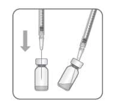 |
|
|---|---|
| B. |
|
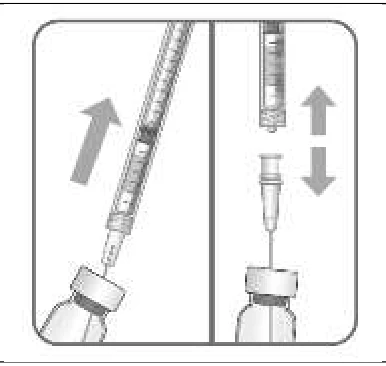 |
|
C. 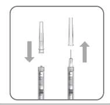 |
Note: Grip at the hub of the injection needle while removing the cap. |
D. 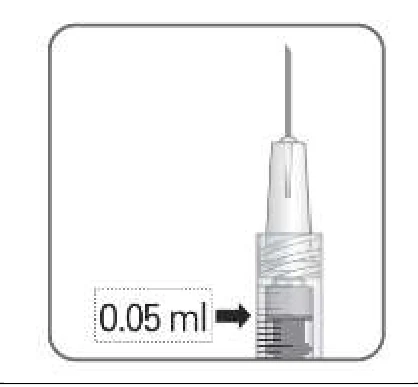 |
Note: Do not wipe the injection needle. Do not pull back on the plunger. |
| E |
|
4.5 Missed Dose
If a planned injection is missed, the patient should return to be assessed by physician as soon as possible and continue dosing depending on physician’s discretion.
5 Overdose
Because MELVIZO (ranibizumab injection) is administered by qualified ophthalmologists experienced in intravitreal injections, the likelihood of an overdose is very low. Cases of accidental overdose (injection of volumes greater than the recommended 0.05 mL MELVIZO) have been reported from the clinical studies and post-marketing data. Adverse reactions most frequently associated with these reported cases were intraocular pressure increased and eye pain. If an overdose occurs, intraocular pressure should be monitored and treated, if deemed necessary by the attending physician.
6 Dosage Forms, Strengths, Composition, and Packaging
To help ensure the traceability of biologic products, including biosimilars, health professionals should recognise the importance of recording both the brand name and the non-proprietary (active ingredient) name as well as other product-specific identifiers such as the Drug Identification Number (DIN) and the batch/lot number of the product supplied.
Table 1 Dosage Forms, Strengths, Composition, and Packaging
MELVIZO (ranibizumab injection) is a sterile, clear colourless to pale yellow and preservative- free aqueous solution for injection.
Packaging
Vial + filter needle + injection needle pack
Single-use glass vial (colourless type I glass) with chlorobutyl rubber stopper containing 2.3 mg ranibizumab in 0.23 mL solution for injection. One pack contains one vial, one filter needle for withdrawal of the vial contents and one injection needle.
Vial only
Single-use glass vial (colourless type I glass) with chlorobutyl rubber stopper containing 2.3 mg ranibizumab in 0.23 mL solution for injection. One pack contains one vial.
For the most recent information in the management of a suspected drug overdose, contact your regional poison control centre or Health Canada's toll-free number, 1-844 POISON-X (1-844-764-7669).
| Route of Administration | Dosage Form/ Strength/Composition | Non-medicinal Ingredients |
| Intravitreal injection | Sterile solution / 10 mg/mL (2.3 mg/0.23 mL/vial) | α,α-trehalose dihydrate; histidine hydrochloride monohydrate; histidine; polysorbate 20; water for injection. |
7 Warnings and Precautions
General
Treatment with MELVIZO (ranibizumab injection) is for intravitreal injection only.
Driving and Operating Machinery
The ranibizumab treatment procedure may induce temporary visual disturbances, which may affect the ability to drive or use machines (see 8 Adverse Reactions). Patients who experience these signs must not drive or use machines until these temporary visual disturbances subside.
Immune
Hypersensitivity
As with all therapeutic proteins, there is a potential for immunogenicity with ranibizumab. Patients should be instructed to report if an intraocular inflammation increases in severity, which may be a clinical sign attributable to intraocular antibody formation. There is a theoretical risk of hypersensitivity reactions including anaphylaxis/anaphylactoid reactions or angioedema which may occur with the use of ranibizumab (see Immunogenicity).
Ranibizumab has not been studied in patients with active systemic infections.
Ophthalmologic
Endophthalmitis and Retinal Detachments
Intravitreal injections, including those with ranibizumab, have been associated with endophthalmitis, intraocular inflammation, hypopyon, rhegmatogenous retinal detachment, retinal tear and iatrogenic traumatic cataract (see 8 Adverse Reactions). Proper aseptic injection techniques must always be used when administering ranibizumab. In addition, patients should be monitored during the week following the injection to permit early treatment if an infection occurs. Patients should be instructed to report any symptoms suggestive of endophthalmitis or any of the aforementioned events without delay.
Retinal Vasculitis and Retinal Occlusive Vasculitis
Retinal vasculitis and retinal occlusive vasculitis have been reported with the use of ranibizumab, typically in the presence of intraocular inflammation or treatment with other intravitreal agents. Discontinue treatment with ranibizumab in patients who develop these events. Patients experiencing any change in vision following treatment should talk to their healthcare professional without delay.
Increases in Intraocular Pressure
Transient increases in intraocular pressure (IOP) have been seen within 60 minutes of injection of ranibizumab (see 8 Adverse Reactions). Sustained IOP increases have also been reported. Both intraocular pressure and the perfusion of the optic nerve head must therefore be monitored and managed appropriately. Monitoring may consist of a check for perfusion of the optic nerve head immediately after the injection and / or tonometry within 30 minutes following the injection.
Ranibizumab has not been studied in patients who have previously received other types of intravitreal injections. Ranibizumab should not be administered concurrently with other anti- VEGF agents (systemic or ocular).
Bilateral treatment
Available data do not suggest an increased risk of systemic adverse events with bilateral treatment. The efficacy of ranibizumab therapy administered to both eyes concurrently has not been studied.
Ranibizumab has not been studied in patients with concurrent eye conditions such as retinal detachment or macular hole.
Additional experience has been gained from post-approval studies on a limited number of patients with prior episodes of RVO and of patients with ischemic branch RVO (BRVO) and central RVO (CRVO). In patients with RVO presenting with clinical signs of irreversible ischemic visual function loss, treatment is not recommended.
Renal
Systemic exposure to ranibizumab may be increased in patients with renal impairment (see 10 10 Clinical Pharmacology – Special Populations and Conditions). The clinical significance of increased systemic exposure to ranibizumab is unknown.
Reproductive Health
Women of Childbearing Potential
Women of childbearing potential should use effective contraception during treatment.
Fertility
There is no fertility data available.
Systemic effects
Thromboembolic events
Although there was a low rate of arterial thromboembolic events (ATEs) observed in the ranibizumab clinical trials, there is a potential risk of ATEs following intravitreal use of VEGF inhibitors. ATEs are defined as nonfatal stroke, nonfatal myocardial infarction, or vascular death (including deaths of unknown cause). The risk of stroke may be greater in patients with known risk factors, including history of prior stroke or transient ischemic attack (see 8 Adverse Reactions). Therefore, these patients should be carefully evaluated by their physician to determine whether treatment with ranibizumab is appropriate, and the benefit outweighs the potential risk. Patients who suffer a thromboembolic event while being treated with ranibizumab should be carefully evaluated by their physician who will assess if continuation of ranibizumab treatment is appropriate, i.e., if the benefit to the patient outweighs the risk.
Non-ocular hemorrhages
Non-ocular hemorrhages have been reported following intravitreal injection of VEGF inhibitors, including ranibizumab in clinical trials for adults (see 8 Adverse Reactions), and there is a potential risk that these may relate to VEGF inhibition.
7.1 Special Populations
7.1.1 Pregnancy
No clinical data concerning exposure to ranibizumab during human pregnancy are available.
Studies in cynomolgus monkeys do not indicate direct or indirect harmful effects with respect to pregnancy or embryonal/fetal development (see 16 Non-Clinical Toxicology). The systemic exposure to ranibizumab is low after ocular administration, but due to its mechanism of action, ranibizumab must be regarded as potentially teratogenic and embryo-/fetotoxic. Therefore, ranibizumab should not be used during pregnancy unless the potential benefit outweighs the potential risk to the fetus. For women who wish to become pregnant and have been treated with ranibizumab, it is recommended to wait at least 3 months after the last dose of ranibizumab before conceiving a child.
7.1.2 Breastfeeding
Based on limited data, ranibizumab is present in human milk following the intravitreal administration of ranibizumab in lactating women and may suppress VEGF levels in breast milk. The effects of VEGF suppression in breast milk on the breastfed infant or the effects of ranibizumab on milk production/excretion are unknown. As a precautionary measure, breast-feeding is not recommended during the use of MELVIZO.
7.1.3 Pediatrics
Pediatrics (< 18 years of age)
Health Canada has not authorized MELVIZO for pediatric use due to insufficient data on safety and efficacy in this sub-population. Limited data from 5 adolescent patients aged 13 to 17 years with visual impairment due to CNV are available.
7.1.4 Geriatrics
Geriatrics (≥ 65 years of age): No dose adjustment is necessary in the elderly.
8 Adverse Reactions
The adverse drug reaction profiles reported in clinical studies that compared MELVIZO to the reference biologic drug Lucentis were comparable. The description of adverse reactions in this section is based on clinical experience with the reference biologic drug.
8.1 Adverse Reaction Overview
Serious adverse events related to the injection procedure included endophthalmitis, rhegmatogenous retinal detachment, retinal tear and iatrogenic traumatic cataract (see 7 Warnings and Precautions).
Other serious ocular events observed among ranibizumab-treated patients included intraocular inflammation (with frequency from 3.3% to 11.7% in the wet AMD trials) and increased intraocular pressure (with frequency from 6.6% to 18.8% in the wet AMD trials) (see 7 Warnings and Precautions).
Overall, ocular and non-ocular events in the DME, RVO, PM and CNV trials were reported with a frequency and severity similar to those seen in the wet-AMD trials.
In the RESOLVE trial in patients with DME, all serious ocular and non-ocular adverse events reported in patients receiving ranibizumab (pooled data) occurred at a frequency of <1%, with the exception of endophthalmitis and hypoglycemia, which both occurred in 2.0% of patients treated with ranibizumab. Serious ocular events other than those listed above observed among ranibizumab treated patients in the RESOLVE trial include retinal artery occlusion, retinal ischemia and vitreous hemorrhage. In the RESTORE trial, the only serious ocular adverse events that occurred in ranibizumab treated patients was cataract, which occurred in 1.7% of patients treated with ranibizumab 0.5 mg and laser. There were no ocular serious adverse events reported in patients treated with ranibizumab monotherapy and no reports of endophthalmitis overall in the study. All serious non-ocular adverse events reported in the ranibizumab arms occurred at a frequency of <1%, with the exception of angina pectoris, coronary artery disease, fall, and pulmonary embolism which each occurred at a frequency of 1.7% in patients treated with ranibizumab monotherapy, as well as hypertension which occurred in 1.7% of patients treated with ranibizumab and laser.
All serious ocular and non-ocular adverse events reported in the RVO trials during the 6-month treatment period in patients receiving ranibizumab (pooled data) occurred at a frequency of <1%. Serious ocular events, other than those listed above for the wet-AMD and DME trials, observed among ranibizumab treated patients in the RVO trials include blindness unilateral, corneal abrasion, corneal edema, iris neovascularization, macular edema, retinal vascular disorder, retinal vascular occlusion and retinal vein occlusion. No serious intraocular inflammation adverse events occurred in the RVO trials.
In the PM trial, all serious ocular and non-ocular adverse events reported in patients receiving ranibizumab also occurred at a frequency of <1%. The serious ocular events observed among ranibizumab treated patients, which were not listed above for the wet-AMD, DME and RVO trials, were corneal erosion and retinoschisis. No serious intraocular inflammation adverse events occurred in the PM trial.
In the CNV trial there were no serious ocular adverse events reported in the study eye or in the fellow treated eye in patients receiving ranibizumab. None of the non-ocular SAEs were suspected to be related to study treatment or ocular injection.
8.2 Clinical Trial Adverse Reactions
Clinical trials are conducted under very specific conditions. The adverse reaction rates observed in the clinical trials; therefore, may not reflect the rates observed in practice and should not be compared to the rates in the clinical trials of another drug. Adverse reaction information from clinical trials may be useful in identifying and approximating rates of adverse drug reactions in real-world use.
Wet AMD Population
A total of 1,315 patients constituted the safety population in the three phase III studies in wet AMD, FVF2598g (MARINA), FVF2587g (ANCHOR) and FVF3192g (PIER) with 24 months exposure to ranibizumab and four hundred and forty (440) patients were treated with the recommended dose of 0.5 mg.
The common ocular and non-ocular adverse events with suspected relationship to ranibizumab treatment occurring in ≥ 1% of patients receiving treatment with ranibizumab 0.5 mg in at least one of the three controlled wet AMD phase III studies FVF2598g (MARINA; 2-year data), FVF2587g (ANCHOR; 2-year data) and FVF3192g (PIER; 2-year data) are summarized in Table 2 and Table 3 below.
The common ocular and non-ocular adverse events, regardless of treatment relationship to ranibizumab, with a difference in incidence rate of ≥ 2% between patients receiving treatment with 0.5 mg ranibizumab and the control group in at least one of the three controlled wet AMD phase III studies FVF2598g (MARINA; 2-year data), FVF2587g (ANCHOR; 2-year data) and FVF3192g (PIER; 2-year data) are summarized in Table 4 below.
Furthermore, the ocular and non-ocular adverse events, regardless of relationship to ranibizumab treatment, occurring in ≥ 1% of patients receiving ranibizumab 0.5 mg in the controlled wet AMD phase III studies FVF2598g (MARINA; 2-year data), FVF2587g (ANCHOR; 2-year data) and FVF3192g (PIER; 2-year data) (pooled data) are summarized in Table 5 and Table 6 below.
Table 2 Ocular Adverse Events in the Study Eye with Suspected Relationship to Ranibizumab Treatment Studies MARINA (FVF2598g), ANCHOR (FVF2587g) and PIER (FVF3192g) Safety Population Adverse Events with Incidence Rate ≥ 1% for Ranibizumab 0.5 mg in at Least One Study
| Preferred Term | % of Patients Study MARINA (Dosage q 1 Month) 2 Years | % of Patients Study ANCHOR (Dosage q 1 Month) 2 Years | % of Patients Study PIER (Dosage q 1 Month for First 3 Months then q 3 Months) 2 Years | ||||||
|---|---|---|---|---|---|---|---|---|---|
| Preferred Term | % of Patients Study MARINA (Dosage q 1 Month) 2 Years | % of Patients Study ANCHOR (Dosage q 1 Month) 2 Years | % of Patients Study PIER (Dosage q 1 Month for First 3 Months then q 3 Months) 2 Years | ||||||
| Preferred Term | % of Patients Study MARINA (Dosage q 1 Month) 2 Years | % of Patients Study ANCHOR (Dosage q 1 Month) 2 Years | % of Patients Study PIER (Dosage q 1 Month for First 3 Months then q 3 Months) 2 Years | ||||||
| Sham (N=236) | Ranibizumab 0.3 mg (N=238) | Ranibizumab 0.5 mg (N=239) | Verteporfin PDT (N=143) | Ranibizumab 0.3 mg (N=137) | Ranibizumab 0.5 mg (N=140) | Sham (N=62) | Ranibizumab 0.3 mg (N=59) | Ranibizumab 0.5 mg (N=61) | |
| Sham (N=236) | Ranibizumab 0.3 mg (N=238) | Ranibizumab 0.5 mg (N=239) | Verteporfin PDT (N=143) | Ranibizumab 0.3 mg (N=137) | Ranibizumab 0.5 mg (N=140) | Sham (N=62) | Ranibizumab 0.3 mg (N=59) | Ranibizumab 0.5 mg (N=61) | |
| Sham (N=236) | Ranibizumab 0.3 mg (N=238) | Ranibizumab 0.5 mg (N=239) | Verteporfin PDT (N=143) | Ranibizumab 0.3 mg (N=137) | Ranibizumab 0.5 mg (N=140) | Sham (N=62) | Ranibizumab 0.3 mg (N=59) | Ranibizumab 0.5 mg (N=61) | |
| Cataract | 0.0% | 0.0% | 1.3% | 0.0% | 0.0% | 1.4% | 0.0% | 0.0% | 0.0% |
| Cataract nuclear | 0.0% | 0.0% | 0.0% | 0.0% | 0.7% | 0.0% | 0.0% | 0.0% | 1.6% |
| Conjunctival hemorrhage | 11.9% | 13.4% | 18.4% | 11.2% | 17.5% | 11.4% | 0.0% | 0.0% | 1.6% |
| Conjunctival hyperemia | 2.1% | 0.4% | 1.7% | 2.1% | 5.1% | 2.1% | 0.0% | 0.0% | 0.0% |
| Conjunctivitis | 1.3% | 1.3% | 1.3% | 0.0% | 0.0% | 0.0% | 0.0% | 0.0% | 0.0% |
| Corneal abrasion | 2.5% | 1.3% | 2.5% | 0.7% | 0.0% | 1.4% | 0.0% | 0.0% | 0.0% |
| Detachment of retinal pigment epithelium | 0.4% | 0.0% | 0.4% | 0.0% | 0.0% | 0.0% | 0.0% | 0.0% | 1.6% |
| Dry eye | 0.8% | 0.4% | 2.1% | 0.7% | 0.0% | 0.7% | 0.0% | 0.0% | 0.0% |
| Eye discharge | 3.0% | 2.9% | 0.4% | 2.1% | 0.7% | 1.4% | 0.0% | 0.0% | 0.0% |
| Eye irritation | 11.4% | 10.1% | 6.7% | 5.6% | 4.4% | 5.7% | 0.0% | 1.7% | 1.6% |
| Eye pain | 23.7% | 26.9% | 28.9% | 18.2% | 22.6% | 17.9% | 0.0% | 0.0% | 0.0% |
| Eye pruritus | 4.7% | 2.9% | 4.2% | 1.4% | 7.3% | 2.9% | 0.0% | 0.0% | 0.0% |
| Eyelid oedema | 1.3% | 0.8% | 1.7% | 1.4% | 2.2% | 1.4% | 0.0% | 0.0% | 0.0% |
| Eyelid pain | 0.4% | 0.8% | 0.4% | 0.7% | 0.0% | 1.4% | 0.0% | 0.0% | 0.0% |
| Foreign body sensation in eyes | 10.2% | 11.8% | 11.3% | 6.3% | 5.1% | 7.9% | 0.0% | 0.0% | 0.0% |
| Hypoaesthesia eye | 0.0% | 0.0% | 0.0% | 0.0% | 0.0% | 0.0% | 0.0% | 0.0% | 1.6% |
| Incorrect route of | 0.0% | 0.0% | 0.0% | 0.0% | 0.7% | 0.0% | 0.0% | 0.0% | 1.6% |
| drug administration | |||||||||
| Injection site hemorrhage | 0.8% | 0.0% | 1.7% | 0.7% | 2.2% | 2.1% | 0.0% | 0.0% | 0.0% |
| Injection site pain | 0.8% | 0.8% | 1.3% | 0.7% | 0.7% | 0.0% | 0.0% | 0.0% | 0.0% |
| Intraocular pressure increased | 3.8% | 18.9% | 18.8% | 5.6% | 17.5% | 15.0% | 1.6% | 5.1% | 6.6% |
| Iridocyclitis | 0.4% | 0.8% | 1.7% | 0.0% | 0.0% | 2.9% | 0.0% | 1.7% | 1.6% |
| Iritis | 3.8% | 5.9% | 5.4% | 0.0% | 2.2% | 6.4% | 1.6% | 0.0% | 1.6% |
| Keratopathy | 0.0% | 0.0% | 0.4% | 0.0% | 0.0% | 0.0% | 0.0% | 0.0% | 1.6% |
| Lacrimation increased | 9.3% | 11.8% | 7.9% | 1.4% | 5.1% | 5.0% | 0.0% | 0.0% | 0.0% |
| Ocular discomfort | 3.0% | 5.9% | 3.8% | 1.4% | 2.9% | 5.0% | 0.0% | 0.0% | 1.6% |
| Ocular hyperemia | 7.2% | 5.0% | 6.3% | 4.2% | 6.6% | 7.9% | 0.0% | 0.0% | 0.0% |
| Punctate keratitis | 0.8% | 0.8% | 1.3% | 0.0% | 0.0% | 0.7% | 0.0% | 0.0% | 0.0% |
| Uveitis | 0.0% | 0.4% | 0.8% | 0.0% | 0.7% | 1.4% | 0.0% | 0.0% | 0.0% |
| Vision blurred | 1.7% | 6.3% | 2.9% | 1.4% | 5.1% | 2.9% | 0.0% | 0.0% | 0.0% |
| Visual acuity reduced | 0.4% | 1.7% | 2.9% | 2.1% | 0.7% | 0.7% | 0.0% | 1.7% | 0.0% |
| Visual disturbance | 0.4% | 3.8% | 6.7% | 0.7% | 2.9% | 1.4% | 0.0% | 0.0% | 0.0% |
| Vitreous detachment | 1.3% | 4.2% | 2.1% | 0.0% | 2.2% | 0.7% | 0.0% | 0.0% | 0.0% |
| Vitreous disorder | 0.0% | 1.3% | 0.4% | 0.0% | 1.5% | 1.4% | 0.0% | 0.0% | 0.0% |
| Vitreous floaters | 2.1% | 18.9% | 17.2% | 1.4% | 9.5% | 4.3% | 0.0% | 1.7% | 1.6% |
| Vitreous hemorrhage | 0.0% | 2.1% | 1.7% | 0.7% | 2.2% | 2.1% | 0.0% | 0.0% | 0.0% |
| Vitritis | 1.3% | 5.5% | 8.4% | 1.4% | 3.6% | 5.0% | 0.0% | 0.0% | 1.6% |
| Total intraocular inflammation+ | 5.1% | 10.9% | 11.7% | 1.4% | 5.8% | 10.7% | 1.6% | 1.7% | 3.3% |
Multiple occurrences of the same event were counted once in the overall incidence + Preferred terms summarized: Anterior chamber inflammation, Hypopyon, Iridocyclitis, Iritis, Uveitis and Vitritis. | |||||||||
Table 3 Non-Ocular Adverse Events with Suspected Relationship to Ranibizumab Treatment Studies MARINA (FVF2598g), ANCHOR (FVF2587g) and PIER (FVF3192g) Safety Population Adverse Events with Incidence Rate ≥ 1% for Ranibizumab 0.5 mg in at Least One Study
| Preferred Term | % of Patients Study MARINA (Dosage q 1 Month) 2 Years | % of Patients Study ANCHOR (Dosage q 1 Month) 2 Years | % of Patients Study PIER (Dosage q 1 Month for First 3 Months then q 3 Months) 2 Years | ||||||
|---|---|---|---|---|---|---|---|---|---|
| Sham (N=236) | Ranibizumab 0.3 mg (N=238) | Ranibizumab 0.5 mg (N=239) | Verteporfin PDT (N=143) | Ranibizumab 0.3 mg (N=137) | Ranibizumab 0.5 mg (N=140) | Sham (N=62) | Ranibizumab 0.3 mg (N=59) | Ranibizumab 0.5 mg (N=61) | |
| INVESTIGATIONS | |||||||||
| Blood creatinine increased | 0.0% | 0.0% | 0.0% | 0.0% | 0.0% | 0.0% | 0.0% | 0.0% | 1.6% |
| Blood pressure diastolic increased | 0.0% | 0.0% | 0.0% | 0.0% | 0.0% | 0.0% | 0.0% | 0.0% | 1.6% |
| Blood urea increased | 0.0% | 0.0% | 0.0% | 0.0% | 0.0% | 0.0% | 0.0% | 0.0% | 1.6% |
| MUSCULOSKELETAL AND CONNECTIVE TISSUE DISORDERS | |||||||||
| Pain in extremity | 0.0% | 0.0% | 0.0% | 0.7% | 0.0% | 0.0% | 0.0% | 0.0% | 1.6% |
| NERVOUS SYSTEM DISORDERS | |||||||||
| Headache | 0.4% | 0.8% | 2.5% | 0.0% | 0.7% | 0.0% | 0.0% | 0.0% | 0.0% |
Multiple occurrences of the same event were counted once in the overall incidence.
Table 4 Ocular (in the study eye) and Non-ocular Adverse Events, regardless Relationship to Treatment, with a Difference in Incidence Rate ≥ 2% between Ranibizumab 0.5 mg and the Control in at Least One Study Studies MARINA (FVF2598g), ANCHOR (FVF2587g) and PIER (FVF3192g) Safety Population
| Preferred Term | % of Patients Study MARINA (Dosage q 1 Month) 2 Years | % of Patients Study ANCHOR (Dosage q 1 Month) 2 Years | % of Patients Study PIER (Dosage q 1 Month for First 3 Months then q 3 Months) 2 Years | ||||||
|---|---|---|---|---|---|---|---|---|---|
| Preferred Term | % of Patients Study MARINA (Dosage q 1 Month) 2 Years | % of Patients Study ANCHOR (Dosage q 1 Month) 2 Years | % of Patients Study PIER (Dosage q 1 Month for First 3 Months then q 3 Months) 2 Years | ||||||
| Preferred Term | % of Patients Study MARINA (Dosage q 1 Month) 2 Years | % of Patients Study ANCHOR (Dosage q 1 Month) 2 Years | % of Patients Study PIER (Dosage q 1 Month for First 3 Months then q 3 Months) 2 Years | ||||||
| Preferred Term | % of Patients Study MARINA (Dosage q 1 Month) 2 Years | % of Patients Study ANCHOR (Dosage q 1 Month) 2 Years | % of Patients Study PIER (Dosage q 1 Month for First 3 Months then q 3 Months) 2 Years | ||||||
| Preferred Term | % of Patients Study MARINA (Dosage q 1 Month) 2 Years | % of Patients Study ANCHOR (Dosage q 1 Month) 2 Years | % of Patients Study PIER (Dosage q 1 Month for First 3 Months then q 3 Months) 2 Years | ||||||
| Sham (N=236) | Ranibizumab 0.3 mg (N=238) | Ranibizumab 0.5 mg (N=239) | Verteporfin PDT (N=143) | Ranibizumab 0.3 mg (N=137) | Ranibizumab 0.5 mg (N=140) | Sham (N=62) | Ranibizumab 0.3 mg (N=59) | Ranibizumab 0.5 mg (N=61) | |
| Sham (N=236) | Ranibizumab 0.3 mg (N=238) | Ranibizumab 0.5 mg (N=239) | Verteporfin PDT (N=143) | Ranibizumab 0.3 mg (N=137) | Ranibizumab 0.5 mg (N=140) | Sham (N=62) | Ranibizumab 0.3 mg (N=59) | Ranibizumab 0.5 mg (N=61) | |
| Sham (N=236) | Ranibizumab 0.3 mg (N=238) | Ranibizumab 0.5 mg (N=239) | Verteporfin PDT (N=143) | Ranibizumab 0.3 mg (N=137) | Ranibizumab 0.5 mg (N=140) | Sham (N=62) | Ranibizumab 0.3 mg (N=59) | Ranibizumab 0.5 mg (N=61) | |
| Sham (N=236) | Ranibizumab 0.3 mg (N=238) | Ranibizumab 0.5 mg (N=239) | Verteporfin PDT (N=143) | Ranibizumab 0.3 mg (N=137) | Ranibizumab 0.5 mg (N=140) | Sham (N=62) | Ranibizumab 0.3 mg (N=59) | Ranibizumab 0.5 mg (N=61) | |
| Sham (N=236) | Ranibizumab 0.3 mg (N=238) | Ranibizumab 0.5 mg (N=239) | Verteporfin PDT (N=143) | Ranibizumab 0.3 mg (N=137) | Ranibizumab 0.5 mg (N=140) | Sham (N=62) | Ranibizumab 0.3 mg (N=59) | Ranibizumab 0.5 mg (N=61) | |
| BLOOD AND LYMPHATIC SYSTEM DISORDERS | |||||||||
| Anaemia | 8.1% | 7.1% | 7.5% | 4.2% | 8.8% | 8.6% | 0.0% | 6.8% | 8.2% |
| CARDIAC DISORDERS | |||||||||
| Angina pectoris | 1.7% | 2.5% | 1.3% | 0.7% | 0.0% | 3.6% | 0.0% | 0.0% | 0.0% |
| Cardiac failure congestive | 4.2% | 4.2% | 2.5% | 3.5% | 4.4% | 4.3% | 3.2% | 3.4% | 6.6% |
| Sinus bradycardia | 0.0% | 0.0% | 0.0% | 0.0% | 1.5% | 0.0% | 0.0% | 0.0% | 3.3% |
| CONGENITAL, FAMILIAL AND GENETIC DISORDERS | |||||||||
| Corneal dystrophy | 2.5% | 4.2% | 2.9% | 0.0% | 2.9% | 2.1% | 0.0% | 0.0% | 0.0% |
| EAR AND LABYRINTH DISORDERS | |||||||||
| Vertigo | 1.7% | 4.2% | 1.7% | 7.0% | 5.8% | 1.4% | 1.6% | 0.0% | 6.6% |
| EYE DISORDERS | |||||||||
| Arcus lipoides | 0.0% | 1.3% | 2.1% | 0.0% | 2.2% | 1.4% | 0.0% | 1.7% | 0.0% |
| Blepharitis | 8.9% | 10.9% | 13.4% | 7.0% | 10.2% | 9.3% | 6.5% | 6.8% | 4.9% |
| Cataract | 6.8% | 7.1% | 5.4% | 7.0% | 9.5% | 13.6% | 1.6% | 6.8% | 6.6% |
| Cataract cortical | 2.1% | 1.3% | 4.6% | 1.4% | 1.5% | 1.4% | 1.6% | 1.7% | 3.3% |
| Cataract nuclear | 5.9% | 4.2% | 3.8% | 1.4% | 5.1% | 5.0% | 3.2% | 1.7% | 8.2% |
| Conjunctival hemorrhage | 66.1% | 77.3% | 75.7% | 50.3% | 71.5% | 70.0% | 29.0% | 50.8% | 52.5% |
| Conjunctival hyperemia | 6.8% | 2.9% | 7.1% | 4.2% | 11.7% | 7.9% | 0.0% | 1.7% | 0.0% |
| Conjunctivitis | 3.0% | 3.8% | 3.3% | 0.0% | 2.2% | 2.1% | 0.0% | 3.4% | 3.3% |
| Conjunctivitis allergic | 1.7% | 2.1% | 3.8% | 0.7% | 0.7% | 1.4% | 1.6% | 0.0% | 3.3% |
| Detachment of retinal pigment epithelium | 15.3% | 11.3% | 9.2% | 3.5% | 2.2% | 4.3% | 3.2% | 8.5% | 16.4% |
| Dry eye | 6.4% | 6.7% | 10.0% | 9.1% | 3.6% | 14.3% | 8.1% | 8.5% | 4.9% |
| Eye irritation | 19.9% | 16.0% | 19.2% | 6.3% | 4.4% | 8.6% | 4.8% | 13.6% | 13.1% |
| Eye pain | 33.5% | 36.1% | 37.2% | 23.1% | 29.9% | 30.0% | 12.9% | 18.6% | 18.0% |
| Eyelid pain | 0.4% | 1.3% | 1.3% | 0.7% | 0.0% | 2.9% | 0.0% | 0.0% | 1.6% |
| Foreign body sensation in eyes | 14.4% | 18.1% | 18.8% | 12.6% | 8.0% | 10.0% | 6.5% | 10.2% | 9.8% |
| Glaucoma | 0.4% | 1.3% | 2.9% | 2.1% | 1.5% | 2.1% | 0.0% | 1.7% | 1.6% |
| Iridocyclitis | 1.3% | 0.8% | 1.7% | 0.0% | 0.0% | 3.6% | 1.6% | 1.7% | 1.6% |
| Iritis | 7.6% | 8.0% | 7.9% | 1.4% | 5.8% | 7.9% | 1.6% | 1.7% | 1.6% |
| Lacrimation increased | 16.1% | 17.2% | 16.3% | 5.6% | 10.2% | 9.3% | 0.0% | 6.8% | 3.3% |
| Maculopathy | 11.4% | 8.4% | 9.6% | 4.9% | 5.1% | 7.9% | 3.2% | 5.1% | 8.2% |
| Ocular discomfort | 4.7% | 7.6% | 7.1% | 2.1% | 4.4% | 6.4% | 0.0% | 0.0% | 8.2% |
| Ocular hyperemia | 10.2% | 10.1% | 10.0% | 5.6% | 7.3% | 12.1% | 1.6% | 8.5% | 6.6% |
| Photophobia | 2.5% | 3.8% | 2.5% | 1.4% | 0.7% | 3.6% | 0.0% | 1.7% | 0.0% |
| Posterior capsule opacification | 5.1% | 6.3% | 7.9% | 2.8% | 3.6% | 5.0% | 0.0% | 3.4% | 3.3% |
| Retinal degeneration | 6.8% | 10.5% | 10.0% | 4.2% | 2.9% | 5.7% | 3.2% | 1.7% | 1.6% |
| Retinal disorder | 9.3% | 11.3% | 12.6% | 2.8% | 8.0% | 6.4% | 0.0% | 0.0% | 1.6% |
| Uveitis | 0.0% | 0.4% | 0.8% | 0.0% | 0.7% | 2.1% | 0.0% | 0.0% | 0.0% |
| Visual disturbance | 11.4% | 13.9% | 15.9% | 5.6% | 10.2% | 7.9% | 1.6% | 3.4% | 4.9% |
| Vitreous detachment | 17.8% | 21.8% | 22.2% | 21.7% | 17.5% | 18.6% | 19.4% | 10.2% | 8.2% |
| Vitreous disorder | 0.0% | 2.1% | 0.8% | 0.0% | 1.5% | 2.1% | 0.0% | 1.7% | 0.0% |
| Vitreous floaters | 9.7% | 31.9% | 29.7% | 4.9% | 19.0% | 22.1% | 3.2% | 11.9% | 13.1% |
| Vitreous hemorrhage | 2.5% | 3.4% | 2.9% | 2.1% | 3.6% | 5.0% | 1.6% | 0.0% | 1.6% |
| Vitritis | 3.4% | 7.1% | 12.6% | 2.1% | 6.6% | 10.7% | 1.6% | 1.7% | 4.9% |
| GASTROINTESTINAL DISORDERS | |||||||||
| Dental caries | 0.8% | 2.1% | 0.8% | 0.0% | 1.5% | 2.1% | 1.6% | 0.0% | 0.0% |
| Diarrhoea | 8.5% | 7.6% | 4.2% | 4.9% | 10.2% | 5.7% | 1.6% | 1.7% | 4.9% |
| Dysphagia | 0.8% | 2.5% | 0.8% | 0.0% | 0.7% | 1.4% | 0.0% | 0.0% | 3.3% |
| Hiatus hernia | 2.1% | 0.8% | 1.3% | 0.7% | 1.5% | 3.6% | 0.0% | 1.7% | 0.0% |
| Nausea | 5.5% | 8.8% | 8.8% | 7.0% | 8.0% | 10.0% | 4.8% | 8.5% | 3.3% |
| GENERAL DISORDERS AND ADMINISTRATION SITE CONDITIONS | |||||||||
| Asthenia | 2.5% | 1.7% | 1.7% | 2.1% | 4.4% | 1.4% | 0.0% | 3.4% | 3.3% |
| Chest pain | 5.5% | 4.2% | 3.8% | 2.1% | 5.1% | 5.0% | 3.2% | 6.8% | 3.3% |
| Injection site hemorrhage | 1.7% | 1.7% | 5.0% | 2.8% | 5.8% | 4.3% | 0.0% | 0.0% | 0.0% |
| INFECTIONS AND INFESTATIONS | |||||||||
| Bronchitis | 8.5% | 9.7% | 10.5% | 9.8% | 8.0% | 12.1% | 4.8% | 5.1% | 6.6% |
| Diverticulitis | 2.1% | 3.8% | 2.9% | 0.7% | 1.5% | 2.9% | 0.0% | 3.4% | 1.6% |
| Gastroenteritis viral | 2.1% | 1.3% | 4.2% | 0.0% | 2.9% | 2.9% | 0.0% | 1.7% | 3.3% |
| Herpes zoster | 2.1% | 5.5% | 4.2% | 2.1% | 2.9% | 0.0% | 1.6% | 1.7% | 4.9% |
| Influenza | 5.1% | 9.7% | 7.9% | 4.2% | 5.8% | 6.4% | 3.2% | 3.4% | 4.9% |
| Localized infection | 2.5% | 2.1% | 0.8% | 0.0% | 0.7% | 1.4% | 0.0% | 0.0% | 3.3% |
| Nasopharyngitis | 13.1% | 13.4% | 15.9% | 12.6% | 24.8% | 16.4% | 9.7% | 8.5% | 6.6% |
| Sinusitis | 5.5% | 7.6% | 8.4% | 10.5% | 10.5% | 8.6% | 4.8% | 5.1% | 1.6% |
| Upper respiratory tract infection | 9.7% | 15.1% | 7.5% | 6.3% | 7.3% | 11.4% | 4.8% | 11.9% | 4.9% |
| Urinary tract infection | 7.6% | 8.8% | 7.1% | 10.5% | 12.4% | 11.4% | 8.1% | 8.5% | 13.1% |
| INJURY, POISONING AND PROCEDURAL COMPLICATIONS | |||||||||
| Corneal abrasion | 3.4% | 2.5% | 2.9% | 0.7% | 2.2% | 3.6% | 1.6% | 1.7% | 0.0% |
| Foot fracture | 0.4% | 1.3% | 0.0% | 0.0% | 0.7% | 2.9% | 1.6% | 0.0% | 0.0% |
| Procedural pain | 1.7% | 1.7% | 1.3% | 2.8% | 2.2% | 5.7% | 1.6% | 5.1% | 0.0% |
| Wrist fracture | 2.5% | 1.7% | 0.4% | 0.0% | 0.7% | 2.1% | 0.0% | 0.0% | 0.0% |
| INVESTIGATIONS | |||||||||
| Blood cholesterol increased | 3.8% | 0.8% | 3.8% | 0.7% | 0.7% | 2.9% | 1.6% | 3.4% | 1.6% |
| Blood glucose increased | 4.7% | 3.4% | 3.8% | 1.4% | 3.6% | 4.3% | 0.0% | 1.7% | 1.6% |
| Intraocular pressure increased | 5.9% | 23.9% | 23.8% | 7.7% | 22.6% | 25.0% | 4.8% | 23.7% | 31.1% |
| White blood cell count increased | 0.0% | 0.0% | 2.5% | 0.0% | 0.7% | 0.7% | 0.0% | 0.0% | 0.0% |
| METABOLISM AND NUTRITION DISORDERS | |||||||||
| Diabetes mellitus | 1.3% | 2.9% | 3.8% | 2.8% | 4.4% | 3.6% | 0.0% | 5.1% | 1.6% |
| Gout | 1.7% | 0.8% | 2.5% | 0.7% | 2.9% | 2.9% | 0.0% | 0.0% | 3.3% |
| Hypokalemia | 3.0% | 1.3% | 1.7% | 4.2% | 4.4% | 2.1% | 0.0% | 0.0% | 8.2% |
| MUSCULOSKELETAL AND CONNECTIVE TISSUE DISORDERS | |||||||||
| Arthralgia | 8.9% | 10.9% | 11.3% | 9.8% | 5.1% | 10.7% | 1.6% | 6.8% | 8.2% |
| Back pain | 9.3% | 10.1% | 9.2% | 13.3% | 10.2% | 4.3% | 3.2% | 1.7% | 6.6% |
| Exostosis | 0.4% | 0.4% | 0.4% | 1.4% | 1.5% | 1.4% | 0.0% | 0.0% | 3.3% |
| Musculoskeletal chest pain | 0.0% | 0.0% | 0.0% | 0.0% | 1.5% | 0.7% | 0.0% | 0.0% | 3.3% |
| Osteoarthritis | 4.2% | 3.8% | 1.7% | 0.7% | 2.9% | 3.6% | 9.7% | 1.7% | 4.9% |
| Pain in extremity | 5.9% | 6.3% | 5.4% | 4.9% | 5.1% | 5.0% | 0.0% | 5.1% | 3.3% |
| NERVOUS SYSTEM DISORDERS | |||||||||
| Dementia | 1.3% | 0.4% | 0.4% | 0.7% | 0.7% | 0.7% | 0.0% | 0.0% | 3.3% |
| Dizziness | 9.7% | 7.6% | 4.6% | 4.9% | 5.1% | 7.9% | 1.6% | 3.4% | 3.3% |
| Headache | 10.2% | 16.0% | 10.0% | 7.0% | 12.4% | 15.0% | 3.2% | 1.7% | 8.2% |
| Sciatica | 0.8% | 1.3% | 1.7% | 0.7% | 2.2% | 2.1% | 0.0% | 1.7% | 3.3% |
| PSYCHIATRIC DISORDERS | |||||||||
| Anxiety | 3.0% | 4.2% | 5.0% | 6.3% | 7.3% | 3.6% | 4.8% | 0.0% | 1.6% |
| RESPIRATORY, THORACIC AND MEDIASTINAL DISORDERS | |||||||||
| Chronic obstructive pulmonary disease | 2.1% | 2.1% | 4.6% | 3.5% | 6.6% | 7.9% | 0.0% | 6.8% | 0.0% |
| Cough | 7.2% | 9.7% | 10.5% | 8.4% | 12.4% | 6.4% | 3.2% | 3.4% | 3.3% |
| Dyspnoea | 2.5% | 5.0% | 2.9% | 3.5% | 3.6% | 5.7% | 3.2% | 6.8% | 1.6% |
| Sinus congestion | 2.5% | 2.1% | 2.1% | 0.0% | 1.5% | 2.1% | 1.6% | 1.7% | 1.6% |
| SKIN AND SUBCUTANEOUS TISSUE DISORDERS | |||||||||
| Ecchymosis | 0.4% | 1.7% | 1.7% | 0.0% | 2.2% | 4.3% | 0.0% | 1.0% | 1.6% |
| Pruritus | 2.1% | 3.8% | 2.5% | 0.7% | 0.0% | 3.6% | 1.6% | 1.7% | 0.0% |
| VASCULAR DISORDERS | |||||||||
| Hypertension | 16.1% | 17.2% | 16.3% | 16.1% | 9.5% | 12.1% | 11.3% | 10.2% | 18.0% |
| Multiple occurrences of the same event were counted once in the overall incidence. | |||||||||
Table 5 Ocular Adverse Events in The Study Eye regardless of Relationship to Treatment by System Organ Class and Preferred Term (2-year Incidence at Least 1.0% in Ranibizumab 0.5 mg Group) in MARINA, ANCHOR, and PIER (Pooled Data) Safety Population
| Preferred Term | Sham N=298 (%) | PDT N=143 (%) | Ranibizumab 0.3 mg N=434 (%) | Ranibizumab 0.5 mg N=440(%) |
|---|---|---|---|---|
| Preferred Term | Sham N=298 (%) | PDT N=143 (%) | Ranibizumab 0.3 mg N=434 (%) | Ranibizumab 0.5 mg N=440(%) |
| Preferred Term | Sham N=298 (%) | PDT N=143 (%) | Ranibizumab 0.3 mg N=434 (%) | Ranibizumab 0.5 mg N=440(%) |
| CONGENITAL, FAMILIAL AND GENETIC DISORDERS | ||||
| Corneal dystrophy | 2.0% | 0.0% | 3.2% | 2.3% |
| EYE DISORDERS | ||||
| Anterior chamber flare | 2.0% | 0.0% | 2.1% | 2.0% |
| Arcus lipoides | 0.0% | 0.0% | 1.6% | 1.6% |
| Blepharitis | 8.4% | 7.0% | 10.1% | 10.9% |
| Cataract | 5.7% | 7.0% | 7.8% | 8.2% |
| Cataract cortical | 2.0% | 1.4% | 1.4% | 3.4% |
| Cataract nuclear | 5.4% | 1.4% | 4.1% | 4.8% |
| Cataract subcapsular | 2.3% | 0.7% | 3.2% | 2.7% |
| Chalazion | 0.3% | 0.7% | 0.7% | 1.6% |
| Choroidal neovascularisation | 16.8% | 12.6% | 2.5% | 2.5% |
| Conjunctival hemorrhage | 58.4% | 50.3% | 71.9% | 70.7% |
| Conjunctival hyperemia | 5.4% | 4.2% | 5.5% | 6.4% |
| Conjunctivitis | 2.3% | 0.0% | 3.2% | 3.0% |
| Conjunctivitis allergic | 1.7% | 0.7% | 1.4% | 3.0% |
| Detachment of retinal pigment epithelium | 12.8% | 3.5% | 8.1% | 8.6% |
| Diplopia | 0.0% | 0.7% | 0.9% | 1.1% |
| Dry eye | 6.7% | 9.1% | 6.0% | 10.7% |
| Eye discharge | 6.7% | 3.5% | 6.2% | 3.0% |
| Eye hemorrhage | 3.4% | 0.0% | 0.7% | 1.1% |
| Eye irritation | 16.8% | 6.3% | 12.0% | 15.0% |
| Eye pain | 29.2% | 23.1% | 31.8% | 32.3% |
| Eye pruritus | 10.4% | 8.4% | 8.5% | 10.2% |
| Eye swelling | 1.3% | 1.4% | 1.2% | 1.4% |
| Eyelid edema | 2.7% | 2.8% | 2.8% | 3.0% |
| Eyelid pain | 0.3% | 0.7% | 0.7% | 1.8% |
| Foreign body sensation in eyes | 12.8% | 12.6% | 13.8% | 14.8% |
| Glaucoma | 0.3% | 2.1% | 1.4% | 2.5% |
| Iridocyclitis | 1.3% | 0.0% | 0.7% | 2.3% |
| Iritis | 6.4% | 1.4% | 6.5% | 7.0% |
| Lacrimation increased | 12.8% | 5.6% | 13.6% | 12.3% |
| Macular degeneration | 62.4% | 70.6% | 43.3% | 42.0% |
| Macular edema | 10.4% | 4.9% | 3.2% | 3.6% |
| Macular scar | 1.7% | 2.1% | 2.3% | 1.8% |
| Maculopathy | 9.7% | 4.9% | 6.9% | 8.9% |
| Meibomianitis | 0.3% | 0.0% | 0.9% | 1.6% |
| Ocular discomfort | 3.7% | 2.1% | 5.5% | 7.0% |
| Ocular hyperemia | 8.4% | 5.6% | 9.0% | 10.2% |
| Photophobia | 2.0% | 1.4% | 2.5% | 2.5% |
| Photopsia | 5.0% | 8.4% | 4.8% | 4.1% |
| Posterior capsule opacification | 4.0% | 2.8% | 5.1% | 6.4% |
| Punctate keratitis | 3.7% | 1.4% | 3.9% | 3.0% |
| Retinal degeneration | 6.0% | 4.2% | 6.9% | 7.5% |
| Retinal detachment | 7.0% | 2.1% | 3.9% | 1.4% |
| Retinal disorder | 7.4% | 2.8% | 8.8% | 9.1% |
| Retinal exudates | 9.7% | 4.9% | 5.5% | 4.8% |
| Retinal hemorrhage | 52.7% | 67.8% | 25.8% | 25.2% |
| Retinal pigmentation | 1.0% | 0.0% | 1.4% | 1.1% |
| Retinal tear | 1.0% | 0.0% | 0.9% | 1.1% |
| Retinal vascular disorder | 2.7% | 0.7% | 0.5% | 1.4% |
| Subretinal fibrosis | 15.1% | 22.4% | 9.2% | 9.5% |
| Trichiasis | 0.7% | 0.7% | 1.2% | 1.6% |
| Uveitis | 0.0% | 0.0% | 0.5% | 1.1% |
| Vision blurred | 7.4% | 9.1% | 11.5% | 6.8% |
| Visual acuity reduced | 17.8% | 16.8% | 11.3% | 8.6% |
| Visual disturbance | 7.4% | 4.9% | 9.2% | 10.5% |
| Vitreous detachment | 17.8% | 21.7% | 18.9% | 19.1% |
| Vitreous disorder | 0.0% | 0.0% | 1.8% | 1.1% |
| Vitreous floaters | 8.1% | 4.9% | 25.1% | 25.0% |
| Vitreous hemorrhage | 2.3% | 2.1% | 3.0% | 3.4% |
| Vitritis | 3.0% | 2.1% | 6.2% | 10.9% |
| GENERAL DISORDERS AND ADMINISTRATION SITE CONDITIONS | ||||
| Injection site hemorrhage | 1.3% | 2.8% | 2.8% | 4.1% |
| IMMUNE SYSTEM DISORDERS | ||||
| Drug hypersensitivity | 2.0% | 4.2% | 1.4% | 1.4% |
| INJURY, POISONING AND PROCEDURAL COMPLICATIONS | ||||
| Corneal abrasion | 3.0% | 0.7% | 2.3% | 2.7% |
| INVESTIGATIONS | ||||
| Intraocular pressure increased | 5.7% | 7.7% | 23.5% | 25.2% |
| SKIN AND SUBCUTANEOUS TISSUE DISORDERS | ||||
| Cutis laxa | 0.7% | 0.7% | 1.4% | 1.1% |
| Dandruff | 0.7% | 0.7% | 0.7% | 1.1% |
Table 6 Non-ocular Adverse Events regardless of Relationship to Treatment by System Organ Class and Preferred Term (2-year Incidence at Least 1.0% in Ranibizumab 0.5 mg Group) in MARINA, ANCHOR, and PIER (Pooled Data) Safety Population
| Preferred Term | Sham N=298 (%) | PDT N=143 (%) | Ranibizumab 0.3 mg N=434 (%) | Ranibizumab 0.5 mg N=440 (%) |
|---|---|---|---|---|
| Preferred Term | Sham N=298 (%) | PDT N=143 (%) | Ranibizumab 0.3 mg N=434 (%) | Ranibizumab 0.5 mg N=440 (%) |
| Preferred Term | Sham N=298 (%) | PDT N=143 (%) | Ranibizumab 0.3 mg N=434 (%) | Ranibizumab 0.5 mg N=440 (%) |
| Preferred Term | Sham N=298 (%) | PDT N=143 (%) | Ranibizumab 0.3 mg N=434 (%) | Ranibizumab 0.5 mg N=440 (%) |
| Preferred Term | Sham N=298 (%) | PDT N=143 (%) | Ranibizumab 0.3 mg N=434 (%) | Ranibizumab 0.5 mg N=440 (%) |
| BLOOD AND LYMPHATIC SYSTEM DISORDERS | ||||
| Anaemia | 6.4% | 4.2% | 7.6% | 8.0% |
| CARDIAC DISORDERS | ||||
| Angina pectoris | 1.3% | 0.7% | 1.4% | 1.8% |
| Atrial fibrillation | 4.0% | 3.5% | 3.2% | 4.5% |
| Bradycardia | 0.7% | 2.1% | 0.9% | 1.1% |
| Cardiac failure congestive | 4.0% | 3.5% | 4.1% | 3.6% |
| Cardiomegaly | 1.7% | 0.7% | 1.2% | 1.1% |
| Coronary artery disease | 3.0% | 0.7% | 2.8% | 2.3% |
| Myocardial infarction | 1.7% | 1.4% | 1.2% | 1.6% |
| EAR AND LABYRINTH DISORDERS | ||||
| Vertigo | 1.7% | 7.0% | 4.1% | 2.3% |
| ENDOCRINE DISORDERS | ||||
| Hypothyroidism | 2.3% | 2.1% | 1.6% | 1.1% |
| GASTROINTESTINAL DISORDERS | ||||
| Abdominal pain | 1.7% | 2.1% | 2.8% | 1.1% |
| Colonic polyp | 1.0% | 2.8% | 1.6% | 1.6% |
| Constipation | 7.0% | 5.6% | 6.7% | 5.2% |
| Dental caries | 1.0% | 0.0% | 1.6% | 1.1% |
| Diarrhoea | 7.0% | 4.9% | 7.6% | 4.8% |
| Diverticulum | 1.3% | 0.7% | 2.1% | 1.6% |
| Dyspepsia | 3.7% | 2.8% | 2.1% | 1.4% |
| Dysphagia | 0.7% | 0.0% | 1.6% | 1.4% |
| Gastritis | 0.7% | 0.7% | 1.2% | 1.6% |
| Gastrooesophageal reflux disease | 5.0% | 8.4% | 6.0% | 3.6% |
| Hemorrhoids | 2.3% | 1.4% | 1.8% | 1.8% |
| Hiatus hernia | 1.7% | 0.7% | 1.2% | 1.8% |
| Inguinal hernia | 0.3% | 0.0% | 0.5% | 1.1% |
| Nausea | 5.4% | 7.0% | 8.5% | 8.4% |
| Toothache | 2.0% | 1.4% | 1.4% | 1.6% |
| Vomiting | 1.3% | 5.6% | 4.4% | 2.0% |
| GENERAL DISORDERS AND ADMINISTRATION SITE CONDITIONS | ||||
| Asthenia | 1.0% | 2.1% | 2.8% | 1.4% |
| Chest pain | 5.0% | 2.1% | 4.8% | 3.9% |
| Fatigue | 3.4% | 4.9% | 3.2% | 3.2% |
| Oedema peripheral | 5.7% | 2.8% | 6.7% | 3.6% |
| Pain | 1.7% | 2.1% | 2.1% | 2.0% |
| Pyrexia | 1.7% | 1.4% | 2.5% | 2.3% |
| HEPATOBILIARY DISORDERS | ||||
| Cholelithiasis | 3.0% | 0.7% | 0.7% | 1.1% |
| IMMUNE SYSTEM DISORDERS | ||||
| Drug hypersensitivity | 2.7% | 1.4% | 0.9% | 2.0% |
| Hypersensitivity | 1.0% | 4.2% | 2.8% | 2.3% |
| Seasonal allergy | 3.4% | 7.0% | 3.7% | 3.9% |
| INFECTIONS AND INFESTATIONS | ||||
| Bronchitis | 8.1% | 9.8% | 9.0% | 10.5% |
| Cellulitis | 2.3% | 0.7% | 2.1% | 1.1% |
| Cystitis | 2.0% | 2.8% | 3.0% | 3.0% |
| Diverticulitis | 1.7% | 0.7% | 3.0% | 2.7% |
| Ear infection | 1.3% | 2.1% | 1.4% | 1.4% |
| Gastroenteritis viral | 1.7% | 0.0% | 1.8% | 3.6% |
| Herpes zoster | 2.0% | 2.1% | 4.1% | 3.0% |
| Influenza | 4.7% | 4.2% | 7.6% | 7.0% |
| Localised infection | 2.0% | 0.0% | 1.4% | 1.4% |
| Lower respiratory tract infection | 0.7% | 1.4% | 1.8% | 1.1% |
| Nasopharyngitis | 12.4% | 12.6% | 16.4% | 14.8% |
| Pneumonia | 5.4% | 4.2% | 6.2% | 4.5% |
| Sinusitis | 5.4% | 10.5% | 8.1% | 7.5% |
| Tooth abscess | 1.7% | 1.4% | 1.2% | 1.6% |
| Tooth infection | 1.3% | 0.0% | 1.6% | 1.1% |
| Upper respiratory tract infection | 8.7% | 6.3% | 12.2% | 8.4% |
| Urinary tract infection | 7.7% | 10.5% | 9.9% | 9.3% |
| INJURY, POISONING AND PROCEDURAL COMPLICATIONS | ||||
| Contusion | 7.4% | 5.6% | 3.9% | 4.1% |
| Excoriation | 1.3% | 0.7% | 1.6% | 1.6% |
| Fall | 4.0% | 2.1% | 3.2% | 3.2% |
| Joint sprain | 0.7% | 0.0% | 1.2% | 1.1% |
| Procedural pain | 1.7% | 2.8% | 2.3% | 2.5% |
| Skin laceration | 2.3% | 3.5% | 3.5% | 1.6% |
| INVESTIGATIONS | ||||
| Blood cholesterol increased | 3.4% | 0.7% | 1.2% | 3.2% |
| Blood glucose increased | 3.7% | 1.4% | 3.2% | 3.6% |
| Blood pressure increased | 6.0% | 2.1% | 4.6% | 5.5% |
| Blood urea increased | 0.3% | 0.7% | 0.9% | 1.1% |
| Blood uric acid increased | 1.3% | 0.0% | 0.7% | 1.8% |
| White blood cell count increased | 0.0% | 0.0% | 0.2% | 1.6% |
| METABOLISM AND NUTRITION DISORDERS | ||||
| Dehydration | 1.0% | 3.5% | 2.8% | 2.3% |
| Diabetes mellitus | 1.0% | 2.8% | 3.7% | 3.4% |
| Gout | 1.3% | 0.7% | 1.4% | 2.7% |
| Hypercholesterolemia | 4.4% | 5.6% | 5.3% | 5.0% |
| Hyperlipidemia | 2.0% | 2.8% | 1.8% | 1.6% |
| Hypokalemia | 2.3% | 4.2% | 2.1% | 2.7% |
| MUSCULOSKELETAL AND CONNECTIVE TISSUE DISORDERS | ||||
| Arthralgia | 7.4% | 9.8% | 8.5% | 10.7% |
| Arthritis | 7.0% | 7.0% | 4.6% | 5.7% |
| Back pain | 8.1% | 13.3% | 9.0% | 7.3% |
| Exostosis | 0.3% | 1.4% | 0.7% | 1.1% |
| Joint swelling | 2.0% | 1.4% | 1.8% | 1.1% |
| Muscle spasms | 2.3% | 2.8% | 2.3% | 1.6% |
| Musculoskeletal pain | 4.4% | 2.8% | 2.8% | 3.0% |
| Musculoskeletal stiffness | 0.0% | 0.7% | 0.2% | 1.1% |
| Myalgia | 1.3% | 2.8% | 1.4% | 1.6% |
| Neck pain | 1.0% | 0.0% | 1.2% | 2.0% |
| Osteoarthritis | 5.4% | 0.7% | 3.2% | 2.7% |
| Osteoporosis | 2.0% | 3.5% | 2.8% | 3.0% |
| Pain in extremity | 4.7% | 4.9% | 5.8% | 5.0% |
| Rotator cuff syndrome | 1.3% | 0.7% | 0.9% | 1.1% |
| NEOPLASMS BENIGN, MALIGNANT AND UNSPECIFIED (INCL CYSTS AND POLYPS) | ||||
| Basal cell carcinoma | 3.7% | 2.1% | 5.1% | 2.3% |
| Skin cancer | 1.0% | 0.0% | 0.9% | 1.1% |
| NERVOUS SYSTEM DISORDERS | ||||
| Balance disorder | 0.3% | 0.0% | 0.2% | 1.1% |
| Carpal tunnel syndrome | 0.3% | 0.7% | 0.9% | 1.4% |
| Cerebrovascular accident | 0.7% | 0.7% | 0.7% | 1.1% |
| Dizziness | 8.1% | 4.9% | 6.2% | 5.2% |
| Headache | 8.7% | 7.0% | 12.4% | 11.4% |
| Nerve compression | 1.0% | 0.0% | 0.5% | 1.1% |
| Sciatica | 0.7% | 0.7% | 1.6% | 2.0% |
| Syncope | 3.0% | 2.1% | 1.4% | 2.7% |
| Transient ischaemic attack | 1.3% | 2.8% | 0.7% | 1.8% |
| PSYCHIATRIC DISORDERS | ||||
| Anxiety | 3.4% | 6.3% | 4.6% | 4.1% |
| Depression | 6.7% | 9.1% | 5.5% | 5.0% |
| Insomnia | 5.0% | 4.2% | 4.1% | 5.2% |
| RENAL AND URINARY DISORDERS | ||||
| Renal cyst | 1.3% | 3.5% | 1.4% | 1.1% |
| Renal failure | 0.3% | 1.4% | 1.8% | 1.1% |
| REPRODUCTIVE SYSTEM AND BREAST DISORDERS | ||||
| Benign prostatic hyperplasia | 1.3% | 2.8% | 2.1% | 2.3% |
| RESPIRATORY, THORACIC AND MEDIASTINAL DISORDERS | ||||
| Asthma | 2.0% | 2.8% | 2.5% | 2.3% |
| Chronic obstructive pulmonary disease | 1.7% | 3.5% | 4.1% | 5.0% |
| Cough | 6.4% | 8.4% | 9.7% | 8.2% |
| Dyspnoea | 2.7% | 3.5% | 4.8% | 3.6% |
| Emphysema | 1.0% | 1.4% | 1.2% | 1.1% |
| Epistaxis | 1.0% | 2.8% | 1.8% | 1.6% |
| Pharyngolaryngeal pain | 0.7% | 2.8% | 2.3% | 1.8% |
| Rhinorrhoea | 1.7% | 1.4% | 2.3% | 1.4% |
| Sinus congestion | 2.3% | 0.0% | 1.8% | 2.0% |
| SKIN AND SUBCUTANEOUS TISSUE DISORDERS | ||||
| Actinic keratosis | 3.0% | 0.7% | 1.8% | 1.4% |
| Dermatitis contact | 0.7% | 2.1% | 0.9% | 1.4% |
| Ecchymosis | 0.0% | 0.0% | 0.7% | 1.6% |
| Pruritus | 1.3% | 0.7% | 2.1% | 2.5% |
| Rash | 3.7% | 5.6% | 2.8% | 3.9% |
| VASCULAR DISORDERS | ||||
| Haematoma | 1.3% | 0.0% | 1.4% | 1.1% |
| Hypertension | 15.1% | 16.1% | 13.8% | 15.2% |
| Hypotension | 2.3% | 4.2% | 2.3% | 1.8% |
Arterial thromboembolic events, as defined by the ANTIPLATELET TRIALISTS’ COLLABORATION (APTC), including vascular deaths, non-fatal myocardial infarctions, non- fatal ischemic strokes and non-fatal haemorrhagic strokes, have been linked to the systemic availability of highly potent vascular endothelial growth factor (VEGF) inhibitors. When the first-year data from all three phase III studies (MARINA, ANCHOR and PIER) were combined, the overall incidence of arterial thromboembolic events was higher for patients treated with ranibizumab 0.5 mg (2.5%) compared with control arm (1.1%). When the second year data from all three phase III (MARINA, ANCHOR and PIER) were combined, the overall incidence of arterial thromboembolic events was similar for patients with ranibizumab 0.5 mg (4.1%) compared with the control arm (3.6%). For (fatal and non-fatal) cerebrovascular accidents (CVAs) the results were not consistent across studies. In the MARINA study, there was a slight numerical imbalance between ranibizumab 0.5 mg (1.3%) and the control arm (0.4%) during the first year, which was still present at the end of the 2-year treatment period (3.3% for 0.5 mg vs. 1.3% for sham treatment). The CVA incidence rate in the MARINA, ANCHOR study and PIER study are shown in Table 7. Although there was a low annual rate of arterial thromboembolic events observed in the ranibizumab (a VEGF inhibitor) clinical trials, there was no imbalance between treatment groups (Table 8). There is a potential risk of arterial thromboembolic events following intravitreal use of VEGF inhibitors.
Table 7 Cerebrovascular Accident (CVA) Rates (Fatal and Non-fatal), Safety Populations (2-year Cumulative Data)
Control
0.3 mg Ranibizumab
0.5 mg Ranibizumab
MARINA – 1 yr
1/236 (0.4%)
1/238 (0.4%)
3/239 (1.3%)
MARINA – 2 yr
3/236 (1.3%)
3/238 (1.3%)
8/239 (3.3%)
ANCHOR – 1 yr
1/143 (0.7%)
1/137 (0.7%)
1/140 (0.7%)
ANCHOR - 2 yr
2/143 (1.4%)
3/137 (2.2%)a
0/140 (0.0%)
PIER – 1 yr
0/62 (0.0%)
0/59 (0.0%)
0/61 (0.0%)
PIER -2 yrb
0/62 (0.0%) (before crossover) 1/39 (2.6%) (after crossover)
0/59 (0.0%)
0/61 (0.0%)
a Includes one serious adverse event of cerebral infarction (Year 1), one adverse event of ischemic stroke (Year 2) and one non-serious adverse event of cerebral ischemia (Year 2)
b After the month 12 visit in the study, patients in the sham-injection group could crossover to the ranibizumab 0.5 mg group for the remainder of the study.
Table 8 Arterial Thromboembolic Events (ATE) as Defined by the Antiplatelet Trialists’ Collaboration (APTC), Safety Populations (2-year Cumulative Data)
Control
0.3 mg Ranibizumab
0.5 mg Ranibizumab
MARINA – 1 yr
2/236 (0.8%)
4/238 (1.7%)
5/239 (2.1%)
MARINA – 2 yr
9/236 (3.8%)
11/238 (4.6%)
11/239 (4.6%)
ANCHOR – 1 yr
3/143 (2.1%)
3/137 (2.2%)
6/140 (4.3%)
ANCHOR 2 yr
6/143 (4.2%)
6/137 (4.4%)
7/140 (5.0%)
PIER – 1 yr
0/62 (0.0%)
0/59 (0.0%)
0/61 (0.0%)
PIER -2 yra
1/62 (1.6%) (before crossover) 1/39 (2.6%) (after crossover)
1/59 (1.7%)
0/61 (0.0%)
a After the month 12 visit in the study, patients in the sham-injection group could crossover to the ranibizumab 0.5 mg group for the remainder of the study.
The SAILOR study, FVF 3689g, was a Phase IIIb, Multicenter Study to Evaluate the Safety and Tolerability of Ranibizumab in Naive and Previously Treated Subjects with Choroidal Neovascularization (CNV) Secondary to Age-Related Macular Degeneration (AMD).
In the study, an interim analysis including data from almost 2400 patients (randomized to the two doses of ranibizumab 0.3 mg and ranibizumab 0.5 mg in a 1:1 ratio) with an average follow-up period of 230 days was performed. The overall incidence of CVAs (fatal and non-fatal) was found to be 1.1% for ranibizumab 0.5 mg compared with 0.3% for ranibizumab 0.3 mg (p=0.021). The rate of deaths from all causes was (1.6% on 0.5 mg vs. 0.6% on 0.3 mg, p=0.029) whereas the differences in the rate of vascular deaths (0.7% on 0.5 mg vs. 0.4% on 0.3 mg) and in the rate of combined APTC events (1.7% on 0.5 mg vs. 1.1% on 0.3 mg) were smaller.
A second interim analysis which was based on an average follow-up time of 335 days showed that the difference in the incidence of CVAs had decreased with increasing exposure to ranibizumab treatment (1.3% on 0.5 mg vs. 0.6% on 0.3 mg). The same applied to the difference in the overall death rate (2.0% on 0.5 mg vs. 1.3% on 0.3 mg).
Upon final analysis of the incidence in strokes in SAILOR study, there exists a trend towards a higher stroke rate in the 0.5 mg group compared to the 0.3 mg group. The respective 95% CIs for the overall stroke rate were wide (0.3% to 1.3% for the 0.3 mg group vs. 0.7% to 2.0% for the 0.5 mg group). The number of strokes was small for both dose groups, and there is not sufficient evidence to conclude (or rule out) that there is a true difference in stroke rates among the treatment groups. Forty-nine deaths were reported, with 20 deaths in the 0.3-mg group (1.7%) and 29 deaths in the 0.5-mg group (2.4%). Of these, 21 were classified as vascular deaths, 11 in the 0.3-mg group (0.9%) and 10 in the 0.5-mg group (0.8%).
DME population
The safety of ranibizumab was studied in a one-year sham-controlled trial (study D2201 - RESOLVE) and in a one year laser-controlled trial (study D2301 - RESTORE) conducted respectively in 102 and 235 ranibizumab-treated patients with visual impairment due to DME (see 14 Clinical Trials – Treatment of visual impairment due to DME). The safety population is grouped by the actual treatment received and is comprised of the safety-evaluable patients from the two studies who received at least one dose of study drug (a total of 496 patients). Overall, ocular and non-ocular events in the RESOLVE and RESTORE trials were reported with a frequency and severity similar to those seen in the wet-AMD trials. The most common ocular adverse events in patients receiving ranibizumab monotherapy (pooled data) were eye pain (14.3%), conjunctival hemorrhage (14.3%), intraocular pressure increased (10.1%), conjunctival hyperemia (5.5%), and foreign body sensation in eyes (5.1%). The most common non-ocular events were nasopharyngitis (9.7%), hypertension (7.4%) and influenza (5.1%).
The common ocular and non-ocular adverse events with suspected relationship to ranibizumab treatment occurring in ≥ 1% of patients receiving treatment with ranibizumab (any group) in at least one of the two studies RESOLVE and RESTORE are summarized in Table 9 and Table 10 below.
The common ocular and non-ocular adverse events, regardless of treatment relationship to ranibizumab, with a difference in incidence rate of > 2% between patients receiving treatment with ranibizumab (any group) and the control group in at least one of the two studies RESOLVE and RESTORE are summarized in Table 11 below.
Table 9 Ocular Adverse Events in the Study Eye with Suspected Relationship to Ranibizumab Treatment Studies RESOLVE and RESTORE Safety Population Adverse Events with Incidence Rate ≥ 1% For Ranibizumab (Any Group) in at Least One Study
% of Patients Study RESOLVE (group A + B)† | % of Patients Study RESTORE | |||||||||||||||||||||
|---|---|---|---|---|---|---|---|---|---|---|---|---|---|---|---|---|---|---|---|---|---|---|
% of Patients Study RESOLVE (group A + B)† | % of Patients Study RESTORE | |||||||||||||||||||||
SYSTEM ORGAN CLASS Preferred term | Ranibizumab 6 mg/mL‡ (N=51) | Ranibizumab 10 mg/mL‡ (N=51) | Ranibizumab Pooled (N=102) | Sham (N=49) | Ranibizumab 0.5 mg (N=115) | Ranibizumab 0.5 mg + Laser (N=120) | Laser (N=110) | |||||||||||||||
SYSTEM ORGAN CLASS Preferred term | Ranibizumab 6 mg/mL‡ (N=51) | Ranibizumab 10 mg/mL‡ (N=51) | Ranibizumab Pooled (N=102) | Sham (N=49) | Ranibizumab 0.5 mg (N=115) | Ranibizumab 0.5 mg + Laser (N=120) | Laser (N=110) | |||||||||||||||
| EYE DISORDERS | ||||||||||||||||||||||
| Conjunctival hemorrhage | 19.6% | 25.5% | 22.5% | 14.3% | 7.0% | 7.5% | 0.0% | |||||||||||||||
| Eye pain | 15.7% | 15.7% | 15.7% | 16.3% | 10.4% | 8.3% | 10.0% | |||||||||||||||
| Vitreous floaters | 2.0% | 13.7% | 7.8% | 0.0% | 0.0% | 0.0% | 0.0% | |||||||||||||||
| Conjunctival hyperemia | 3.9% | 2.0% | 2.9% | 2.0% | 7.0% | 3.3% | 5.5% | |||||||||||||||
Foreign body sensation in eyes | 5.9% | 3.9% | 4.9% | 2.0% | 3.5% | 5.8% | 1.8% | |||||||||||||||
| Lacrimation increased | 3.9% | 5.9% | 4.9% | 0.0% | 1.7% | 2.5% | 0.9% | |||||||||||||||
| Eye irritation | 5.9% | 2.0% | 3.9% | 2.0% | 1.7% | 0.0% | 1.8% | |||||||||||||||
| Eye pruritus | 3.9% | 3.9% | 3.9% | 0.0% | 0.9% | 0.8% | 1.8% | |||||||||||||||
| Endophthalmitis | 2.0% | 3.9% | 2.9% | 0.0% | 0.0% | 0.0% | 0.0% | |||||||||||||||
| Ocular hyperemia | 2.0% | 3.9% | 2.9% | 8.2% | 0.9% | 0.8% | 0.0% | |||||||||||||||
| Vision blurred | 2.0% | 3.9% | 2.9% | 0.0% | 1.7% | 0.8% | 1.8% | |||||||||||||||
| Vitreous hemorrhage | 2.0% | 3.9% | 2.9% | 0.0% | 0.0% | 0.0% | 0.0% | |||||||||||||||
| Eye discharge | 2.0% | 0.0% | 1.0% | 0.0% | 2.6% | 2.5% | 0.9% | |||||||||||||||
| Visual impairment | 0.0% | 0.0% | 0.0% | 0.0% | 2.6% | 1.7% | 0.0% | |||||||||||||||
| Eyelid edema | 0.0% | 0.0% | 0.0% | 6.1% | 1.7% | 2.5% | 0.9% | |||||||||||||||
| Vitreous disorder | 2.0% | 2.0% | 2.0% | 0.0% | 0.0% | 0.0% | 0.0% | |||||||||||||||
| Blepharitis allergic | 0.0% | 2.0% | 1.0% | 0.0% | 0.0% | 0.0% | 0.0% | |||||||||||||||
| Conjunctival edema | 2.0% | 0.0% | 1.0% | 0.0% | 0.0% | 0.0% | 0.0% | |||||||||||||||
| Corneal disorder | 0.0% | 2.0% | 1.0% | 0.0% | 0.0% | 0.0% | 0.0% | |||||||||||||||
| Corneal erosion | 2.0% | 0.0% | 1.0% | 2.0% | 0.9% | 0.8% | 0.0% | |||||||||||||||
| Erythema of eyelid | 2.0% | 0.0% | 1.0% | 0.0% | 0.9% | 0.0% | 0.0% | |||||||||||||||
| Eye disorder | 2.0% | 0.0% | 1.0% | 0.0% | 0.0% | 0.0% | 0.0% | |||||||||||||||
| Lenticular opacities | 2.0% | 0.0% | 1.0% | 0.0% | 0.0% | 0.0% | 0.0% | |||||||||||||||
| Retinal artery | 0.0% | 2.0% | 1.0% | 0.0% | 0.0% | 0.0% | 0.0% | |||||||||||||||
| occlusion | ||||||||||||||||||||||
| Retinal disorder | 2.0% | 0.0% | 1.0% | 0.0% | 0.0% | 0.0% | 0.0% | |||||||||||||||
| Retinal exudates | 2.0% | 0.0% | 1.0% | 0.0% | 0.0% | 0.0% | 0.0% | |||||||||||||||
| Ulcerative keratitis | 2.0% | 0.0% | 1.0% | 0.0% | 0.9% | 0.0% | 0.0% | |||||||||||||||
| GENERAL DISORDERS AND ADMINISTRATION SITE CONDITIONS | ||||||||||||||||||||||
| Sensation of foreign body | 2.0% | 0.0% | 1.0% | 0.0% | 0.0% | 0.0% | 0.0% | |||||||||||||||
| INFECTIONS AND INFESTATIONS | ||||||||||||||||||||||
| Hypopyon | 2.0% | 0.0% | 1.0% | 0.0% | 0.0% | 0.0% | 0.0% | |||||||||||||||
| INJURY, POISONING AND PROCEDURAL COMPLICATIONS | ||||||||||||||||||||||
Post procedural complication | 3.9% | 2.0% | 2.9% | 0.0% | 0.0% | 0.0% | 0.0% | |||||||||||||||
| Corneal abrasion | 0.0% | 2.0% | 1.0% | 0.0% | 0.0% | 0.8% | 0.0% | |||||||||||||||
| Foreign body in eye | 0.0% | 2.0% | 1.0% | 0.0% | 0.0% | 0.0% | 0.0% | |||||||||||||||
| Medication error | 2.0% | 0.0% | 1.0% | 0.0% | 0.0% | 0.0% | 0.0% | |||||||||||||||
| INVESTIGATIONS | ||||||||||||||||||||||
Intraocular pressure increased | 11.8% | 27.5% | 19.6% | 0.0% | 0.9% | 0.8% | 0.0% | |||||||||||||||
†The RESOLVE study (D2201) consisted of an exploratory part (Group A) and a confirmatory part (Group B) (see 14 Clinical Trials - Treatment of visual impairment due to DME). For the purpose of the safety analyses, only data on the overall population (Group A+B) is presented.
‡Patients in the 6 mg/ml group received an actual dose of 0.3 mg or 0.6 mg ranibizumab, and patients in the 10 mg/ml group received an actual dose of 0.5 mg or 1.0 mg ranibizumab.
Table 10 Non-ocular Adverse Events with Suspected Relationship to Ranibizumab Treatment Studies RESOLVE and RESTORE Safety Population Adverse Events with Incidence Rate ≥ 1% for Ranibizumab (any group) in at Least One Study
% of Patients Study RESOLVE (group A + B)† | % of Patients Study RESTORE | |||||||||||||||||||
|---|---|---|---|---|---|---|---|---|---|---|---|---|---|---|---|---|---|---|---|---|
% of Patients Study RESOLVE (group A + B)† | % of Patients Study RESTORE | |||||||||||||||||||
SYSTEM ORGAN CLASS Preferred term | Ranibizumab 6 mg/ml‡ (N=51) | Ranibizumab 10 mg/ml‡ (N=51) | Ranibizumab Pooled (N=102) | Sham (N=49) | Ranibizumab 0.5 mg (N=115) | Ranibizumab 0.5 mg + Laser (N=120) | Laser (N=110) | |||||||||||||
SYSTEM ORGAN CLASS Preferred term | Ranibizumab 6 mg/ml‡ (N=51) | Ranibizumab 10 mg/ml‡ (N=51) | Ranibizumab Pooled (N=102) | Sham (N=49) | Ranibizumab 0.5 mg (N=115) | Ranibizumab 0.5 mg + Laser (N=120) | Laser (N=110) | |||||||||||||
| CARDIAC DISORDERS | ||||||||||||||||||||
| Myocardial infarction | 0.0% | 2.0% | 1.0% | 2.0% | 0.0% | 0.0% | 0.0% | |||||||||||||
| GASTROINTESTINAL DISORDERS | ||||||||||||||||||||
| Nausea | 0.0% | 2.0% | 1.0% | 0.0% | 0.9% | 0.0% | 0.0% | |||||||||||||
| GENERAL DISORDERS AND ADMINISTRATION SITE CONDITIONS | ||||||||||||||||||||
| Facial pain | 0.0% | 2.0% | 1.0% | 0.0% | 0.0% | 0.0% | 0.0% | |||||||||||||
| Sensation of foreign body | 2.0% | 0.0% | 1.0% | 0.0% | 0.0% | 0.0% | 0.0% | |||||||||||||
| INJURY, POISONING AND PROCEDURAL COMPLICATIONS | ||||||||||||||||||||
| Procedural hypertension | 2.0% | 0.0% | 1.0% | 0.0% | 0.0% | 0.0% | 0.0% | |||||||||||||
| INVESTIGATIONS | ||||||||||||||||||||
| Heart rate irregular | 3.9% | 2.0% | 2.9% | 0.0% | 0.0% | 0.0% | 0.0% | |||||||||||||
| Blood pressure increased | 2.0% | 2.0% | 2.0% | 2.0% | 0.0% | 0.0% | 0.0% | |||||||||||||
| RESPIRATORY, THORACIC AND MEDIASTINAL DISORDERS | ||||||||||||||||||||
| Pulmonary embolism | 0.0% | 0.0% | 0.0% | 0.0% | 1.7% | 0.0% | 0.0% | |||||||||||||
| VASCULAR DISORDERS | ||||||||||||||||||||
| Hypertension | 3.9% | 2.0% | 2.9% | 4.1% | 0.9% | 0.0% | 0.9% | |||||||||||||
| Accelerated hypertension | 0.0% | 2.0% | 1.0% | 0.0% | 0.0% | 0.0% | 0.0% | |||||||||||||
†The RESOLVE study (D2201) consisted of an exploratory part (Group A) and a confirmatory part (Group B) (see 14 Clinical Trials - Treatment of visual impairment due to DME). For the purpose of the safety analyses, only data on the overall population (Group A+B) is presented.
‡Patients in the 6 mg/ml group received an actual dose of 0.3 mg or 0.6 mg ranibizumab, and patients in the 10 mg/ml group received an actual dose of 0.5 mg or 1.0 mg ranibizumab.
Table 11 Ocular (in the study eye) and Non-ocular Adverse Events, regardless Relationship to Treatment, with a Difference in Incidence Rate > 2% between Ranibizumab (any group) and the Control, and at a Higher Rate in the Ranibizumab Group, in at Least One Study Studies RESOLVE and RESTORE Safety Population
% of Patients Study RESOLVE (group A + B)† | % of Patients Study RESTORE | |||||||||||||||||||||
|---|---|---|---|---|---|---|---|---|---|---|---|---|---|---|---|---|---|---|---|---|---|---|
% of Patients Study RESOLVE (group A + B)† | % of Patients Study RESTORE | |||||||||||||||||||||
% of Patients Study RESOLVE (group A + B)† | % of Patients Study RESTORE | |||||||||||||||||||||
SYSTEM ORGAN CLASS Preferred term | Ranibizumab 6 mg / ml‡ (N=51) | Ranibizumab 10 mg / ml‡ (N=51) | Ranibizumab Pooled (N=102) | Sham (N=49) | Ranibizumab 0.5 mg (N=115) | Ranibizumab 0.5 mg + Laser (N=120) | Laser (N=110) | |||||||||||||||
SYSTEM ORGAN CLASS Preferred term | Ranibizumab 6 mg / ml‡ (N=51) | Ranibizumab 10 mg / ml‡ (N=51) | Ranibizumab Pooled (N=102) | Sham (N=49) | Ranibizumab 0.5 mg (N=115) | Ranibizumab 0.5 mg + Laser (N=120) | Laser (N=110) | |||||||||||||||
SYSTEM ORGAN CLASS Preferred term | Ranibizumab 6 mg / ml‡ (N=51) | Ranibizumab 10 mg / ml‡ (N=51) | Ranibizumab Pooled (N=102) | Sham (N=49) | Ranibizumab 0.5 mg (N=115) | Ranibizumab 0.5 mg + Laser (N=120) | Laser (N=110) | |||||||||||||||
| BLOOD AND LYMPHATIC SYSTEM DISORDER | ||||||||||||||||||||||
| Anemia | 5.9% | 3.9% | 4.9% | 0.0% | 0.9% | 1.7% | 2.7% | |||||||||||||||
| EYE DISORDERS | ||||||||||||||||||||||
| Conjunctival hemorrhage | 19.6% | 25.5% | 22.5% | 14.3% | 7.0% | 8.3% | 0.0% | |||||||||||||||
| Vitreous floaters | 2.0% | 15.7% | 8.8% | 0.0% | 0.0% | 0.0% | 0.0% | |||||||||||||||
| Lacrimation increased | 7.8% | 7.8% | 7.8% | 0.0% | 1.7% | 3.3% | 0.9% | |||||||||||||||
| Conjunctival hyperemia | 0.0% | 0.0% | 0.0% | 0.0% | 7.8% | 5.0% | 5.5% | |||||||||||||||
Foreign body sensation in eyes | 5.9% | 5.9% | 5.9% | 2.0% | 4.3% | 6.7% | 1.8% | |||||||||||||||
| Corneal disorder | 5.9% | 3.9% | 4.9% | 0.0% | 0.0% | 0.0% | 0.0% | |||||||||||||||
| Vitreous hemorrhage | 2.0% | 7.8% | 4.9% | 0.0% | 0.9% | 0.8% | 1.8% | |||||||||||||||
| Eye irritation | 5.9% | 2.0% | 3.9% | 2.0% | 1.7% | 0.0% | 2.7% | |||||||||||||||
| Retinal disorder | 5.9% | 2.0% | 3.9% | 0.0% | 0.0% | 0.0% | 0.0% | |||||||||||||||
| Visual disturbance | 5.9% | 2.0% | 3.9% | 2.0% | 0.0% | 0.0% | 0.0% | |||||||||||||||
| Diabetic retinopathy | 2.0% | 0.0% | 1.0% | 2.0% | 0.9% | 5.0% | 2.7% | |||||||||||||||
| Endophthalmitis | 2.0% | 3.9% | 2.9% | 0.0% | 0.0% | 0.0% | 0.0% | |||||||||||||||
| Visual impairment | 0.0% | 0.0% | 0.0% | 0.0% | 3.5% | 1.7% | 0.9% | |||||||||||||||
| Lenticular opacities | 2.0% | 3.9% | 2.9% | 0.0% | 0.0% | 0.0% | 0.9% | |||||||||||||||
| Retinal hemorrhage | 2.0% | 3.9% | 2.9% | 6.1% | 0.0% | 0.0% | 0.9% | |||||||||||||||
| Eye discharge | 2.0% | 0.0% | 1.0% | 0.0% | 2.6% | 3.3% | 0.9% | |||||||||||||||
| GASTROINTESTINAL DISORDERS | ||||||||||||||||||||||
| Vomiting | 3.9% | 0.0% | 2.0% | 0.0% | 0.9% | 0.8% | 3.6% | |||||||||||||||
| Constipation | 0.0% | 0.0% | 0.0% | 0.0% | 0.9% | 3.3% | 0.9% | |||||||||||||||
| GENERAL DISORDERS AND ADMINISTRATION SITE CONDITIONS | ||||||||||||||||||||||
| Fatigue | 0.0% | 3.9% | 2.0% | 0.0% | 0.9% | 0.8% | 0.9% | |||||||||||||||
| Sensation of foreign body | 3.9% | 0.0% | 2.0% | 0.0% | 0.0% | 0.0% | 0.0% | |||||||||||||||
| INFECTIONS AND INFESTATIONS | ||||||||||||||||||||||
| Nasopharyngitis | 9.8% | 9.8% | 9.8% | 2.0% | 9.6% | 10.0% | 14.5% | |||||||||||||||
| Influenza | 3.9% | 5.9% | 4.9% | 2.0% | 5.2% | 1.7% | 5.5% | |||||||||||||||
| Cystitis | 3.9% | 0.0% | 2.0% | 0.0% | 0.9% | 1.7% | 0.0% | |||||||||||||||
| Urinary tract infection | 3.9% | 0.0% | 2.0% | 2.0% | 3.5% | 0.8% | 0.0% | |||||||||||||||
| Rhinitis | 0.0% | 0.0% | 0.0% | 2.0% | 2.6% | 0.0% | 0.0% | |||||||||||||||
| INJURY, POISONING AND PROCEDURAL COMPLICATIONS | ||||||||||||||||||||||
Post procedural complication | 3.9% | 2.0% | 2.9% | 0.0% | 0.0% | 0.0% | 0.0% | |||||||||||||||
| INVESTIGATIONS | ||||||||||||||||||||||
Intraocular pressure increased | 11.8% | 29.4% | 20.6% | 2.0% | 0.0% | 0.0% | 0.0% | |||||||||||||||
| Heart rate irregular | 3.9% | 2.0% | 2.9% | 0.0% | 0.0% | 0.0% | 0.0% | |||||||||||||||
| Blood creatinine increased | 0.0% | 0.0% | 0.0% | 0.0% | 2.6% | 0.0% | 0.0% | |||||||||||||||
| METABOLISM AND NUTRITION DISORDERS | ||||||||||||||||||||||
| Hypoglycemia | 3.9% | 3.9% | 3.9% | 0.0% | 1.7% | 2.5% | 3.6% | |||||||||||||||
| Hyperglycemia | 3.9% | 0.0% | 2.0% | 0.0% | 0.9% | 1.7% | 0.9% | |||||||||||||||
| MUSCULOSKELETAL AND CONNECTIVE TISSUE DISORDERS | ||||||||||||||||||||||
| Osteoarthritis | 3.9% | 0.0% | 2.0% | 0.0% | 0.0% | 0.0% | 0.9% | |||||||||||||||
| Pain in extremity | 0.0% | 2.0% | 1.0% | 0.0% | 2.6% | 0.0% | 0.0% | |||||||||||||||
| PSYCHIATRIC DISORDERS | ||||||||||||||||||||||
| Depression | 3.9% | 0.0% | 2.0% | 0.0% | 0.9% | 0.8% | 0.9% | |||||||||||||||
| Nervousness | 3.9% | 0.0% | 2.0% | 0.0% | 0.0% | 0.0% | 0.0% | |||||||||||||||
†The RESOLVE study (D2201) consisted of an exploratory part (Group A) and a confirmatory part (Group B) (see 14 Clinical Trials - Treatment of visual impairment due to DME). For the purpose of the safety analyses, only data on the overall population (Group A+B) is presented.
‡Patients in the 6 mg/ml group received an actual dose of 0.3 mg or 0.6 mg ranibizumab, and patients in the 10 mg/ml group received an actual dose of 0.5 mg or 1.0 mg ranibizumab
There was no significant imbalance in the incidence rate of arterial thromboembolic events in the RESOLVE and RESTORE studies between the ranibizumab and control arms. In the RESOLVE study, 3 patients reported arterial thromboembolic events in the 10 mg/mL ranibizumab arm (5.9%) and 2 in sham arm (4.1%). One of the 3 events in the treatment arm (retina artery occlusion) was classified as an ATE, however the event was reported to occur due to the pressure caused by the intraocular injection, not due to an arterial thromboembolic event. In the RESTORE study, arterial thromboembolic events were reported in 4 patients in the ranibizumab arm (3.5%), 4 patients in the ranibizumab + laser arm (3.3%), and in 3 patients in the laser arm (2.7%).
A meta-analysis of pooled safety data from completed, randomized, double masked global studies showed a higher incidence rate of non-serious, non-ocular wound infection/inflammation in DME patients treated with ranibizumab 0.5 mg (1.85/100 patient years) compared to control (0.27/100 patient years). The relationship to ranibizumab remains unknown.
RVO population
The safety of ranibizumab was studied in two 12-month trials (BRAVO and CRUISE) conducted respectively in 264 and 261 ranibizumab-treated patients with visual impairment due to macular edema secondary to Branch RVO (BRVO) and Central RVO (CRVO) (see 14 Clinical Trials – Treatment of visual impairment due to macular edema secondary to RVO). The safety population comprises all patients from the BRAVO and CRUISE studies who received at least 1 injection of study drug. Ocular and non-ocular events in the BRAVO and CRUISE trials were reported with a frequency and severity similar to those seen in the wet-AMD trials, with no new safety signals identified from the RVO population. The most commonly reported ocular events in the ranibizumab groups during the 6-month treatment period were conjunctival hemorrhage, retinal exudates, and eye pain. The most common non-ocular adverse event reported overall during the treatment period was hypertension (8.1% in the sham group vs. 6.0% and 5.0% in the 0.3 mg and 0.5 mg groups, respectively). Overall, the cumulative 12-month safety profile of ranibizumab in both studies was consistent with that observed at month 6.
The ocular and non-ocular adverse events occurring in ≥ 1% of patients receiving ranibizumab in the controlled RVO phase III studies BRAVO and CRUISE (pooled data) are summarized in Table 12 and Table 13 below.
Table 12 Ocular Adverse Events in the Study Eye Regardless of Relationship to Treatment, during the 6-Month Treatment Period, by Primary System Organ Class and Preferred Term (at Least 1.0% in Ranibizumab Group) in BRAVO and CRUISE (pooled data) Safety Population
SYSTEM ORGAN CLASS Preferred term | Sham N=260 (%) | Ranibizumab 0.3 mg N=266 (%) | Ranibizumab 0.5 mg N=259 (%) |
|---|---|---|---|
SYSTEM ORGAN CLASS Preferred term | Sham N=260 (%) | Ranibizumab 0.3 mg N=266 (%) | Ranibizumab 0.5 mg N=259 (%) |
| EYE DISORDERS | |||
| Conjunctival hemorrhage | 37.3% | 51.5% | 47.9% |
| Retinal exudates | 12.7% | 25.9% | 20.8% |
| Eye pain | 12.3% | 16.5% | 17.4% |
| Retinal vascular disorder | 9.2% | 11.3% | 12.4% |
| Retinal hemorrhage | 11.2% | 12.0% | 11.2% |
| Maculopathy | 7.3% | 13.5% | 10.8% |
| Retinal depigmentation | 4.2% | 6.4% | 8.9% |
| Myodesopsia | 2.3% | 9.8% | 6.9% |
| Foreign body sensation in eye | 5.0% | 3.8% | 6.9% |
| Ocular vascular disorder | 5.0% | 6.4% | 6.6% |
| Eye irritation | 6.2% | 5.3% | 6.6% |
| Ocular hyperemia | 2.7% | 6.8% | 5.0% |
| Vision blurred | 3.1% | 3.4% | 4.6% |
| Vitreous detachment | 2.3% | 2.6% | 3.9% |
| Vitreous hemorrhage | 5.8% | 4.1% | 3.5% |
| Dry eye | 2.7% | 2.3% | 2.7% |
| Ocular discomfort | 2.3% | 1.1% | 2.3% |
| Retinal disorder | 1.2% | 1.1% | 2.3% |
| Retinal pigmentation | 3.5% | 3.0% | 2.3% |
| Lacrimation increased | 2.7% | 3.8% | 1.9% |
| Macular edema | 6.2% | 3.4% | 1.9% |
| Cataract | 0.4% | 1.1% | 1.5% |
| Punctate keratitis | 0.8% | 1.9% | 1.5% |
| Optic atrophy | 0.4% | 0.0% | 1.5% |
| Photopsia | 1.2% | 1.5% | 1.5% |
| Conjunctivitis | 0.0% | 0.0% | 1.2% |
| Keratitis | 0.0% | 0.4% | 1.2% |
| Eye pruritus | 2.3% | 2.6% | 1.2% |
| Visual acuity reduced | 1.2% | 0.0% | 1.2% |
| Metamorphopsia | 1.2% | 1.9% | 1.2% |
| Iritis | 2.7% | 1.1% | 0.8% |
| Eye discharge | 1.2% | 1.1% | 0.8% |
| Papilloedema | 1.9% | 1.1% | 0.8% |
| Optic disc vascular disorder | 3.1% | 4.1% | 0.8% |
| Retinal degeneration | 0.0% | 1.1% | 0.8% |
| Diplopia | 0.4% | 1.5% | 0.8% |
| Visual impairment | 1.2% | 2.3% | 0.8% |
| Conjunctival hyperemia | 0.4% | 1.5% | 0.4% |
| Blepharitis | 1.2% | 1.1% | 0.4% |
| Eye swelling | 0.0% | 1.5% | 0.4% |
| INJURY, POISONING AND PROCEDURAL COMPLICATIONS | |||
| Corneal abrasion | 1.5% | 1.5% | 0.4% |
| INVESTIGATIONS | |||
| Intraocular pressure increased | 2.3% | 6.8% | 6.6% |
Table 13 Non-ocular Adverse Events regardless of Relationship to Treatment, during the 6-month Treatment Period, by System Organ Class and Preferred Term (at Least 1.0% in Ranibizumab Monotherapy Group) in BRAVO and CRUISE (Pooled Data) Safety Population
System Organ Class Preferred term | Sham N=260 (%) | Ranibizumab 0.3 mg N=266 (%) | Ranibizumab 0.5 mg N=259 (%) |
|---|---|---|---|
System Organ Class Preferred term | Sham N=260 (%) | Ranibizumab 0.3 mg N=266 (%) | Ranibizumab 0.5 mg N=259 (%) |
System Organ Class Preferred term | Sham N=260 (%) | Ranibizumab 0.3 mg N=266 (%) | Ranibizumab 0.5 mg N=259 (%) |
| BLOOD AND LYMPHATIC SYSTEM DISORDERS | |||
| Anemia | 1.2% | 1.1% | 1.2% |
| EAR AND LABYRINTH DISORDERS | |||
| Vertigo | 2.7% | 1.1% | 0.4% |
| GASTROINTESTINAL DISORDERS | |||
| Nausea | 1.5% | 0.8% | 1.2% |
| Vomiting | 1.5% | 0.4% | 1.2% |
| Gastroesophageal reflux disease | 0.4% | 1.1% | 0.8% |
| Diarrhea | 2.7% | 1.9% | 0.4% |
| GENERAL DISORDERS AND ADMINISTRATION SITE CONDITIONS | |||
| Pain | 0.8% | 1.1% | 0.8% |
| Fatigue | 0.8% | 1.1% | 0.0% |
| IMMUNE SYSTEM DISORDERS | |||
| Hypersensitivity | 0.4% | 0.8% | 1.5% |
| Seasonal allergy | 1.9% | 1.5% | 0.4% |
| INFECTIONS AND INFESTATIONS | |||
| Nasopharyngitis | 3.8% | 5.3% | 5.4% |
| Influenza | 1.9% | 1.5% | 3.1% |
| Sinusitis | 1.9% | 5.3% | 3.1% |
| Upper respiratory tract infection | 1.5% | 2.6% | 2.3% |
| Cystitis | 0.4% | 0.4% | 1.2% |
| Urinary tract infection | 1.5% | 1.9% | 0.8% |
| Bronchitis | 1.5% | 1.1% | 0.4% |
| Pneumonia | 1.5% | 1.5% | 0.4% |
| INJURY, POISONING AND PROCEDURAL COMPLICATIONS | |||
| Fall | 2.3% | 0.8% | 1.9% |
| Contusion | 1.9% | 0.8% | 1.5% |
| Upper limb fracture | 0.0% | 1.1% | 0.0% |
| INVESTIGATIONS | |||
| Blood pressure increased | 0.8% | 0.8% | 1.2% |
| METABOLISM AND NUTRITION DISORDERS | |||
| Hypercholesterolemia | 1.2% | 1.5% | 0.8% |
| MUSCULOSKELETAL AND CONNECTIVE TISSUE DISORDERS | |||
| Back pain | 0.8% | 1.5% | 2.7% |
| Arthralgia | 0.8% | 1.1% | 2.3% |
| Osteoporosis | 0.4% | 0.0% | 1.2% |
| Arthritis | 0.4% | 1.1% | 0.8% |
| Pain in extremity | 0.8% | 1.1% | 0.8% |
| Neck pain | 0.4% | 1.1% | 0.0% |
| Osteoarthritis | 0.4% | 1.5% | 0.0% |
| NERVOUS SYSTEM DISORDERS | |||
| Headache | 3.5% | 4.9% | 2.7% |
| Sinus headache | 0.4% | 0.0% | 1.2% |
| Dizziness | 3.5% | 2.3% | 0.8% |
| PSYCHIATRIC DISORDERS | |||
| Depression | 0.4% | 0.8% | 1.2% |
| Anxiety | 1.5% | 1.5% | 0.8% |
| RESPIRATORY, THORACIC AND MEDIASTINAL DISORDERS | |||
| Cough | 1.5% | 1.1% | 1.5% |
| Sinus congestion | 0.4% | 0.8% | 1.5% |
| SKIN AND SUBCUTANEOUS TISSUE DISORDERS | |||
| Hyperhidrosis | 0.0% | 0.0% | 1.2% |
| VASCULAR DISORDERS | |||
| Hypertension | 8.1% | 6.0% | 5.0% |
Sinusitis occurred in 5/260 (1.9%) of patients on sham and in 8/259 (3.1%) of patients on 0.5 mg of ranibizumab in the pooled BRAVO and CRUISE trials.
There was no significant imbalance in the incidence rate of arterial thromboembolic events during the 6-month treatment period of the BRAVO and CRUISE studies between the ranibizumab and control arms. In the BRAVO study, the rate of APTC arterial thromboembolic events during the 6-month treatment period was similar between treatment groups, with 1 subject (0.8%) in the sham group experiencing a non-fatal hemorrhagic cerebrovascular accident, no subjects in the 0.3-mg group experiencing an arterial thromboembolic event, and 2 subjects (1.5%) in the 0.5-mg group experiencing one arterial thromboembolic event each (non-fatal myocardial infarction and fatal hemorrhagic cerebrovascular accident). In the CRUISE study, the rate of APTC arterial thromboembolic events during the 6-month treatment period were also balanced between treatment groups, with 1 subject (0.8%) in the sham group, 1 subject (0.8%) in the 0.3-mg group, and 1 subject (0.8%) in the 0.5-mg group experiencing one such event each (non-fatal myocardial infarction). Low rates of these events (<2.5%) were observed at 12 months in both 0.3 and 0.5-mg groups.
The long term safety profile of ranibizumab observed in the BRIGHTER and CRYSTAL 24-month studies was consistent with the known ranibizumab safety profile (see 14 Clinical Trials, Post-marketing studies).
PM population
The safety data of ranibizumab as studied in the 12-month clinical study (RADIANCE), which included 224 ranibizumab-treated patients with PM (see 14 Clinical Trials – Treatment of visual impairment due to CNV secondary to PM). The safety population includes all randomized patients who received at least 1 application of study treatment (ranibizumab (sham) and/or vPDT (sham)) and had at least 1 post-baseline safety assessment during the study. Ocular and non-ocular events in this trial were reported with a frequency and severity consistent with those seen in the wet-AMD trials. Up to Month 12, the most frequently reported ocular adverse events following ranibizumab treatment were conjunctival hemorrhage, punctate keratitis, intraocular pressure increased, eye pain, and injection site hemorrhage. The most common non-ocular adverse events up to Month 12 in the ranibizumab groups were nasopharyngitis, headache, hypertension, upper respiratory tract infection, urinary tract infection, back pain, influenza and abdominal pain.
The ocular and non-ocular adverse events occurring in ≥ 1% of patients receiving ranibizumab in the controlled PM phase III study are summarized in Table 14 and Table 15 below.
Table 14 Ocular adverse events of the study eye up to Month 12, regardless of relationship to treatment, by primary system organ class and preferred term (at least 1.0% in ranibizumab groups [Group I and II]) in study RADIANCE (Safety Set)
| Ranibizumab 0.5 mg | Verteporfin PDT | ||||||||||||||||||||||
|---|---|---|---|---|---|---|---|---|---|---|---|---|---|---|---|---|---|---|---|---|---|---|---|
| Ranibizumab 0.5 mg | Verteporfin PDT | ||||||||||||||||||||||
SYSTEM ORGAN CLASS Preferred term | Group I by stabilization N=106 n (%) | Group II by disease activity N=118 n (%) | Group III With Ranibizumab 0.5 mg from Month 3 N=38 n (%) | Group III without Ranibizumab 0.5 mg from Month 3 N=15 n (%) | |||||||||||||||||||
SYSTEM ORGAN CLASS Preferred term | Group I by stabilization N=106 n (%) | Group II by disease activity N=118 n (%) | Group III With Ranibizumab 0.5 mg from Month 3 N=38 n (%) | Group III without Ranibizumab 0.5 mg from Month 3 N=15 n (%) | |||||||||||||||||||
| EYE DISORDERS | 39 (36.8) | 36 (30.5) | 12 (31.6) | 4 (26.7) | |||||||||||||||||||
| Conjunctival hemorrhage | 12 (11.3) | 12 (10.2) | 2 (5.3) | 0 | |||||||||||||||||||
| Punctate keratitis | 8 (7.5) | 3 (2.5) | 2 (5.3) | 0 | |||||||||||||||||||
| Vitreous floaters | 5 (4.7) | 1 (0.8) | 0 | 0 | |||||||||||||||||||
| Dry eye | 4 (3.8) | 2 (1.7) | 0 | 1 (6.7) | |||||||||||||||||||
| Eye pain | 4 (3.8) | 4 (3.4) | 1 (2.6) | 1 (6.7) | |||||||||||||||||||
| Blepharitis | 2 (1.9) | 2 (1.7) | 0 | 0 | |||||||||||||||||||
| Conjunctivitis | 2 (1.9) | 1 (0.8) | 0 | 0 | |||||||||||||||||||
| Eyelid edema | 2 (1.9) | 0 | 0 | 0 | |||||||||||||||||||
| Retinal tear | 2 (1.9) | 1 (0.8) | 0 | 0 | |||||||||||||||||||
| Cataract | 1 (0.9) | 2 (1.7) | 0 | 1 (6.7) | |||||||||||||||||||
| Conjunctivitis allergic | 1 (0.9) | 5 (4.2) | 1 (2.6) | 0 | |||||||||||||||||||
| Ocular hyperemia | 1 (0.9) | 2 (1.7) | 1 (2.6) | 0 | |||||||||||||||||||
| Retinal hemorrhage | 1 (0.9) | 3 (2.5) | 0 | 0 | |||||||||||||||||||
| Metamorphopsia | 0 | 3 (2.5) | 0 | 0 | |||||||||||||||||||
GENERAL DISORDERS AND ADMINISTRATION SITE CONDITIONS | 4 (3.8) | 3 (2.5) | 2 (5.3) | 0 | |||||||||||||||||||
| Injection site hemorrhage | 3 (2.8) | 3 (2.5) | 2 (5.3) | 0 | |||||||||||||||||||
| INVESTIGATIONS | 3 (2.8) | 7 (5.9) | 4 (10.5) | 0 | |||||||||||||||||||
| Intraocular pressure increased | 3 (2.8) | 7 (5.9) | 4 (10.5) | 0 | |||||||||||||||||||
PDT = photodynamic therapy
Table 15 Non-ocular adverse events up to Month 12, regardless of relationship to treatment, by primary system organ class and preferred term (at least 1.0% in ranibizumab groups [Groups I and II]) in study RADIANCE (Safety Set)
| Ranibizumab 0.5 mg | Verteporfin PDT | ||||||||||||||||||||||
|---|---|---|---|---|---|---|---|---|---|---|---|---|---|---|---|---|---|---|---|---|---|---|---|
| Ranibizumab 0.5 mg | Verteporfin PDT | ||||||||||||||||||||||
| Ranibizumab 0.5 mg | Verteporfin PDT | ||||||||||||||||||||||
SYSTEM ORGAN CLASS Preferred term | Group I by stabilization N=106 n (%) | Group II by disease activity N=118 n (%) | Group III with Ranibizumab 0.5 mg from Month 3 N=38 n (%) | Group III without Ranibizumab 0.5 mg from Month 3 N=15 n (%) | |||||||||||||||||||
SYSTEM ORGAN CLASS Preferred term | Group I by stabilization N=106 n (%) | Group II by disease activity N=118 n (%) | Group III with Ranibizumab 0.5 mg from Month 3 N=38 n (%) | Group III without Ranibizumab 0.5 mg from Month 3 N=15 n (%) | |||||||||||||||||||
SYSTEM ORGAN CLASS Preferred term | Group I by stabilization N=106 n (%) | Group II by disease activity N=118 n (%) | Group III with Ranibizumab 0.5 mg from Month 3 N=38 n (%) | Group III without Ranibizumab 0.5 mg from Month 3 N=15 n (%) | |||||||||||||||||||
| EAR AND LABYRINTH DISORDERS | 2 (1.9) | 4 (3.4) | 0 | 1 (6.7) | |||||||||||||||||||
| Tinnitus | 0 | 2 (1.7) | 0 | 1 (6.7) | |||||||||||||||||||
| GASTROINTESTINAL DISORDERS | 11 (10.4) | 16 (13.6) | 3 (7.9) | 1 (6.7) | |||||||||||||||||||
| Abdominal pain | 3 (2.8) | 1 (0.8) | 0 | 0 | |||||||||||||||||||
| Nausea | 2 (1.9) | 1 (0.8) | 1 (2.6) | 0 | |||||||||||||||||||
| Toothache | 2 (1.9) | 1 (0.8) | 0 | 0 | |||||||||||||||||||
| Vomiting | 2 (1.9) | 1 (0.8) | 0 | 0 | |||||||||||||||||||
| Dental caries | 0 | 2 (1.7) | 0 | 1 (6.7) | |||||||||||||||||||
| Hemorrhoids | 0 | 2 (1.7) | 0 | 0 | |||||||||||||||||||
| Tooth disorder | 0 | 2 (1.7) | 0 | 0 | |||||||||||||||||||
| GENERAL DISORDERS AND ADMINISTRATION SITE CONDITIONS | 5 (4.7) | 4 (3.4) | 1 (2.6) | 0 | |||||||||||||||||||
| Fatigue | 0 | 2 (1.7) | 0 | 0 | |||||||||||||||||||
| IMMUNE SYSTEM DISORDERS | 1 (0.9) | 2 (1.7) | 0 | 0 | |||||||||||||||||||
| Seasonal allergy | 0 | 2 (1.7) | 0 | 0 | |||||||||||||||||||
| INFECTIONS AND INFESTATIONS | 25 (23.6) | 24 (20.3) | 8 (21.1) | 5 (33.3) | |||||||||||||||||||
| Nasopharyngitis | 12 (11.3) | 12 (10.2) | 1 (2.6) | 2 (13.3) | |||||||||||||||||||
| Upper respiratory tract infection | 3 (2.8) | 4 (3.4) | 1 (2.6) | 0 | |||||||||||||||||||
| Urinary tract infection | 3 (2.8) | 3 (2.5) | 0 | 0 | |||||||||||||||||||
| Bacteriuria | 2 (1.9) | 0 | 0 | 0 | |||||||||||||||||||
| Influenza | 2 (1.9) | 4 (3.4) | 1 (2.6) | 0 | |||||||||||||||||||
| Pharyngitis | 2 (1.9) | 0 | 0 | 0 | |||||||||||||||||||
| Bronchitis | 1 (0.9) | 4 (3.4) | 1 (2.6) | 0 | |||||||||||||||||||
| METABOLISM AND NUTRITION DISORDERS | 3 (2.8) | 6 (5.1) | 1 (2.6) | 0 | |||||||||||||||||||
| Diabetes mellitus | 2 (1.9) | 1 (0.8) | 1 (2.6) | 0 | |||||||||||||||||||
| Hypercholesterolemia | 1 (0.9) | 2 (1.7) | 0 | 0 | |||||||||||||||||||
| Hyperglycemia | 0 | 2 (1.7) | 0 | 0 | |||||||||||||||||||
| Hyperlipidemia | 0 | 2 (1.7) | 0 | 0 | |||||||||||||||||||
MUSCULOSKELETAL AND CONNECTIVE TISSUE DISORDERS | 12 (11.3) | 9 (7.6) | 1 (2.6) | 1 (6.7) | |||||||||||||||||||
| Back pain | 2 (1.9) | 4 (3.4) | 0 | 0 | |||||||||||||||||||
| Intervertebral disc protrusion | 2 (1.9) | 0 | 0 | 0 | |||||||||||||||||||
| Osteoporosis | 2 (1.9) | 0 | 0 | 0 | |||||||||||||||||||
| Pain in extremity | 2 (1.9) | 1 (0.8) | 0 | 1 (6.7) | |||||||||||||||||||
| Arthralgia | 1 (0.9) | 2 (1.7) | 0 | 0 | |||||||||||||||||||
| Tendonitis | 1 (0.9) | 2 (1.7) | 0 | 0 | |||||||||||||||||||
| NERVOUS SYSTEM DISORDERS | 13 (12.3) | 16 (13.6) | 1 (2.6) | 0 | |||||||||||||||||||
| Headache | 8 (7.5) | 11 (9.3) | 1 (2.6) | 0 | |||||||||||||||||||
| Migraine | 2 (1.9) | 1 (0.8) | 1 (2.6) | 0 | |||||||||||||||||||
| Sciatica | 1 (0.9) | 2 (1.7) | 0 | 0 | |||||||||||||||||||
| RESPIRATORY, THORACIC AND MEDIASTINAL DISORDERS | 4 (3.8) | 5 (4.2) | 2 (5.3) | 0 | |||||||||||||||||||
| Cough | 1 (0.9) | 2 (1.7) | 1 (2.6) | 0 | |||||||||||||||||||
| SKIN AND SUBCUTANEOUS TISSUE DISORDERS | 2 (1.9) | 5 (4.2) | 0 | 0 | |||||||||||||||||||
| Urticaria | 0 | 2 (1.7) | 0 | 0 | |||||||||||||||||||
| VASCULAR DISORDERS | 5 (4.7) | 6 (5.1) | 3 (7.9) | 0 | |||||||||||||||||||
| Hypertension | 3 (2.8) | 5 (4.2) | 3 (7.9) | 0 | |||||||||||||||||||
PDT = photodynamic therapy
CNV population
The safety of ranibizumab was studied in a 12-month clinical study (MINERVA), which included 171 ranibizumab-treated patients with visual impairment due to CNV (see 14 Clinical Trials – Treatment of visual impairment due to CNV). The safety profile in these patients was consistent with that seen in previous clinical trials with ranibizumab. The safety population includes those adult patients who received at least one application of study treatment and had at least one post-baseline safety assessment. Safety and tolerability of ranibizumab 0.5 mg were compared to safety and tolerability of sham injections up to Month 2 and over time up to Month 12 (overall safety).
Up to Month 12, the most commonly reported ocular adverse events were conjunctival hemorrhage (7 (5.9%) ranibizumab patients and 6 (11.5%) sham with ranibizumab patients), choroidal neovascularization (3 (2.5%) ranibizumab patients and 0 sham with ranibizumab patients), visual acuity reduced (3 (2.5%) ranibizumab patients and 1 (1.9%) sham with ranibizumab patients) and conjunctivitis (2 (1.7%) ranibizumab patients and 3 (5.8%) sham with ranibizumab patients). The most commonly reported non-ocular adverse event up to Month 12 was nasopharyngitis (14 (11.8%) ranibizumab patients and 9 (17.3%) sham with ranibizumab patients).
The ocular and non-ocular adverse events occurring in ≥ 1% of patients receiving ranibizumab in the controlled CNV phase III study are summarized in Table 16 and Table 17 below.
Table 16 Ocular adverse events of the study eye up to Month 12, regardless of relationship to treatment, by primary system organ class and preferred term (at least 1.0% in ranibizumab groups) in study MINERVA (Safety Set)
SYSTEM ORGAN CLASS Preferred term | Ranibizumab 0.5 mg N=119 n (%) | Sham with ranibizumab 0.5 mg N=52 n (%) | Sham without ranibizumab 0.5 mg N=7 n (%) |
|---|---|---|---|
SYSTEM ORGAN CLASS Preferred term | Ranibizumab 0.5 mg N=119 n (%) | Sham with ranibizumab 0.5 mg N=52 n (%) | Sham without ranibizumab 0.5 mg N=7 n (%) |
| EYE DISORDERS | 25 (21.0) | 16 (30.8) | 3 (42.9) |
| Conjunctival hemorrhage | 7 (5.9) | 6 (11.5) | 0 |
| Choroidal neovascularisation | 3 (2.5) | 0 | 1 (14.3) |
| Visual acuity reduced | 3 (2.5) | 1 (1.9) | 1 (14.3) |
| Ocular hypertension | 2 (1.7) | 0 | 0 |
| Retinal hemorrhage | 2 (1.7) | 1 (1.9) | 0 |
| Vision blurred | 2 (1.7) | 1 (1.9) | 0 |
| Vitreous detachment | 2 (1.7) | 0 | 0 |
| Vitreous floaters | 2 (1.7) | 0 | 0 |
| Eye irritation | 1 (0.8) | 1 (1.9) | 0 |
| Eye pain | 1 (0.8) | 1 (1.9) | 0 |
| Foreign body sensation in eyes | 1 (0.8) | 1 (1.9) | 1 (14.3) |
| Ocular discomfort | 1 (0.8) | 1 (1.9) | 0 |
| Ocular hyperemia | 1 (0.8) | 1 (1.9) | 1 (14.3) |
| Conjunctival edema | 0 | 1 (1.9) | 0 |
| Diplopia | 0 | 1 (1.9) | 0 |
| Dry eye | 0 | 1 (1.9) | 0 |
| Eye discharge | 0 | 1 (1.9) | 0 |
| Eye hemorrhage | 0 | 1 (1.9) | 0 |
| Eyelid cyst | 0 | 1 (1.9) | 0 |
| Eyelid edema | 0 | 2 (3.8) | 0 |
| Lacrimation increased | 0 | 1 (1.9) | 0 |
| Retinal pigment epithelial tear | 0 | 1 (1.9) | 0 |
| Uveitis | 0 | 1 (1.9) | 0 |
| Visual impairment | 0 | 1 (1.9) | 0 |
| IMMUNE SYSTEM DISORDERS | 0 | 1 (1.9) | 0 |
| Reaction to preservatives | 0 | 1 (1.9) | 0 |
| INFECTIONS AND INFESTATIONS | 3 (2.5) | 4 (7.7) | 0 |
| Conjunctivitis | 2 (1.7) | 3 (5.8) | 0 |
| Adenoviral conjunctivitis | 1 (0.8) | 1 (1.9) | 0 |
| INVESTIGATIONS | 3 (2.5) | 1 (1.9) | 0 |
| Intraocular pressure increased | 2 (1.7) | 1 (1.9) | 0 |
MUSCULOSKELETAL AND CONNECTIVE TISSUE DISORDERS | 0 | 1 (1.9) | 0 |
| Sjogren's syndrome | 0 | 1 (1.9) | 0 |
SKIN AND SUBCUTANEOUS TISSUE DISORDERS | 0 | 1 (1.9) | 0 |
| Scab | 0 | 1 (1.9) | 0 |
Table 17 Non-ocular adverse events up to Month 12, regardless of relationship to treatment, by primary system organ class and preferred term (at least 1.0% in ranibizumab groups) in study MINERVA (Safety Set)
| Ranibizumab 0.5 mg N=119 n (%) | Sham with ranibizumab 0.5 mg N=52 n (%) | Sham without ranibizumab 0.5 mg N=7 n (%) | |
|---|---|---|---|
| Ranibizumab 0.5 mg N=119 n (%) | Sham with ranibizumab 0.5 mg N=52 n (%) | Sham without ranibizumab 0.5 mg N=7 n (%) | |
| Ranibizumab 0.5 mg N=119 n (%) | Sham with ranibizumab 0.5 mg N=52 n (%) | Sham without ranibizumab 0.5 mg N=7 n (%) | |
| Ranibizumab 0.5 mg N=119 n (%) | Sham with ranibizumab 0.5 mg N=52 n (%) | Sham without ranibizumab 0.5 mg N=7 n (%) | |
SYSTEM ORGAN CLASS Preferred term | |||
| GASTROINTESTINAL DISORDERS | 13 (10.9) | 2 (3.8) | 0 |
| Diarrhea | 4 (3.4) | 0 | 0 |
| Toothache | 3 (2.5) | 0 | 0 |
| Abdominal pain upper | 2 (1.7) | 0 | 0 |
| Dyspepsia | 2 (1.7) | 0 | 0 |
| Nausea | 1 (0.8) | 1 (1.9) | 0 |
| Faecaloma | 0 | 1 (1.9) | 0 |
| GENERAL DISORDERS AND ADMINISTRATION SITE CONDITIONS | 1 (0.8) | 1 (1.9) | 0 |
| Pyrexia | 0 | 1 (1.9) | 0 |
| IMMUNE SYSTEM DISORDERS | 2 (1.7) | 2 (3.8) | 0 |
| Seasonal allergy | 1 (0.8) | 1 (1.9) | 0 |
| Allergy to arthropod bite | 0 | 1 (1.9) | 0 |
| INFECTIONS AND INFESTATIONS | 37 (31.1) | 17 (32.7) | 1 (14.3) |
| Nasopharyngitis | 14 (11.8) | 9 (17.3) | 1 (14.3) |
| Influenza | 9 (7.6) | 0 | 0 |
| Bronchitis | 3 (2.5) | 1 (1.9) | 0 |
| Cystitis | 3 (2.5) | 2 (3.8) | 0 |
| Otitis media | 2 (1.7) | 0 | 0 |
| Rhinitis | 2 (1.7) | 0 | 0 |
| Sinusitis | 2 (1.7) | 3 (5.8) | 0 |
| Urinary tract infection | 2 (1.7) | 1 (1.9) | 0 |
| Dermatophytosis | 0 | 1 (1.9) | 0 |
| Latent tuberculosis | 0 | 1 (1.9) | 0 |
| Tonsillitis | 0 | 1 (1.9) | 0 |
| Urosepsis | 0 | 1 (1.9) | 0 |
| INJURY, POISONING AND PROCEDURAL COMPLICATIONS | 4 (3.4) | 4 (7.7) | 0 |
| Foot fracture | 1 (0.8) | 1 (1.9) | 0 |
| Ligament sprain | 1 (0.8) | 1 (1.9) | 0 |
| Head injury | 0 | 1 (1.9) | 0 |
| Laceration | 0 | 1 (1.9) | 0 |
| Muscle strain | 0 | 1 (1.9) | 0 |
| INVESTIGATION | 10 (8.4) | 3 (5.8) | 0 |
| Bacterial test positive | 2 (1.7) | 0 | 0 |
| Blood pressure increased | 2 (1.7) | 0 | 0 |
| Blood alkaline phosphatase increased | 1 (0.8) | 1 (1.9) | 0 |
| Alanine aminotransferase increased | 0 | 1 (1.9) | 0 |
| Hepatic enzyme increased | 0 | 1 (1.9) | 0 |
| Prostatic specific antigen increased | 0 | 1 (1.9) | 0 |
| Transaminases increased | 0 | 1 (1.9) | 0 |
| MUSCULOSKELETAL AND CONNECTIVE TISSUE DISORDERS | 9 (7.6) | 4 (7.7) | 1 (14.3) |
| Back pain | 6 (5.0) | 0 | 1 (14.3) |
| Arthralgia | 1 (0.8) | 1 (1.9) | 0 |
| Musculoskeletal pain | 0 | 1 (1.9) | 0 |
| Osteoarthritis | 0 | 1 (1.9) | 0 |
| Polyarthritis | 0 | 1 (1.9) | 0 |
| Rheumatic disorder | 0 | 1 (1.9) | 0 |
| Tenosynovitis | 0 | 1 (1.9) | 0 |
NEOPLASM BENIGN, MALIGNANT AND UNSPECIFIED (incl cysts and polyps) | 7 (5.9) | 1 (1.9) | 0 |
| Hepatocellular carcinoma | 0 | 1 (1.9) | 0 |
| NERVOUS SYSTEM DISORDERS | 9 (7.6) | 3 (5.8) | 1 (14.3) |
| Dizziness | 3 (2.5) | 0 | 0 |
| Sciatica | 2 (1.7) | 0 | 0 |
| Headache | 1 (0.8) | 3 (5.8) | 1 (14.3) |
| PSYCHIATRIC DISORDERS | 2 (1.7) | 2 (3.8) | 0 |
| Depression | 2 (1.7) | 1 (1.9) | 0 |
| Fear of injection | 0 | 1 (1.9) | 0 |
| Sleep disorder | 0 | 1 (1.9) | 0 |
RENAL AND URINARY DISORDERS | 0 | 1 (1.9) | 0 |
| Oliguria | 0 | 1 (1.9) | 0 |
REPRODUCTIVE SYSTEM AND BREAST DISORDERS | 2 (1.7) | 1 (1.9) | 0 |
| Benign prostatic hyperplasia | 1 (0.8) | 1 (1.9) | 0 |
| RESPIRATORY, THORACIC AND MEDIASTINAL DISORDERS | 3 (2.5) | 5 (9.6) | 0 |
| Chronic obstructive pulmonary disease | 0 | 1 (1.9) | 0 |
| Cough | 0 | 1 (1.9) | 0 |
| Oropharyngeal pain | 0 | 1 (1.9) | 0 |
| Paranasal cyst | 0 | 1 (1.9) | 0 |
| Pharyngeal inflammation | 0 | 1 (1.9) | 0 |
SKIN AND SUBCUTANEOUS TISSUE DISORDERS | 2 (1.7) | 1 (1.9) | 0 |
| Actinic keratosis | 0 | 1 (1.9) | 0 |
| VASCULAR DISORDERS | 6 (5.0) | 1 (1.9) | 1 (14.3) |
| Hypertension | 5 (4.2) | 1 (1.9) | 1 (14.3) |
In the study MINERVA (Safety Set), only 1 patient (0.8%) in the ranibizumab arm experienced a non-ocular risk of non-myocardial arterial thromboembolic events up to Month 2.
8.3 Less Common Clinical Trial Adverse Reactions
Adults
The adverse events with suspected relationship to ranibizumab treatment listed below are events that occurred at an incidence of < 1.0%. The safety data described below also includes procedure and potential drug related ocular (in the study eye) and non-ocular adverse events.
Cardiac disorders: Arrhythmia, atrial fibrillation, coronary artery occlusion, myocardial infarction.
Ear and labyrinth disorders: Ear pain.
Eye disorders: Abnormal sensation in eye, angle closure glaucoma, anterior chamber disorder, anterior chamber inflammation, anterior chamber flare, blepharitis, blindness, cataract, cataract subcapsular, conjunctival edema, conjunctival hyperemia, conjunctivitis, conjunctivitis allergic, corneal deposits, corneal edema, corneal epithelium defect, corneal erosion, corneal striae, dellen, detachment of retinal pigment epithelium, diplopia, endophthalmitis, episcleritis, erythema of eyelid, eye allergy, eye discharge, eye hemorrhage, eye irritation, eye swelling, eyelid edema, eyelid irritation, eyelid pain, eyelid ptosis, glaucoma, hyphema, iridocyclitis, iris adhesions, iritis, keratitis, keratopathy, lenticular pigmentation, lid margin discharge, macular degeneration, maculopathy, metamorphosia, ocular discomfort, photophobia, photopsia, pigmentary glaucoma, posterior capsule opacification, pupillary reflex impaired, retinal artery occlusion, retinal cyst, retinal disorder, retinal scar, retinal tear, scleral hyperemia, subretinal fibrosis, uveitis, visual acuity reduced, visual impairment, vitreous detachment, vitreous degeneration, vitreous hemorrhage, vitreous opacities, vitreous prolapse.
Gastrointestinal disorders: Intestinal obstruction, nausea.
General disorders and administration site conditions: Asthenia, facial pain, influenza like illness, injection site irritation, injection site pain, pain.
Hepatobiliary disorders: Hepatic function abnormal.
Immune system disorders: Drug hypersensitivity.
Infections and infestations: Adenoviral conjunctivitis, endophthalmitis, hypopyon, influenza, sinusitis.
Injury, poisoning and procedural complications: Cataract traumatic, contusion, corneal abrasion, eye injury, scratch.
Investigations: Intraocular pressure decreased.
Metabolism and nutrition disorders: Hypoglycemia.
Musculoskeletal and connective tissue disorders: Arthralgia, back pain, pain in jaw.
Nervous System disorders: Cerebral hemorrhage, dizziness, hyperaesthesia, intracranial pressure increased, presyncope.
Psychiatric disorders: Anticipatory anxiety, anxiety.
Respiratory, thoracic and mediastinal disorders: Cough, dyspnea, increased upper airway secretion, rhinorrhea, wheezing.
Skin and subcutaneous tissue disorders: Dermatitis allergic, lichenoid ketatosis, urticaria.
Vascular disorders: Arterial thrombosis limb.
8.4 Abnormal Laboratory Findings: Hematologic, Clinical Chemistry, and Other Quantitative Data
Wet AMD population:
There were no findings to suggest a relationship between ranibizumab and the development of clinically significant abnormalities.
RVO population:
There were no findings to suggest a relationship between ranibizumab and the development of clinically significant abnormalities. There was no imbalance among treatment groups regarding the hematology and biochemistry post-baseline values.
PM population:
There were no findings to suggest a relationship between ranibizumab and the development of clinically significant abnormalities. There was no imbalance among treatment groups regarding the hematology and biochemistry post-baseline values.
CNV population:
Overall, there were no meaningful shifts in laboratory parameters over 12 months in the MINERVA study. There were no patients with newly occurring notable values or with reported laboratory abnormalities which were classified as serious adverse events.
8.5 Post-Market Adverse Reactions
The common ocular and non-ocular adverse drug reactions been reported with an incidence rate of ≥ 1.0% of the total adverse reactions reported in Phase IV clinical trials and with marketed use of ranibizumab in wet AMD are summarized in Table 18 below.
Table 18 Ranibizumab Post-Market Ocular and Non-ocular Adverse Reactions: Phase IV Studies [SECURE (CRFB002A2402) and EVEREST (CBPD952A2308)], Literature Cases and Spontaneous Reports. Adverse Reactions with Incidence Rate ≥ 1% of Total Adverse Reactions Reported (N=4074)
| Preferred Term | Studies SECURE and EVEREST | Literature Cases | Spontaneous Reports |
|---|---|---|---|
| EYE DISORDERS | |||
| Visual acuity reduced | 5 (0.1%) | 31 (0.8%) | 226 (5.5%) |
| Eye pain | 2 (0.0%) | 0 (0.0%) | 182 (4.5%) |
| Visual impairment | 1 (0.0%) | 2 (0.0%) | 125 (3.1%) |
| Endophthalmitis | 5 (0.1%) | 12 (0.3%) | 77 (1.9%) |
| Ocular hyperemia | 0 (0.0%) | 0 (0.0%) | 93 (2.3%) |
| Vision blurred | 0 (0.0%) | 0 (0.0%) | 80 (2.0%) |
| Myodesopsia | 0 (0.0%) | 0 (0.0%) | 57 (1.4%) |
| Foreign body sensation in eyes | 0 (0.0%) | 0 (0.0%) | 51 (1.2%) |
| Eye hemorrhage | 0 (0.0%) | 1 (0.0%) | 45 (1.1%) |
| Lacrimation increased | 0 (0.0%) | 0 (0.0%) | 45 (1.1%) |
| Retinal pigment epithelial tear | 0 (0.0%) | 11 (0.3%) | 34 (0.8%) |
| Eye irritation | 0 (0.0%) | 0 (0.0%) | 43 (1.1%) |
| GASTROINTESTINAL DISORDERS | |||
| Nausea | 0 (0.0%) | 0 (0.0%) | 39 (1.0%) |
| GENERAL DISORDERS AND ADMINISTRATION SITE CONDITIONS | |||
| Drug ineffective | 0 (0.0%) | 3 (0.1%) | 82 (2.0%) |
| INVESTIGATIONS | |||
| Blood pressure increased | 0 (0.0%) | 0 (0.0%) | 50 (1.2%) |
| NERVOUS SYSTEM DISORDERS | |||
| Cerebrovascular accident | 0 (0.0%) | 1 (0.0%) | 86 (2.1%) |
| Headache | 0 (0.0%) | 0 (0.0%) | 85 (2.1%) |
| Dizziness | 1 (0.0%) | 0 (0.0%) | 51 (1.2%) |
| SKIN AND SUBCUTANEOUS DISORDERS | |||
| Allergic reactions+ | 1 (0.0%) | 1 (0.0%) | 71 (1.7%) |
+ Preferred terms summarized: Rash, Erythema, Urticaria, Pruritus and Pruritus generalized
9 Drug Interactions
9.2 Drug Interactions Overview
For the adjunctive use of verteporfin photodynamic therapy (PDT) and ranibizumab injection in wet AMD and PM, see 14.1 Clinical Trials by Indication.
For the adjunctive use of laser photocoagulation and ranibizumab in DME, see 14.1 Clinical Trials by Indication and 4 Dosage and Administration – Treatment of Visual Impairment Due to DME sections.
For the adjunctive use of laser photocoagulation and ranibizumab in BRVO, see 14.1 Clinical Trials by Indication and 4 Dosage and Administration – Treatment of Visual Impairment Due to macular edema secondary to RVO sections.
9.4 Drug-Drug Interactions
Interactions with other drugs have not been established.
9.5 Drug-Food Interactions
Interactions with food have not been established.
9.6 Drug-Herb Interactions
Interactions with herbal products have not been established.
9.7 Drug-Laboratory Test Interactions
Interactions with laboratory tests have not been established.
10 Clinical Pharmacology
10.1 Mechanism of Action
Ranibizumab is a humanised recombinant monoclonal antibody fragment targeted against human vascular endothelial growth factor-A (VEGF-A). Ranibizumab is designed to penetrate all retinal layers. It binds with high affinity to all active VEGF-A isoforms (e.g. VEGF110, VEGF121 and VEGF165), thereby preventing binding of VEGF-A to its receptors VEGFR-1 and VEGFR-2. Binding of VEGF-A to its receptors leads to endothelial cell proliferation and neovascularisation, as well as vascular leakage, which are thought to contribute to the progression of the neovascular form of age-related macular degeneration, of macular edema causing visual impairment in diabetes and retinal vein occlusion, and of choroidal neovascularization secondary to pathologic myopia.
10.2 Pharmacodynamics
The pharmacology of ranibizumab has been evaluated in several in vitro assays and in vivo animal studies. Ranibizumab binds with high affinity to the human VEGF isoforms (KD: ≤ 192 pM for VEGF110, VEGF121, and VEGF165), inhibits VEGF-induced HUVEC proliferation (IC50: ≤ 0.56 nM) and tissue factor expression (IC50: 0.31 nM), and does not bind to complement C1q and Fc gamma receptors that mediate complement-dependent cytotoxicity and antibody-dependent cellular cytotoxicity, respectively. Ranibizumab also inhibits VEGF-induced changes in vascular permeability in a guinea pig skin model (IC50: ≤ 1.2 nM). In the non-human primate model of laser-induced CNV, intravitreal injection of ranibizumab alone at 0.5 mg/eye can prevent formation of clinically significant CNV membranes and decreases leakage of already formed CNV membranes. Ranibizumab in combination with verteporfin PDT causes a reduction in CNV leakage compared to verteporfin PDT alone, irrespective of the order of treatment.
10.3 Pharmacokinetics
Following monthly intravitreal administration of ranibizumab injection to patients with neovascular AMD, serum concentrations of ranibizumab were generally low. Maximum serum levels (Cmax), measured after single administration and estimated using population pharmacokinetics (PK) for repeated administration, were generally below the ranibizumab concentration necessary to inhibit the biological activity of VEGF by 50% (11-27 ng/mL, as assessed in an in vitro cellular proliferation assay). Following single administration, Cmax was dose proportional over the dose range of 0.05 to 1.0 mg/eye. Serum ranibizumab concentrations in RVO patients were similar to those observed in wet AMD patients. Although the PK data suggest that serum ranibizumab levels remain below the level necessary to inhibit the biological activity of VEGF by 50%, an assessment of additional time points around the Cmax would be required to confirm that serum ranibizumab levels do not exceed this threshold at any timepoint upon monthly intravitreal injection of 0.5 mg ranibizumab in humans.
Based on analysis of limited population pharmacokinetics data from patients with wet AMD treated with the 0.5 mg dose, serum ranibizumab Cmax, attained approximately 1 day after dosing, is predicted to generally range between 0.79 and 2.90 ng/mL, and Cmin is predicted to generally range between 0.07 and 0.49 ng/mL.
Special Populations and Conditions
- Pediatrics: No formal studies have been conducted to examine the pharmacokinetics of ranibizumab in pediatric patients.
- Geriatrics (65 years or above): No dose adjustment is required in the elderly.
- Sex: No special considerations are needed.
- Hepatic Insufficiency: No formal studies have been conducted to examine the pharmacokinetics of ranibizumab in patients with hepatic impairment.
Renal Insufficiency: No formal studies have been conducted to examine the pharmacokinetics of ranibizumab in patients with renal impairment. In a population
- pharmacokinetic analysis of neovascular AMD patients, 68% (136 of 200) had renal impairment (46.5% mild [50-80 mL/min], 20% moderate [30-50 mL/min], and 1.5% severe [<30 mL/min]). In RVO patients, 48.2% (253 of 525) had renal impairment (36.4% mild, 9.5% moderate and 2.3% severe). Systemic clearance was slightly lower in patients with mild and moderate renal impairment. Three patients with severe renal impairment had a clearance that was reduced by approximately 42%. The clinical significance of these findings is unknown.
11 Storage, Stability, and Disposal
Vial
Store in a refrigerator (2°C - 8°C). DO NOT FREEZE.
Keep the vial in the outer carton in order to protect from light.
Prior to use, the unopened vial may be stored at room temperatures up to a maximum of 30°C for a period up to 60 days.
Any unused product or waste material should be disposed of in accordance with local requirements.
12 Special Handling Instructions
Do not use if particles, discolouration or cloudiness are evident.
Vials are for single use only.
In the absence of compatibility studies, this medicinal product must not be mixed with other medicinal products.
Part 2: Scientific Information
13 Pharmaceutical Information
Drug Substance
Proper name:
Ranibizumab
Chemical name:
Immunoglobulin G1, anti-(human vascular endothelial growth factor) Fab fragment (human-mouse monoclonal rhuFab V2 γ1- chain), disulfide with human-mouse monoclonal rhuFab V2 κ- chain
Molecular formula and molecular mass:
The molecular weight of the light and of the heavy chain deduced from the amino acid sequence is 23,433 and 24,957 Da, respectively. The molecular weight of the Fab fragment is 48 kDa and is produced by an E. coli expression system in a nutrient medium containing the antibiotic tetracycline. Tetracycline is not detectable in the final product.
Structural formula:
Ranibizumab is a humanised monoclonal antibody fragment produced in Escherichia coli cells by recombinant DNA technology. It consists of a 214-residue light chain linked by a disulfide bond at its C-terminus to the 231-residue N-terminal segment of the heavy chain. The expected amino acid sequences of the heavy and light chains are shown below. Ranibizumab is not glycosylated.
Physicochemical properties:
a sterile, clear colourless to pale yellow and preservative- free aqueous solution for injection.
Pharmaceutical standard:
In-house standard
Heavy Chain of MELVIZO
001 EVQLVESGGG LVQPGGSLRL SCAASGYDFT HYGMNWVRQA PGKGLEWVGW INTYTGEPTY
061 AADFKRRFTF SLDTSKSTAY LQMNSLRAED TAVYYCAKYP YYYGTSHWYF DVWGQGTLVT
121 VSSASTKGPS VFPLAPSSKS TSGGTAALGC LVKDYFPEPV TVSWNSGALT SGVHTFPAVL
181 QSSGLYSLSS VVTVPSSSLG TQTYICNVNH KPSNTKVDKK VEPKSCDKTH L 231
Light Chain of MELVIZO
001 DIQLTQSPSS LSASVGDRVT ITCSASQDIS NYLNWYQQKP GKAPKVLIYF TSSLHSGVPS
061 RFSGSGSGTD FTLTISSLQP EDFATYYCQQ YSTVPWTFGQ GTKVEIKRTV AAPSVFIFPP
121 SDEQLKSGTA SVVCLLNNFY PREAKVQWKV DNALQSGNSQ ESVTEQDSKD STYSLSSTLT
181 LSKADYEKHK VYACEVTHQG LSSPVTKSFN RGEC 214
Cysteine residue: Underlined letters
Variable region: Bold letters
Product Characteristics
The drug substance (DS) manufacturing process of MELVIZO involves pre-culture, main culture, harvest of the cell culture fluid (primary downstream process), purification, and dispensing, resulting in highly purified DS of MELVIZO.
The drug product (DP) manufacturing process of MELVIZO involves drug substance (DS) thawing, bioburden reduction filtration and DS pooling/mixing, sterile filtration, and aseptic filling/stoppering/crimping.
14 Clinical Trials
14.1 Clinical Trials by Indication
Treatment of Wet AMD
In wet AMD, the clinical safety and efficacy of ranibizumab injection have been assessed in three randomized, double-masked, sham1- or active-controlled studies in patients with neovascular AMD. A total of 1,323 patients (879 active and 444 control) were enrolled in these studies.
1 The sham ranibizumab injection control procedure involved anesthetising the eye in a manner identical to a ranibizumab intravitreal injection. The tip of a needleless syringe was then pressed against the conjunctiva and the plunger of the needleless syringe depressed.
In study FVF2598g (MARINA), patients with minimally classic or occult with no classic choroidal neovascularisation (CNV) received monthly intravitreal injections of ranibizumab 0.3 mg or 0.5 mg or sham injections. A total of 716 patients were enrolled in this study (sham, 238; ranibizumab 0.3 mg, 238; ranibizumab 0.5 mg, 240). Data are available up to the end of Month 24.
In study FVF2587g (ANCHOR), patients with predominantly classic CNV lesions received either: 1) monthly intravitreal injections of ranibizumab 0.3 mg and sham photodynamic therapy (PDT); 2) monthly intravitreal injections of ranibizumab 0.5 mg and sham PDT; or 3) sham intravitreal injections and active verteporfin PDT. Sham or active verteporfin PDT was given with the initial ranibizumab injection and every 3 months thereafter if fluorescein angiography showed persistence or recurrence of vascular leakage. A total of 423 patients were enrolled in this study (sham, 143; ranibizumab 0.3 mg, 140; ranibizumab 0.5 mg, 140). Data are available up to the end of Month 24.
Study FVF3192g (PIER) was a randomized, double-masked, sham-controlled, two-year study designed to assess the safety and efficacy of ranibizumab in patients with neovascular AMD (with or without a classic CNV component). Data are available up to the end of Month 12. Patients received ranibizumab 0.3 mg or 0.5 mg intravitreal injections or sham injections once a month for 3 consecutive doses, followed by a dose administered once every 3 months. A total of 184 patients were enrolled in this study (ranibizumab 0.3 mg, 60; ranibizumab 0.5 mg, 61; sham, 63); 171 (93%) completed 12 months of this study.
In both studies MARINA and ANCHOR the primary efficacy endpoint was the proportion of patients who maintained vision, defined as losing fewer than 15 letters of visual acuity at 12 months compared with baseline. Almost all ranibizumab-treated patients (90-96%) depending on study and dose maintained their visual acuity (see Table 19 and Figure 1). In study FVF3192g (PIER), the primary efficacy endpoint was mean change in BCVA from baseline at Month 12 (see Figure 2). In both dose groups, visual acuity was maintained, on average.
Table 19 Overview of the Primary Endpoints by Study, Randomized Subjects, with Last Observation Carried Forward (LOCF) Method to Impute Missing Data
| Study MARINA (Outcome at 12 and 24 months) | ||||||
|---|---|---|---|---|---|---|
| Outcome Measure | Month | Sham (N=238) | Ranibizumab 0.3mg (N=238) | Estimated Difference between Sham and Ranibizumab 0.3mg | Ranibizumab 0.5mg (N=240) | Estimated Difference between Sham and Ranibizumab 0.5mg |
Loss of < 15 letters in visual acuity (%) (95% CI) | 12 | 62.2 (56.0, 68.3) | 94.5 (91.7, 97.4) | 32.4 (25.5, 39.2) | 94.6 (91.7, 97.4) | 32.4 (25.6, 39.2) |
| 24 | 52.9 (46.6, 59.3) | 92.0 (88.6, 95.5) | 39.1 (31.9, 46.3) | 90.0 (86.2, 93.8) | 37.1 (29.7, 44.4) | |
| Study ANCHOR (Outcome at 12 and 24 months) | ||||||
|---|---|---|---|---|---|---|
| Outcome Measure | Month | PDT (N=143) | Ranibizumab 0.3mg (N=140) | Estimated Difference between PDT and Ranibizumab 0.3mg | ranibizumab 0.5mg (N=139) | Estimated Difference between PDT and Ranibizumab 0.5mg |
Loss of < 15 letters in visual acuity (%) (95% CI) | 12 | 64.3 (56.5, 72.2) | 94.3 (90.4, 98.1) | 30.0 (21.2, 38.7) | 96.4 (93.3, 99.5) | 32.1 (23.6, 40.5) |
| 24 | 65.7 (58.0, 73.5) | 90.0 (85.0, 95.0) | 24.3 (15.0, 33.5) | 89.9 (84.9, 94.9) | 24.2 (14.9, 33.4) | |
| Study PIER (Outcome at 12 months) | ||||||
|---|---|---|---|---|---|---|
| Outcome Measure | Month | Sham (N=63) | Ranibizumab 0.3mg (N=60) | Estimated Difference between Sham and Ranibizumab 0.3mg | Ranibizumab 0.5mg (N=61) | Estimated Difference between Sham and Ranibizumab 0.5mg |
| Mean change (SD) in BCVA# from Baseline (Letters) (95% CI) | 12 | -16.3 (22.3) (-21.9, -10.7) | -1.6 (15.1) (-5.4, 2.3) | 14.8 (7.9, 21.6) | -0.2 (13.1) (-3.5, 3.2) | 16.2 (9.6, 22.7) |
#Best Corrected Visual Acuity
Randomized subjects = all subjects randomized. Note for Study ANCHOR, ranibizumab 0.5 mg group, 140 subjects were randomized, however one subject did not have a BCVA baseline value, therefore, data from 139 patients are included in the results for this group.
Patient withdrawal rates (control, 0.3 mg ranibizumab, 0.5 mg ranibizumab):
- MARINA 1 year (discontinued study on or prior to Month 12): 8.8%, 2.5%, 2.5%
- MARINA 2 year (discontinued from study): 20.2%, 11.8%, 10.4%
- ANCHOR 1 year (discontinued study on or prior to Month 12): 7.0%, 7.1%, 3.6%
- ANCHOR 2 year (discontinued from study): 23.1%, 16.4%, 17.1%
- PIER 1 year (discontinued study on or prior to Month 12): 12.7%, 1.7%, 3.3%
The sham ranibizumab injection control procedure involved anesthetising the eye in a manner identical to a ranibizumab intravitreal injection. The tip of a needleless syringe was then pressed against the conjunctiva and the plunger of the needleless syringe depressed.
Figure 1 Mean Change in Visual Acuity from Baseline to Month 24 in Study FVF2598g (MARINA) and Study FVF2587g (ANCHOR), Randomized Subjects
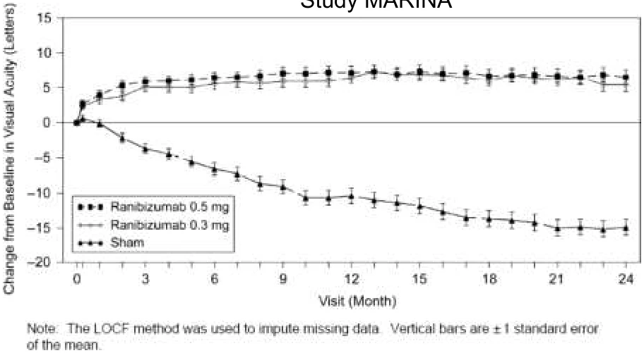
Ranibizumab 0.5 (N=240): +6.6
Ranibizumab 0.3 (N=238): +5.4
Sham (N=238): -14.9
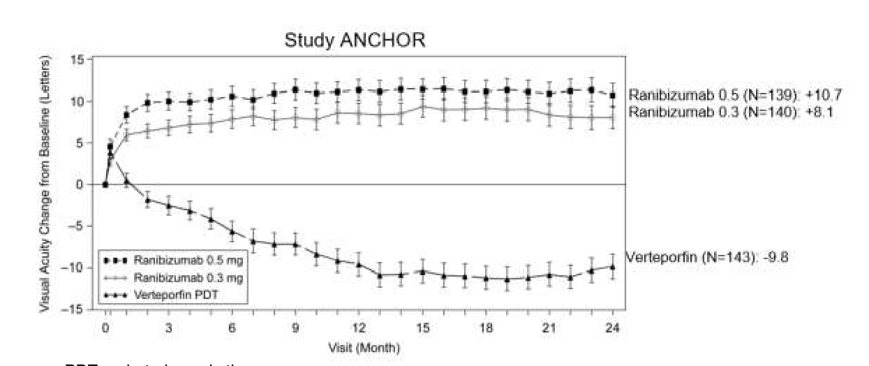
PDT = photodynamic therapy
Note: The LOCF method was used to impute missing data. Vertical bars are ± 1 standard error of the mean
| Study MARINA |
|---|
Randomized subjects = all subjects randomized. Note for Study ANCHOR, ranibizumab 0.5 mg, 140 subjects were randomized, however one subject did not have a BCVA baseline value, therefore, data from 139 patients are included in the results for this group.
Patient withdrawal rates (control, 0.3 mg ranibizumab, 0.5 mg ranibizumab):
- MARINA 1 year (discontinued study on or prior to Month 12): 8.8%, 2.5%, 2.5%
- MARINA 2 year (discontinued from study): 20.2%, 11.8%, 10.4%
- ANCHOR 1 year (discontinued study on or prior to Month 12): 7.0%, 7.1%, 3.6%
- ANCHOR 2 year (discontinued from study): 23.1%, 16.4%, 17.1%
The sham ranibizumab injection control procedure involved anesthetising the eye in a manner identical to a ranibizumab intravitreal injection. The tip of a needleless syringe was then pressed against the conjunctiva and the plunger of the needleless syringe depressed.
Figure 2 Mean Change in Visual Acuity from Baseline to Month 12 in Study FVF3192g (PIER), Randomized Subjects
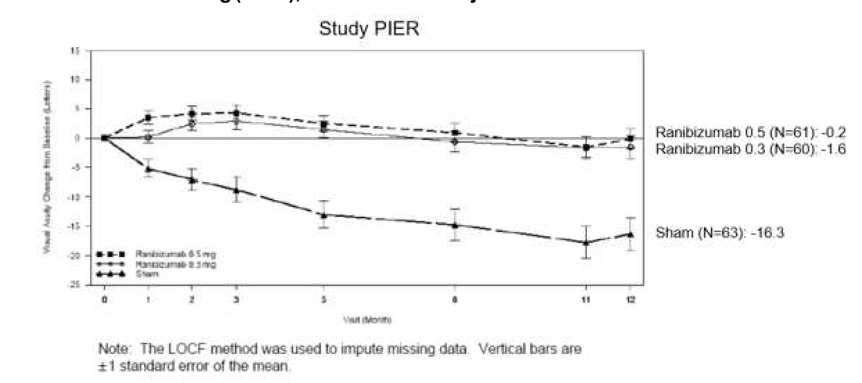
Randomized subjects = all subjects randomized
Patient withdrawal rates (control, 0.3 mg ranibizumab, 0.5 mg ranibizumab):
- PIER 1 year (discontinued study on or prior to Month 12): 12.7%, 1.7%, 3.3%
The sham ranibizumab injection control procedure involved anesthetising the eye in a manner identical to a ranibizumab intravitreal injection. The tip of a needleless syringe was then pressed against the conjunctiva and the plunger of the needleless syringe depressed.
Thirty-four percent (34%) to 40% of ranibizumab-treated patients in studies MARINA and ANCHOR, (0.5 mg) experienced a clinically significant, sustained improvement in vision, defined as gaining 15 or more letters at 24 months (p<0.01), regardless of lesion type. Twenty- five percent (25%) to 36% of ranibizumab-treated patients (0.3 mg) experienced a clinically significant, sustained improvement in vision (Table 20). In both studies, mean changes in BCVA from baseline at Month 24 demonstrated an improvement of vision by 6.6-11.3 letters (0.5 mg) and 5.4-8.5 letters (0.3 mg) respectively. The gain in BCVA was essentially achieved after the first 3 injections with ranibizumab (at Month 3) and maintained until Month 24 in both studies.
In PIER, almost all ranibizumab-treated patients (90%) maintained their visual acuity at Month
In study FVF3192g (PIER), the proportion of patients who lost fewer than 15 letters of BCVA at Month 12 was 90% on 0.5 mg and 83% on 0.3mg.
Table 20 Overview of the Main Secondary Endpoints by Study, Randomized Subjects, with Last Observation Carried Forward (LOCF) Method to Impute Missing Data
| Study MARINA (Outcomes at 12 and 24 months) | ||||||
|---|---|---|---|---|---|---|
| Outcome Measure | Month | Sham (N= 238) | Ranibizumab 0.3mg (N=238) | Estimated Difference between Sham and Ranibizumab 0.3mg | Ranibizumab 0.5mg (N=240) | Estimated Difference between Sham and Ranibizumab 0.5mg |
| Gain of ≥ 15 letters in visual acuity (%) (95% CI) | 12 | 4.6 (2.0, 7.3) | 24.8 (19.3, 30.3) | 20.2 (14.1, 26.3) | 33.8 (27.8, 39.7) | 29.1 (22.6, 35.7) |
| 24 | 3.8 (1.4, 6.2) | 26.1 (20.5, 31.6) | 22.3 (16.2, 28.3) | 33.3 (27.4, 39.3) | 29.6 (23.1, 36.0) | |
Mean change (SD) in BCVA# from Baseline (Letters) (95% CI) | 12 | -10.5 (16.6) (-12.6, -8.3) | 6.5 (12.7) (4.9, 8.1) | 17.0 (14.3, 19.6) | 7.2 (14.4) (5.4, 9.1) | 17.7 (14.9, 20.5) |
| 24 | -14.9 (18.7) (-17.3, -12.5) | 5.4 (15.2) (3.5, 7.4) | 20.3 (17.3, 23.4) | 6.6 (16.5) (4.5, 8.7) | 21.4 (18.3, 24.6) | |
| Study ANCHOR (Outcomes at 12 and 24 months) | ||||||
| Outcome Measure | Month | PDT (N=143) | Ranibizumab 0.3mg (N=140) | Estimated Difference between PDT and Ranibizumab 0.3mg | Ranibizumab 0.5mg (N=139) | Estimated Difference between PDT and Ranibizumab 0.5mg |
| Gain of ≥ 15 letters in visual acuity (%) (95% CI) | 12 | 5.6 (1.8, 9.4) | 35.7 (27.8, 43.7) | 30.1 (21.3, 38.9) | 40.3 (32.1, 48.4) | 34.7 (25.7, 43.7) |
| 24 | 6.3 (2.3, 10.3) | 34.3 (26.4, 42.1) | 28.0 (19.2, 36.8) | 41.0 (32.8, 49.2) | 34.7 (25.6, 43.8) | |
Mean change (SD) in BCVA# from Baseline (Letters) (95% CI) | 12 | -9.5 (16.4) (-12.3, -6.8) | 8.5 (14.6) (6.1, 11.0) | 18.1 (14.4, 21.7) | 11.3 (14.6) (8.9, 13.8) | 20.9 (17.2, 24.5) |
| 24 | -9.8 (17.6) (-12.7, -6.9) | 8.1 (16.2) (5.4, 10.7) | 17.9 (13.9, 21.8) | 10.7 (16.5) (7.9, 13.5) | 20.5 (16.5, 24.5) | |
| Study PIER (Outcomes at 12 months) | ||||||
| Outcome Measure | Month | Sham (N=63) | Ranibizumab 0.3mg (N=60) | Estimated Difference between Sham and Ranibizumab 0.3mg | Ranibizumab 0.5mg (N=61) | Estimated Difference between Sham and Ranibizumab 0.5mg |
| Loss of ≥ 15 letters in visual acuity (%) (95% CI) | 12 | 49.2 (36.9, 61.6) | 83.3 (73.9, 92.8) | 34.1 (18.6, 49.7) | 90.2 (82.7, 97.6) | 41.0 (26.5, 55.4) |
# Best Corrected Visual Acuity
Randomized subjects = all subjects randomized. Note for Study ANCHOR, ranibizumab 0.5 mg, 140 subjects were randomized, however one subject did not have a BCVA baseline value, therefore, data from 139 patients are included in the results for this group.
Patient withdrawal rates (control, 0.3 mg ranibizumab, 0.5 mg ranibizumab):
- MARINA 1 year (discontinued study on or prior to Month 12): 8.8%, 2.5%, 2.5%
- MARINA 2 year (discontinued from study): 20.2%, 11.8%, 10.4%
- ANCHOR 1 year (discontinued study on or prior to Month 12): 7.0%, 7.1%, 3.6%
- ANCHOR 2 year (discontinued from study): 23.1%, 16.4%, 17.1%
- PIER 1 year (discontinued study on or prior to Month 12): 12.7%, 1.7%, 3.3%
The sham ranibizumab injection control procedure involved anesthetising the eye in a manner identical to a ranibizumab intravitreal injection. The tip of a needleless syringe was then pressed against the conjunctiva and the plunger of the needleless syringe depressed.
Patients in the groups treated with ranibizumab had minimal observable CNV lesion growth, on average. At Month 12, the mean change in the total area of the CNV lesion was 0.1 to 0.3 disc area for ranibizumab versus 2.3 to 2.6 disc area for the control arms. Results from both trials indicated that continued ranibizumab-treatment may be of benefit also in patients who lost 15 letters of best-corrected visual acuity (BCVA) in the first year of treatment.
The size of the lesion did not significantly affect the results. In general, patients with poor visual acuity (<20/200) at the onset of treatment experienced a benefit of treatment. However, neovascular AMD that has evolved into lesions characterised by subretinal fibrosis and advanced geographic atrophy is not likely to respond to ranibizumab.
In MARINA and ANCHOR, at month 12 patients treated with ranibizumab reported, on average, a statistically (p<0.01) and clinically meaningful improvement in their ability to perform activities related to near vision (such as reading; MARINA: 0.5 mg ranibizumab: + 10.4 point increase; 0.3 mg ranibizumab: + 9.4 point increase; ANCHOR: 0.5 mg ranibizumab: + 9.1 point increase; 0.3 mg ranibizumab: + 6.6 point increase), distance vision (such as driving; MARINA: 0.5 mg ranibizumab: + 7.0 point increase; 0.3 mg ranibizumab: + 6.7 point increase; ANCHOR: 0.5 mg ranibizumab: + 9.3 point increase; 0.3 mg ranibizumab: + 6.4 point increase) and vision– specific dependency (such as seeing faces; MARINA: 0.5 mg ranibizumab: + 6.8 point increase; 0.3 mg ranibizumab: + 3.6 point increase; ANCHOR: 0.5 mg ranibizumab: + 8.9 point increase; mg ranibizumab: + 7.6 point increase), as measured by the National Eye Institute (NEI) Visual Functioning Questionnaire-25 (VFQ-25). Sham-treated patients reported a decrease in their ability to perform these activities (MARINA: near vision:
- 2.6 point decrease; distance vision: -5.9 point decrease; vision-specific dependency: -4.7 point decrease) and verteporfin PDT-treated patients reported a small increase or decrease (ANCHOR: near vision: + 3.7 point increase; distance vision: + 1.7 point increase; vision-specific dependency: - 1.4 point decrease).
In MARINA, this increase from baseline in each of the three VFQ-25 subscales at Month 12 was maintained at Month 24 for ranibizumab-treated patients, while in the sham-injection group the mean change from baseline decreased further from Month 12 to Month 24 in each of these subscales. Therefore, the treatment benefit of ranibizumab over the sham control at Month 24 was greater than that at Month 12.
In the verteporfin PDT group, the mean improvement from baseline in the near activities and distance activities subscale scores at Month 12 were lost at Month 24, while the mean decrease from baseline in the vision-specific dependency subscale score at Month 12 was maintained at Month 24. These changes between Months 12 and 24 within each treatment group resulted in either maintained or greater treatment benefit of ranibizumab over verteporfin PDT compared with Month 12, while the treatment benefit of ranibizumab in the vision-specific dependency subscale was smaller at Month 24 compared with Month 12 (p-values ranging from 0.0023 to 0.0006).
Treatment of visual impairment due to DME
Clinical efficacy of ranibizumab in patients with visual impairment secondary to diabetic macular edema (DME) was assessed in the randomized, double-masked, controlled study D2301 (RESTORE). Clinical safety of ranibizumab has been assessed in the randomized, double-masked, controlled studies D2301 (RESTORE) and D2201 (RESOLVE).
Table 21 Summary of Patient Demographics for Clinical Trials in Visual Impairment due to DME
| Study # | Trial Design | Dosage, Route of Administration and Duration | Study Subjects (n=number) | Mean Age (Range) | Gender |
|---|---|---|---|---|---|
| D2301† (RESTORE) | Randomized, double-masked, multicenter, laser-controlled study. | Ranibizumab 0.5mg intravitreal injection (+ sham laser), prn‡; ranibizumab 0.5mg intravitreal injection + laser, prn; or Sham injection + laser, prn 12 month study. | Ranibizumab 0.5mg : n= 116 ranibizumab 0.5mg + laser: n=118 Sham injection + laser: n=111 | 63.5 (37-87 years) | Male: 58.3% Female: 41.7% |
| D2201 (RESOLVE) | Randomized, double-masked, multicenter, sham-controlled study. | Ranibizumab 0.3mg intravitreal injection (dose doubling permitted), prn; ranibizumab 0.5mg intravitreal injection (dose doubling permitted), prn; or Sham injection, prn 12 month study. | Ranibizumab 0.3mg: n=51 ranibizumab 0.5mg: n=51 Sham injection: n=49 | 63.6 (32-85 years) | Male: 53.6% Female: 46.4% |
† a. There is only limited experience in the treatment of subjects with DME due to Type I diabetes.
b. There is limited experience in patients older than 75 years of age with DME.
c. Patients with the HbA1c > 10% were not included in the clinical trial.
‡ prn: pro re nata (as needed)
In study D2301 (RESTORE), a total of 345 patients with visual impairment due to macular edema were enrolled to receive either initial intravitreal injection of ranibizumab 0.5 mg as monotherapy and sham laser photocoagulation, combined ranibizumab 0.5 mg and laser photocoagulation, or sham injection and laser photocoagulation monotherapy. Treatment with ranibizumab was started with monthly intravitreal injections. Treatment was suspended when visual acuity stability was observed over the last three consecutive visits. The treatment was reinitiated when there was a reduction in BCVA due to DME progression. Laser photocoagulation was administered at baseline, and then as needed based on ETDRS criteria.
The primary efficacy endpoint was mean average change in BCVA from Month 1 to Month 12 compared to baseline. The 12-month results demonstrate statistically significant superiority of ranibizumab as monotherapy or adjunctive to laser photocoagulation compared to laser control, on both primary and secondary endpoints of visual acuity, and on the effect on central retinal thickness (CRT). Clinical significance of the effect on central retinal thickness in this population is unknown. A rapid improvement in BCVA was observed as early as the first follow-up visit and was maintained through the 12-month period (Figure 3). The mean average change in BCVA over 12 months showed an improvement of 5.4 and 4.9 letters, respectively, for ranibizumab and ranibizumab adjunctive to laser compared to laser monotherapy, and in the laser arm, a maintenance of the baseline BCVA of about + 1 letter over the 12 months study period.
The results of the primary endpoint are detailed below in Table 22 and illustrated in Figure 3.
Table 22 Outcomes at Month 12 in Study D2301 (RESTORE), Full Analysis Set, with Last Observation Carried Forward (LOCF)
| Primary Endpoint | |||||
|---|---|---|---|---|---|
| Outcome Measure | Ranibizumab 0.5 mg (n=116) | Ranibizumab 0.5 mg + Laser (n=118) | Laser (n=111) | Estimated Difference between Laser and Ranibizumab 0.5 mg | Estimated Difference between Laser and Ranibizumab 0.5 mg + Laser |
Mean average change in BCVA from month 1 to month 12 compared to baseline (letters) (SD) (95% CI)b | 6.1 (6.43) (4.9, 7.3) | 5.9 (7.92) (4.4, 7.3) | 0.8 (8.56) (-0.8, 2.4) | 5.4 (3.5, 7.4) | 4.9 (2.8, 7.0) |
b p<0.0001
Figure 3 Mean BCVA Change from Baseline over Time in Study D2301 (RESTORE)
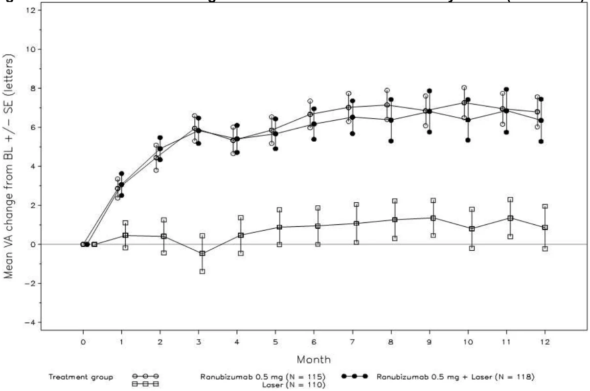
The results for the key secondary efficacy endpoints based on BCVA were consistent with those obtained for the primary endpoint and showed statistically significant treatment differences in ranibizumab treated patients compared to laser. At Month 12, the mean BCVA improvement compared to baseline for patient treated with ranibizumab and ranibizumab adjunctive to laser was 6.8 and 6.4 letters, compared to 0.9 letters in the laser treated patients. The proportion of ranibizumab treated patients who gained at least 15 letters from baseline at 12 months was 22.6% (ranibizumab) and 22.9% (ranibizumab + laser) compared to 8.2% in laser control.
The improvement in the visual acuity outcomes was also accompanied by a rapid and sustained decrease in the macular edema as assessed by the central retinal thickness. Clinical significance of the effect on central retinal thickness in this population is unknown.
In study D2201 (RESOLVE), a total of 151 patients with macular center involvement causing visual impairment were enrolled to receive either: 1) initial intravitreal injection of ranibizumab
0.3 mg (6 mg/mL formulation) and then monthly injection until treatment success or futility was observed (51 patients); 2) initial intravitreal injection of ranibizumab 0.5 mg (10 mg/mL formulation) and then monthly injection until treatment success or futility was observed (51 patients); 3) initial sham injection and then monthly sham injections when needed following the same treatment criteria (49 patients). The initial ranibizumab dose could be doubled at any time during the study after the first injection if the investigator evaluated that response to treatment was not sufficiently achieved. The dose doubling was achieved by doubling of the injection volume from 0.05 to 0.1 mL. Laser photocoagulation rescue treatment was allowed at any time in the study in the active and control arms after month 3 of the study, based on the investigators’ opinion.
Treatment of visual impairment due to macular edema secondary to RVO
Clinical safety and efficacy of ranibizumab in patients with visual impairment due to macular edema secondary to RVO have been assessed in the randomized, double-masked, controlled studies BRAVO and CRUISE.
In the BRAVO study, a total of 397 patients with visual impairment due to macular edema secondary to Branch RVO were enrolled to receive either 0.3 mg or 0.5 mg intravitreal ranibizumab or sham1 injections. Laser photocoagulation standard of care was allowed in all three arms and was administered, based on protocol defined criteria, as rescue treatment beginning at Month 3. A proportion of 57.6% of patients in the sham-control arm and 20.1% and 21.4% in the ranibizumab-treated arms (0.3 mg and 0.5 mg, respectively) received at least one laser treatment during the first 6 months of the study.
After 6 months, the patients in the sham-control arm were crossed-over to 0.5 mg ranibizumab. The proportion of patients receiving at least one laser treatment during the entire 12 months study period was as follows: 61.4% in the sham/0.5 mg ranibizumab arm, 41.0% in the 0.3 mg ranibizumab arm, and 34.4% in the 0.5 mg ranibizumab arm.
The primary endpoint was the mean change from baseline in best-corrected visual acuity (BCVA) at Month 6: treatment with 0.5 mg ranibizumab, on average, showed a statistically significant improvement of 18.3 letters compared to a gain of 7.3 letters in the control group (p<0.0001). Improvement in BCVA was rapid (as early as day 7, p<0.0001), and sustained over the next months through Month 12 (secondary endpoint), with a mean improvement from baseline in BCVA score at Month 12 of 18.3 letters for the 0.5 mg ranibizumab group, compared to 12.1 letters for the sham/0.5 mg group.
Statistically significant improvements were also observed in key secondary endpoints at Month 6, and sustained to Month 12. At month 6, 61.1% of the patients in the 0.5 mg ranibizumab group gained ≥15 letters in visual acuity score from baseline compared with 28.8% of patients in the sham group, which represents an estimated difference of 31.3% between the two groups. At month 12, 60.3% of the patients in the 0.5 mg ranibizumab group gained ≥15 letters in visual acuity score from baseline compared with 43.9% of patients in the sham/0.5 mg ranibizumab arm. For the crossover sham/0.5-mg group, improvements in visual acuity were also observed during the 6-month observation period.
Table 23 BCVA Primary Efficacy Outcome at Month 6 (BRAVO), Randomized Patients, with Last Observation Carried Forward Method (LOCF) to Impute Missing Data
Sham (n=132) | Ranibizumab 0.5 mg (n=131) | Estimated Difference between Sham and Ranibizumab 0.5 mg | |
|---|---|---|---|
Primary endpoint Mean change in BCVA from baseline ETDRSa (letters) (SD) (95% CI)b | +7.3 (13.0) (5.1, 9.5) | +18.3 (13.2) (16.0,20.6) | 10.6 (7.6,13.6) |
a: Early Treatment Diabetic Retinopathy Study
b: p<0.0001
Figure 4 Mean Change from Baseline BCVA over Time to Month 6 (BRAVO)
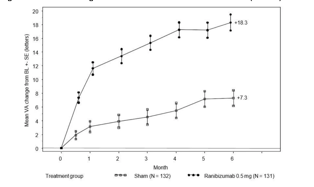
In the CRUISE study, a total of 392 patients with visual impairment due to macular edema secondary to Central RVO were enrolled to receive either 0.3 mg or 0.5 mg intravitreal ranibizumab or sham injections. After 6 months, the patients in the sham-control arm were crossed-over to 0.5 mg ranibizumab.
The primary endpoint was the mean change from baseline in BCVA at Month 6: treatment with
0.5 mg ranibizumab, on average, showed a statistically significant improvement of 14.9 letters compared to 0.8 letter gain in the control group (p<0.0001). Improvement in BCVA was rapid (as early as day 7, p<0.0001), and sustained over the next months through month 12, with a mean improvement from baseline in BCVA score at Month 12 of 13.9 letters for the 0.5 mg ranibizumab group, compared to 7.3 letters for the sham/0.5 mg group.
Statistically significant improvements were also observed in key secondary endpoints at Month 6 and sustained through Month 12. At month 6, 47.7% of the patients in the 0.5 mg ranibizumab group gained ≥15 letters in visual acuity score from baseline compared with 16.9% of patients in the sham group, which represents an estimated difference of 30.3% between the two groups. At month 12, 50.8% of the patients in the 0.5 mg ranibizumab group gained ≥15 letters in visual acuity score from baseline compared with 33.1% of patients in the sham/0.5 mg ranibizumab arm. For the crossover sham/0.5 mg group, improvements in visual acuity were also observed during the 6-month observation period.
Table 24 BCVA Primary Efficacy Outcome at Month 6 (CRUISE), Randomized Patients, with Last Observation Carried Forward Method (LOCF) to Impute Missing Data
| Primary endpoint | Sham (n=130) | Ranibizumab 0.5 mg (n=130) | Estimated Difference between Sham and Ranibizumab 0.5 mg |
|---|---|---|---|
| Mean change in BCVA from baseline ETDRSa (letters) (SD) (95% CI)b | +0.8 (16.2) (-2.0, 3.6) | +14.9 (13.2) (12.6, 17.2) | 13.8 (10.3, 17.4) |
a: Early Treatment Diabetic Retinopathy Study
b: p<0.0001
Figure 5 Mean Change from Baseline BCVA over Time to Month 6 (CRUISE)
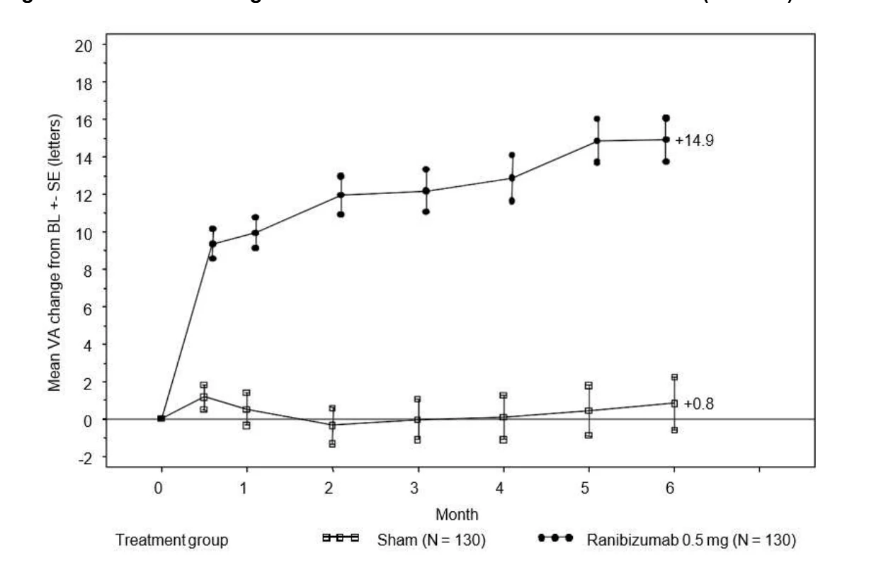
In both studies, the improvement in the visual acuity outcomes were accompanied by a rapid and sustained decrease in the macular edema as assessed by the central retinal thickness, both at Month 6 and Month 12.
In both studies, the improvement in visual acuity outcomes seen with ranibizumab treatment at both 6 and 12 months was accompanied by patient-reported benefits as measured by the National Eye Institute Visual Function Questionnaire (NEI VFQ-25), and particularly in the sub-scales related to near and distance activities, as pre-specified secondary efficacy endpoints.
In BRAVO and CRUISE, patients treated with ranibizumab achieved statistically significant improvements at Month 6 in near activities (BRAVO: ranibizumab 0.5 mg: +13.7; sham: +7.3; p=0.0002; CRUISE: ranibizumab 0.5 mg: +9.3; sham: +5.1; p=0.0099) and distance activities (BRAVO: ranibizumab 0.5 mg: +11.3; sham: +6.3; p=0.0014; CRUISE: ranibizumab 0.5 mg:
+6.7; sham: +2.8; p=0.0199). In both studies, this increase from baseline in each of the two VFQ-25 subscales at Month 6 was sustained at Month 12.
Post-marketing studies
The long term (24 month) clinical safety and efficacy of ranibizumab in patients with visual impairment due to macular edema secondary to RVO were assessed in the BRIGHTER (BRVO) and CRYSTAL (CRVO) studies. In both studies, subjects received a 0.5 mg ranibizumab PRN dosing regimen driven by individualized stabilization criteria.
BRIGHTER was a 3-arm, randomized, active-controlled study that compared 0.5 mg ranibizumab given as monotherapy or in combination with adjunctive laser photocoagulation, to laser photocoagulation alone. After 6 months, subjects in the laser monotherapy arm could receive 0.5 mg ranibizumab. CRYSTAL was a single-arm study with 0.5 mg ranibizumab monotherapy.
The key outcome measures from BRIGHTER and CRYSTAL are shown in the Table below.
Table 25 Outcomes at Month 6 and 24 (BRIGHTER and CRYSTAL)
| BRIGHTER | CRYSTAL | ||||||||
|---|---|---|---|---|---|---|---|---|---|
Ranibizumab 0.5 mg N=180 | Ranibizumab 0.5 mg + Laser N=178 | Laser* N=90 | Ranibizumab 0.5 mg (N=356) | ||||||
| Mean change in BCVA at Month 6 (letters) (SD) | +14.8 (10.7) | +14.8 (11.13) | +6.0 (14.27) | +12.0 (13.95) | |||||
| Mean change in BCVA at Month 24 (letters) (SD) | +15.5 (13.91) | +17.3 (12.61) | +11.6 (16.09) | +12.1 (18.60) | |||||
* Starting at Month 6 treatment with ranibizumab 0.5 mg was allowed (24 patients were treated with laser only).
The effect of visual improvement was observed in all patients treated with 0.5 mg ranibizumab monotherapy regardless of their disease duration in both BRIGHTER and CRYSTAL.
Treatment of visual impairment due CNV secondary to PM
The clinical safety and efficacy of ranibizumab in patients with visual impairment due to CNV in PM have been assessed based on the 12-month data of the randomized, double-masked, controlled pivotal study F2301 (RADIANCE) which was designed to evaluate two different dosing regimens of 0.5 mg ranibizumab given as intravitreal injection in comparison to verteporfin photodynamic therapy PDT (vPDT). PM is characterized by abnormal and progressive elongation of the eyeball, usually to an axial length greater than 26 mm and refractive error of more than -6.0 Diopters, with concomitant degenerative changes in the posterior segment of the eye such as posterior staphyloma, chorioretinal atrophy, Bruch’s membrane (lacquer) cracks, subretinal hemorrhage, retinal detachment, and CNV.
Table 26 Summary of Patient Demographics for Clinical Trials in Visual Impairment due to CNV Secondary to PM
| Study # | Trial Design | Dosage, Route of Administration and Duration | Study Subjects (n=number) | Mean Age (Range) | Gender |
|---|---|---|---|---|---|
| F2301 (RADIANCE) | Randomized, double- masked, multicenter, active- controlled study. | Group I: Ranibizumab 0.5 mg intravitreal injection (VA stabilization) Group II: Ranibizumab 0.5 mg intravitreal injection (disease activity) Group III: vPDT† 12 month study | Group I: n= 106 Group II: n=116 Group III: n=55 | 55.5 (18- 87 years) | Male: 24.5% Female: 75.5% |
†Patients in the vPDT were allowed to receive ranibizumab treatment as of Month 3.
The 277 patients were randomized to one of the following arms:
- Group I (ranibizumab 0.5 mg, dosing regimen driven by “stability” criteria defined as no change in best corrected visual acuity (BCVA) compared to two preceding monthly evaluations)
- Group II (ranibizumab 0.5 mg, dosing regimen driven by “disease activity” criteria defined as vision impairment attributable to intra-or-subretinal fluid or active leakage due to the CNV lesion as assessed by OCT and/or FA)
- Group III (vPDT - patients were allowed to receive ranibizumab treatment as of Month 3)
Over the 12 months of the study patients received on average 4.6 injections (median 4.0, range 1- 12) in Group I and 3.5 (median 2.0, range 1-12) injections in Group II. In Group II (in which patients received the recommended treatment regimen based on disease activity, see 4 Dosage and Administration), 50.9% of patients required 1 or 2 injections, 34.5% required 3 to 5 injections and 14.7% required 6 to 12 injections over the 12-month study period. In Group II, 62.9% of patients did not require injections in the second 6 months of the study.
The primary efficacy endpoint was the mean average change in BCVA from baseline to Month
1 through Month 3. Both ranibizumab treatment arms demonstrated statistically significant superior efficacy compared with vPDT: the mean average BCVA score of the study eye from Month 1 to Month 3 exceeded baseline by 10.5 letters (Group I; min-max: -19.3 to +31.0) and
10.6 letters (Group II; min-max: -8.3 to +32.0) in the ranibizumab groups and by 2.2 letters in the vPDT group (min-max: -24.7 to +24.3). The treatment benefit was maintained for the 12- month duration of the study. The mean average change in BCVA from baseline to Month 1 through Month 12 was 12.8 letters and 12.5 letters in Group I and Group II, respectively.
Table 27 Primary Efficacy Outcome at Month 3 in Study RADIANCE, Randomized Patients, with Modified Last Observation Carried Forward (LOCF) Method
| Group I Ranibizumab 0.5 mg “Visual Acuity Stability” (n=105) | Group II Ranibizumab 0.5 mg “Disease Activity” (n=116) | Group III vPDT (n=55)† | |
|---|---|---|---|
Primary endpoint Mean average BCVA change from Month 1 to Month 3 compared to baselinea (letters) (SD) | +10.5 (8.16) | +10.6 (7.26) | +2.2 (9.47) |
† Comparative control up to Month 3. Patients randomized to vPDT were allowed to receive ranibizumab treatment as of Month 3, as per Investigator discretion (in Group III, 38 patients received ranibizumab from Month 3 onwards)
a: p<0.00001 comparison with vPDT control
Results for secondary endpoint were consistent with those of the primary endpoint. The time course of mean change BCVA from baseline through Month 12 showed a rapid improvement, most of it reached by Month 2. The improvement in BCVA continued through Month 12 achieving a mean change in BCVA of about 14 letters gain in both ranibizumab arms (Figure 6). At Month 3, the proportion of patients who gained ≥10 letters (or reached a BCVA of ≥84 letters) from baseline was 61.9% and 65.5% in Group I and II, compared to 27.3% in the vPDT group; and the proportion of patients who gained ≥15 letters (or ≥84 letters total) was 38.1% and 43.1% in Group I and II, compared to 14.5% in the vPDT group. At Month 12, the proportion of patients who gained ≥10 letters (or ≥84 letters total) from baseline was 69.5% and 69.0% in Group I and II; and the proportion of patients who gained ≥15 letters (or ≥84 letters total) from baseline was 53.3% and 51.7% in Group I and II, respectively.
Figure 6 Mean Change from Baseline BCVA over Time up to Month 12 (RADIANCE)
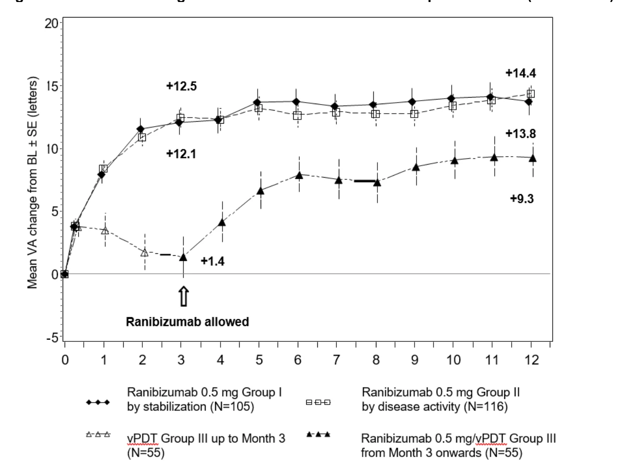
BL = baseline; SE = standard error of the mean.
Patients randomized to vPDT were allowed to receive ranibizumab from Month 3 onwards.
There are limited data regarding treatment with ranibizumab in PM patients with extrafoveal lesions.
The improvement of vision was accompanied by a reduction in central retinal thickness; however, the clinical significance of this is yet to be determined.
Treatment of visual impairment due to CNV
The clinical safety and efficacy of ranibizumab in patients with visual impairment due to CNV secondary to etiologies other than wet AMD and PM have been assessed based on a Phase 3 multi-center study G2301 (MINERVA), which was randomized, double-masked, sham controlled for 2 months followed by an open-label extension of 10 months.
Table 28 Summary of Patient Demographics for Clinical Trials in Visual Impairment due to CNV
| Study # | Trial Design | Dosage, Route of Administration and Duration | Study Subjects (n=number) | Mean Age (Range) | Gender |
|---|---|---|---|---|---|
| G2301 (MINERVA) | Randomized, double- masked, sham- controlled, multicenter, study. | Arm 1: Ranibizumab 0.5 mg intravitreal injection Arm 2: Sham intravitreal injection (up to Month 2) As of Month 2, treatment was open-label 12 month study | Arm 1: n= 119 Arm 2: n=59 | 53.7 years (19 to 86 years) | Male: 49.4% Female: 50.6% |
In this study, 178 adult patients were randomized in a 2:1 ratio to one of the following arms, stratified by the presence of angioid streaks at baseline (yes/no):
- ranibizumab 0.5 mg at baseline followed by an individualized dosing regimen based on evidence of disease activity.
- sham injection at baseline followed by an individualized treatment regimen based on evidence of disease activity.
Starting at Month 2, all patients received open-label individualised treatment with ranibizumab based on disease activity. The primary endpoint was assessed by the best corrected visual acuity (BCVA) change from baseline to Month 2.
The primary outcome measure, demonstrated statistically superior efficacy in ranibizumab- treated patients compared to patients randomized to sham (Table 29).
Table 29 Change in Visual Acuity at Month 2 in study MINERVA
| Ranibizumab 0.5 mg (N=119) | Sham (N=59) | |
|---|---|---|
| N | 118 | 57 |
| Mean BCVA change from baseline to Month 2 (letters) (Least Squares Mean) a | +9.5 (0.95) | -0.4 (1.16) |
| 95% CI for LS mean | (7.6,11.4) | (-2.8, 1.9) |
| Difference in LS means (Ranibizumab minus Sham) (SE) | 9.94 (1.502) | |
| 95% CI for difference | (6.97, 12.91) | |
| One-sided p-value for treatment difference (1) | < 0.001 |
n is the number of patients with data available in the analysis
(1) Analyzed using MMRM, which contains scheduled visit, the type of underlying pathophysiologic mechanism (angioid streaks vs. others) and treatment group as fixed effect factors, centered baseline BCVA as a continuous covariate and treatment group by visit and visit by centered baseline BCVA interactions.
The mean BCVA change from baseline over time to Month 12 is shown in Figure 7.
Figure 7 Mean BCVA Change from Baseline over Time up to Month 12 (MINERVA)
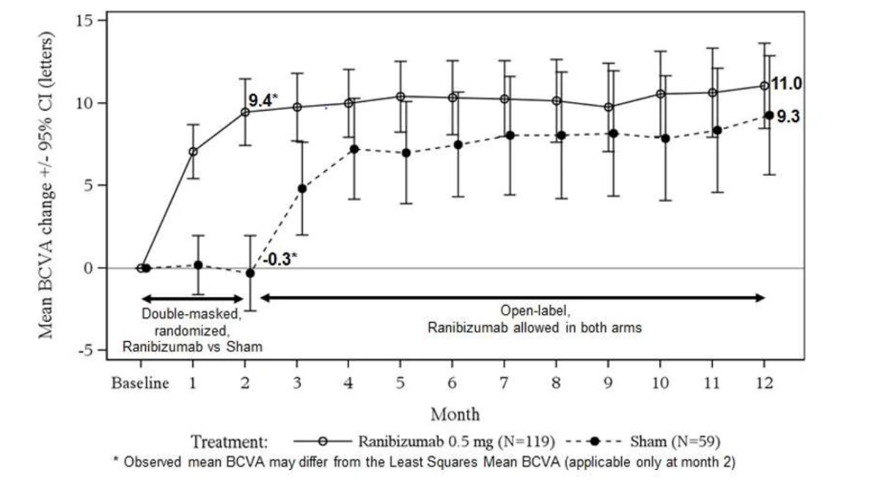
A difference was observed in the change from baseline to Month 2 in BCVA (13.0 letters vs 4.2 letters) for the 2 age groups (patients <= 60 years vs patients > 60 years, respectively).
A subgroup analysis for the primary variable was conducted using the following subgroups of type of underlying ocular pathophysiological mechanism (baseline etiology) in the study eye: (i) angioid streaks; (ii) post-inflammatory retinochoroidopathy; (iii) idiopathic chorioretinopathy;
(iv) central serous chorioretinopathy (CSC) and (v) miscellaneous (any etiology that does not belong to the above subgroups). The subgroups were based on data entered in the eCRF, and the definition of each baseline etiology subgroup was determined prior to database lock. Within the “Miscellaneous” baseline etiology subgroup, there were 15 different etiologies causing CNV. In total, the study enrolled patients with 19 different etiologies. Results from the subgroup analysis for the primary variable by baseline etiology are shown in Table 30, and suggest that the treatment effect varies by baseline etiology.
Table 30 Overall Results on Change in Visual Acuity and per Baseline Etiology at Month 2 (MINERVA)
| Overall and per Baseline Etiology | Change from Baseline to Month 2 in BCVA (Letters) | Treatment Difference over Sham (Letters) | |||
|---|---|---|---|---|---|
| Ranibizumab | Sham | ||||
| N | LS mean | n | LS mean | ||
| Overall | 118* | 9.5 | 57* | -0.4 | 9.9 |
| Angioid streaks | 18 | 11.0 | 9 | -3.5 | 14.6 |
| Post-inflammatory retinochoroidopathy | 18 | 7.0 | 9 | 0.4 | 6.5 |
| Central serous chorioretinopathy | 17 | 6.6 | 6 | 1.6 | 5.0 |
| Idiopathic chorioretinopathy | 37 | 12.5 | 25 | 1.1 | 11.4 |
| Miscellaneous etiologiesa | 28 | 7.5 | 8 | -3.0 | 10.6 |
a comprises CNV etiologies which do not fall under the other subgroups
* number of patients with data available in the analysis
The mean number of ranibizumab injections given in the study eye over 12 months was 5.8 (range 1-12) in the ranibizumab arm versus 5.4 (range 1-10) over 10 months in those patients in the sham with ranibizumab group. In the sham arm, 7 out of 59 patients did not receive any treatment with ranibizumab in the study eye during the 12-month period.
Immunogenicity
In the wet AMD studies, the pre-treatment incidence of immunoreactivity to ranibizumab was 0% - 3% across treatment groups. After monthly dosing with ranibizumab for 12 to 24 months, low titres of antibodies to ranibizumab were detected in approximately 1% - 6% of patients. The immunogenicity data reflect the percentage of patients whose test results were considered positive for antibodies to ranibizumab in an electrochemiluminescence assay and are highly dependent on the sensitivity and specificity of the assay. The clinical significance of immunoreactivity to ranibizumab is unclear at this time, although some patients with the highest levels of immunoreactivity were noted to have iritis or vitritis.
15 Microbiology
No microbiological information is required for this drug product.
16 Non-Clinical Toxicology
General Toxicology
The non-clinical safety of ranibizumab was assessed primarily in cynomolgus monkeys, because of the close homology between the cynomolgus monkey and human VEGF. All repeat-dose toxicology studies were conducted in cynomolgus monkeys, whilst rabbits were employed for single-dose local tolerance studies.
The toxicology program was designed to support ITV administration and included 4-, 13-, and 26-week repeat-dose ITV toxicity studies in cynomolgus monkeys. Because transient ocular inflammation was observed in the toxicity studies, a 16-week study was conducted to investigate the non-clinical safety of different ITV dosing regimens (various dose escalation and dose frequency regimens), as well as the effect of oral and topical corticosteroid treatment on ocular inflammation. The non-clinical safety of ranibizumab/verteporfin/PDT combination treatments was assessed in cynomolgus monkeys with laser-induced CNV. Human tissue cross-reactivity, hemolytic potential, and blood compatibility were also investigated.
Bilateral intravitreal administration of ranibizumab to cynomolgus monkeys at doses between
0.25 mg/eye and 2.0 mg/eye once every 2 weeks for up to 26 weeks resulted in dose-dependent ocular effects.
Intraocularly, there were dose-dependent increases in anterior chamber flare and cells with a peak 2 days after injection. The severity of the inflammatory response generally diminished with subsequent injections or during recovery. In the posterior segment, there were vitreal cell infiltration and floaters, which also tended to be dose-dependent and generally persisted to the end of the treatment period. In the 26-week study, the severity of the vitreous inflammation increased with the number of injections. However, evidence of reversibility was observed after recovery. The nature and timing of the posterior segment inflammation is suggestive of an immune-mediated antibody response, which may be clinically irrelevant. Cataract formation was observed in some animals after a relatively long period of intense inflammation, suggesting that the lens changes were secondary to severe inflammation. A transient increase in post-dose intraocular pressure was observed following intravitreal injections, irrespective of dose.
Microscopic ocular changes were related to inflammation and did not indicate degenerative processes. Granulomatous inflammatory changes were noted in the optic disc of some eyes. These posterior segment changes diminished, and in some instances resolved, during the recovery period. Following intravitreal administration, no signs of systemic toxicity were detected. Serum and vitreous antibodies to ranibizumab were found in a subset of treated animals.
Carcinogenicity and mutagenicity: No carcinogenicity and mutagenicity data are available.
Reproductive and Developmental Toxicology: The potential of ranibizumab to affect embryo-fetal and/or placental development has been investigated in pregnant cynomolgus monkeys given bilateral ITV injections of ranibizumab every 14 days from Day 20 until Day 62 of gestation. The selected ITV doses were 0.125 and 1.0 mg/eye, administered in a 50 mcL volume, and were chosen as to give predicted maximum maternal serum levels (Cmax) about 10- and 100-fold, respectively, higher than the median Cmax in humans given monthly unilateral ITV injection of 0.5 mg ranibizumab/eye. The dose of 1.0 mg/eye was considered to be the highest dose that could be administered to pregnant animals, based on the ocular inflammation observed in the previously performed 4-26 weeks toxicity studies in non-pregnant monkeys. In those studies, no systemic toxicity was observed up to the highest investigated dose of 2.0 mg/eye. The dose-escalation regimen used in non-pregnant monkeys was considered to be inappropriate for an embryo-fetal development (EFD) study.
In the EFD study, fetal (cord blood) serum was sampled at caesarian section (on gestation day 100 ± 1), 32 ± 1 days (i.e. approximately 7-9 half-lives, based on an "apparent" serum ranibizumab half-life of 3.5-4.5 days in monkeys) after the last administration of ranibizumab (on gestation day 62). With one exception, fetal serum ranibizumab concentrations were below the limit of quantitation, irrespective of the dose. The exception was in a high dose (1.0 mg/eye) animal that was positive for anti-ranibizumab antibodies and that had an unusually high maternal serum ranibizumab concentration (1990 ng/mL, presumably due to the presence of anti-ranibizumab antibodies) after the last dose on gestation day 62. In this animal, fetal (cord blood) serum ranibizumab concentrations at caesarian section were 230 pg/mL, which is approximately twice the "minimum quantifiable concentration" (MQC) of the analyte (equal to the lower limit of quantitation multiplied by the minimum dilution factor required for accurate quantitation of the analyte in the sample matrix) of 100 pg/mL.
In this animal, anti-ranibizumab antibodies may have acted as a (Fc region containing) carrier protein for ranibizumab, thereby decreasing its maternal serum clearance, and enabling its placental transfer. However, the EFD study in monkeys was not designed to address this question and the collected data do not allow for an unambiguous answer.
In pregnant monkeys, ITV ranibizumab treatment did not elicit developmental toxicity or teratogenicity, and had no effect on weight or structure of the placenta. However, based on its pharmacological effect, ranibizumab should be regarded as potentially teratogenic and embryo-foetotoxic. The absence of ranibizumab-mediated effects on the embryo-fetal development is plausibly related to the inability of the Fab fragment to cross the placenta. The embryo-fetal development investigations were performed in healthy pregnant animals and disease (such as e.g. diabetes) may modify the permeability of the placenta towards a Fab fragment (see recommendations in 7.1 Special Populations).
Repeated-Dose Toxicity Studies
| Species /Strain | No./Sex /Group | Route of Admin. | Nominal Doses (mg/eye) | Study Duration | Dosing Regimen | Findings |
|---|---|---|---|---|---|---|
Cynomolgus monkey | 2-4/M 2-4/F | ITV (bilateral) | 0 0.5 2.0 | 4 weeks dosing / 4 weeks recovery | Once every 14 days | No test material-related systemic (non-ocular) effects. Ocular findings: Dose-dependent, transient ocular inflammation, characterized by anterior chamber cells and flare and by vitreous cells. Evidence of scleral weakening due to repeated ITV dosing and vitreous fluid sampling at identical sites. Retinal perivascular infiltrates and/or sheathing that may have been test material-related in 2 animals at the highest dose. The intensity of the inflammatory responses partially or completely diminished upon recovery. Increase in intraocular pressure, most plausibly due to the increase in intraocular volume related to the injection procedure. No test material-related-related changes in fluorescein angiography nor effects on scotopic/photopic electroretinography (ERG). Anti-drug antibody in the serum, but not in the vitreous, of 4/16 ranibizumab-treated animals. |
Cynomolgus monkey | 4-6/M 4-6/F | ITV (bilateral) | 0 0.25 0.5/0.75 0.5/2.0 | 13 weeks dosing / 4 weeks recovery | Once every 14 days | No test-material-related systemic (non-ocular) effects. Ocular findings (to minimize the degree of transient ocular inflammation a dose-ramping design was used): Dose-dependent, transient ocular inflammation, characterized by anterior chamber cells and flare and by vitreous cells, that diminished with subsequent injections. The intensity of the inflammatory response diminished during the recovery. Retinal perivascular sheathing in 9/24 eyes given 2.0 mg and in 3/16 eyes given 0.75 mg ranibizumab. White exudate over the surface of the optic disc in 3 cases and into the macular/foveal region in 1 case. Optic disc changes, characterized by a primarily perivascular inflammatory cell infiltrate. Small vitreal floaters in eyes of both vehicle and ranibizumab-treated animals. Increase in intraocular pressure, most plausibly due to the increase in intraocular volume related to the injection procedure. No effects on scotopic/photopic ERG or on cortical visual evoked potentials. Vitreous and serum anti-drug antibodies in 3/28 and 15/28, respectively, ranibizumab-treated animals. |
Repeated-Dose Toxicity Studies (cont.)
| Species /Strain | No./Sex /Group | Route of Admin. | Nominal Doses (mg/eye) | Study Duration | Dosing Regimen | Findings |
|---|---|---|---|---|---|---|
Cynomolgus monkey | 4-6/M 4-6/F | ITV (bilateral) | 0 0.5 0.5/1.0 0.5/1.0/2.0 | 26 weeks dosing / 8 weeks recovery | Once every 14 days | No test material-related systemic (non-ocular) effects. Ocular findings (to minimize the degree of transient ocular inflammation a dose-ramping design was used): Dose-dependent, transient ocular inflammation, characterized by anterior chamber cells and flare and by vitreous cells, that diminished with subsequent injections. The intensity of the inflammatory response diminished when dosing was stopped or during the recovery. Two types of inflammatory responses in the posterior portion of the eye: single to multifocal perivenous retinal hemorrhages, typically with white centers in the far peripheral retina, and focal to multifocal, white, perivascular sheathing around peripheral retinal venules. Ocular inflammation tended to increase in severity with subsequent doses suggesting that the 2-week dosing interval did not allow the eye to fully recover before the next dose was administered. Cataracts after relatively long periods of intense inflammation only and in the 1.0 or 2.0 mg/eye groups only, suggesting that they were secondary to severe inflammation. No observations suggesting degenerative processes in any ocular structure. Colour fundus photographs revealed venous dilatation and tortuosity, venous beading, possible peripapillary retinal thickening, macular thickening, possible papillary swelling, avascular papillary tuft, and small preretinal droplets. With the exception of preretinal droplet (considered artifacts related to the injection procedure) these findings were associated with the observed inflammation and tended to be dose related. Retinal function, as assessed by ERG, was not affected Antibodies to ranibizumab in the serum of 15/28 ranibizumab-treated animals. |
Repeated-Dose Toxicity Studies (cont.)
| Species /Strain | No./Sex /Group | Route of Admin. | Nominal Doses (mg/eye) | Study Duration | Dosing Regimen | Findings |
|---|---|---|---|---|---|---|
Cynomolgus monkey | 4/M 4/F | ITV (bilateral) | 0.25/0.5/2.0 /2.0 or 0.25/2.0/2.0 or 0.5/2.0/2.0 | 9 weeks dosing / 7 weeks recovery | ITV injections on Days 1, 15, 29, and 57; or on Days 1, 29, and 57 | No test material-related systemic (non-ocular) effects. Ocular findings: Transient anterior chamber inflammatory that was most intense after the first injection. Subsequent doses at the same dose level or with two-fold increase or administered 2 weeks apart resulted in a lesser inflammatory reaction. When the dose was increased four-fold from the previous dosing or dosing was 4 weeks from the previous dose, inflammation was not diminished. Systemic and topical corticosteroids administered both prior to and after dosing did not mitigate the inflammatory response. Increase in intraocular pressure, most plausibly due to the increase in intraocular volume related to the injection procedure. Changes of the posterior segment of the eye: Acute focal or multifocal, perivenous retinal hemorrhages with or without white centers in the venules of the far peripheral retina following the first dose; resolved within 1 week, and were diminished or did not reoccur with subsequent treatments. Focal or multifocal, white, perivascular sheathing around peripheral retinal venules. Repeated dosing at 2.0 mg/eye resulted in more prominent sheathing. Infiltrates in various ocular structures among all groups. Systemic and topical corticosteroids given both before and after dosing did not appreciably mitigate the inflammatory response. Low to moderate anti- ranibizumab antibody titers in the serum of 11/24 animals |
Local Tolerance Studies
| Species /Strain | No./Sex /Group | Route of Admin. | Nominal Doses (mg/eye) | Dosing Regimen | Findings |
|---|---|---|---|---|---|
Rabbit Hra(NZW) SPF | 9/M | ITV (unilateral, left eye) | 2.0 (right eye: vehicle) | Single dose (necropsy 1, 3 and 7 days post dose) | No signs of active inflammation such as flare or inflammatory cells in the anterior segment. Vitreous floaters in 1/9 and iris inflammation in 1/9 ranibizumab-treated eyes 1 day following dose administration. Decreased intraocular pressure in 4/9 ranibizumab-treated eyes, which may have been associated with a mild, transient cyclitis. Microscopical changes limited to subacute inflammation in the vitreous, visible as an infiltration of neutrophils and mononuclear cells in the vitreous adjacent to but not including the retina, ciliary body, or iris. The severity increased slightly between Day 2 and Day 8. |
Rabbit Hra(NZW) SPF | 9/M | ITV (unilateral, left eye) | 2.5 (right eye: Lot M4-TOX8 left eye: Lot M4-TOX14) | Single dose (necropsy 1, 3 and 7 days post dose) | On Day 4, vitreous floaters in 3/6 eyes given ranibizumab Lot M4-TOX14 and 1/6 eyes given ranibizumab Lot M4-TOX8. On Day 2 vitreous flare in 1/9 eyes given ranibizumab Lot M4-TOX8. Findings are indicative of low grade cyclitis, part of which may be induced by the ITV injection. Microscopical observations consisted of inflammatory cell infiltrates into various locations in the globe. Lot M4-TOX8 and Lot M4-TOX14 caused very similar overall responses with slight differences in the time course and the extent of the responses. |
Rabbit Hra(NZW) SPF | 9/M | ITV (unilateral, left eye) | 2.0 (right eye: Lot M4-TOX14 left eye: Lot M4-TOX61) | Single dose (necropsy 1, 3 and 7 days post dose) | Transient inflammation of the iris and vitreous flare 1 day post-dose, associated with the ITV injection. Low intraocular pressure on Days 2 and 4 in some animals given ranibizumab Lot M4-TOX14 or Lot M3-TOX61. Vitreous floaters on Days 2, 4, and 8 in 1/9, 2/6, and 1/3 eyes given Lot M4-TOX14, respectively. Vitreous flare on Days 2 and 8 in 2/9 and 1/3 eyes given Lot M4-TOX14, respectively. Vitreous findings coupled with low intraocular pressures are indicative of low-grade cyclitis, part of which may have been induced by ITV injection. Inflammatory cell infiltrates observed in several intraocular locations and in the conjunctiva/eyelids of the eyes given Lot M4-TOX14 or Lot M3-TOX61. In other observations from M4-TOX14, the cellular composition of the infiltrates did not differ between the two lots. |
Other Toxicity Studies
| Study Type | Species /Strain | No./Sex /Group | Route of Admin. | Nominal Doses | Study Duration | Dosing Regimen | Findings |
|---|---|---|---|---|---|---|---|
| Safety in combination with i.v. verteporfin/ PDT following laser-induced CNV | Cynomolgus monkey | 2-7/ M&F | ITV (unilateral, vehicle in contralateral eye) | 0.5→2.0 mg/eye (verteporfin: 6 mg/m2) | 42-63 days | Once every 14 days; either before, after, or at the same time as PDT | Combination treatment with ranibizumab and PDT, by any regimen, either in normal eyes or eyes with laser-induced CNV lesions, did not alter the inflammatory response induced by ranibizumab. Anti-drug antibody in the serum, but not in the vitreous, of 2/21 animals. |
| Tissue cross-reactivity | Human | -- | In vitro | 0.01, 0.025 or 0.4 mg/mL rhuMAb VEGF (full length antibody counterpart of ranibizumab) | -- | -- | No target antigen-specific or cross-reactive binding of rhuMAb VEGF was observed in any of the normal human tissues examined. |
| Hemolytic potential/ Blood and vitreous fluid compatibility | Human; Cynomolgus monkey | -- | In vitro | 0, 2.5, 7.5, or 20 mg/mL (final conc.) | -- | -- | Ranibizumab did not cause hemolysis of human erythrocytes, and were compatible with cynomolgus monkey and human serum and plasma, and with human vitreous fluid. The weak positive hemolytic response for ranibizumab vehicle and the ranibizumab samples from one, but not a second, cynomolgus donor was not considered to be caused by the ranibizumab protein. It is possible that the red blood cells from the animal with the weak positive response were atypically sensitive to the ranibizumab Vehicle in this test. |
17 Supporting Product Monographs
- 1. LUCENTIS 10 mg/mL solution for ranibizumab intravitreal injection, submission control No. 276291, Product Monograph, Novartis Pharmaceuticals Canada Inc. May 27, 2024