


Product Monograph - Lotemax
SUMMARY PRODUCT INFORMATION
Route of Administration | Dosage Form / Strength | Clinically Relevant Nonmedicinal Ingredients |
| Ophthalmic | Suspension, 0.5% w/v | Benzalkonium Chloride For a complete listing see Dosage Forms, Composition and Packaging section. |
INDICATIONS AND CLINICAL USE
Lotemax™ (loteprednol etabonate) Ophthalmic Suspension is indicated for:
- Treatment of post-operative inflammation following cataract surgery
Pediatrics (< 18 years of age):
Lotemax™ should not be used in pediatric patients. The safety and efficacy of Lotemax™ have not been studied in pediatric patients.
Geriatrics:
No overall differences in safety or effectiveness have been observed between elderly and younger patients.
CONTRAINDICATIONS
- Suspected or confirmed infection of the eye: viral diseases of the cornea and conjunctiva including epithelial herpes simplex keratitis (dendritic keratitis), vaccinia, and varicella; untreated ocular infection of the eye; mycobacterial infection of the eye and fungal diseases of ocular structures.
- Hypersensitivity to this drug or any ingredient in the formulation or container, or to other corticosteroids. For a complete listing, see the Dosage Forms, Composition and Packaging section.
WARNINGS AND PRECAUTIONS
General
For ophthalmic use only.
Lotemax™ is indicated for short-term treatment only (up to 14 days). The initial prescription and renewal of Lotemax™ should be made by a physician only after appropriate ophthalmologic examination is performed. If signs and symptoms fail to improve after two days, the patient should be re-evaluated. If Lotemax™ is used for 10 days or longer, intraocular pressure should be closely monitored. See WARNINGS and PRECAUTIONS – Ophthalmologic.
The use of steroids after cataract surgery may delay wound healing.
Prolonged use of corticosteroids may result in cataract and/or glaucoma formation. Lotemax™ should not be used in the presence of glaucoma or elevated intraocular pressure, unless absolutely necessary and close ophthalmologic monitoring is undertaken. Extreme caution should be exercised, and duration of treatment should be kept as short as possible. See WARNINGS and PRECAUTIONS – Ophthalmologic.
Lotemax™ should not be used in cases of existing (suspected or confirmed) ocular viral, fungal, or mycobacterial infections. Lotemax™ may suppress the host response and thus increase the hazard of secondary ocular infections. The use of Lotemax™ in patients with a history of herpes simplex requires great caution and close monitoring. See WARNINGS and PRECAUTIONS – Ophthalmologic.
Lotemax™ contains benzalkonium chloride. See DOSAGE and ADMINISTRATION.
Lotemax™ has not been studied in pregnant or nursing women, but has been found to be teratogenic in animals. Lotemax™ should not be used in pregnant or nursing women unless the benefits to the mother clearly outweigh the risk to the foetus or the nursing child. See WARNINGS and PRECAUTIONS – Special Populations.
Carcinogenesis and Mutagenesis
Long-term animal studies have not been conducted to evaluate the carcinogenic potential of loteprednol etabonate. Loteprednol etabonate was not genotoxic in vitro in the Ames test, the mouse lymphoma tk assay, or in a chromosome aberration test in human lymphocytes, or in vivo in the single dose mouse micronucleus assay. See TOXICOLOGY.
Ophthalmologic
Lotemax™ should be used as a short term treatment. If Lotemax™ is used for 10 days or longer, intraocular pressure should be monitored. The initial prescription and renewal of Lotemax™ should be made by a physician only after appropriate ophthalmologic examination is performed with the aid of magnification, such as slit lamp biomicroscopy and, where appropriate, fluorescein staining. If signs and symptoms fail to improve after
two days, the patient should be re-evaluated.
Prolonged use of corticosteroids may result in glaucoma with damage to the optic nerve, defects in visual acuity and fields of vision, and in posterior subcapsular cataract formation. Lotemax™ should not be used in the presence of glaucoma or elevated intraocular pressure, unless absolutely necessary and careful and close appropriate ophthalmologic monitoring (including intraocular pressure and lens clarity) is undertaken.
Fungal infections of the cornea are particularly prone to develop coincidentally with long-term local steroid application. Fungus invasion must be considered in any persistent corneal ulceration where a steroid has been used or is in use. Fungal cultures should be taken when appropriate.
Prolonged use of corticosteroids may suppress the host response and thus increase the hazard of secondary ocular infections. In those diseases causing thinning of the cornea or sclera, perforations have been known to occur with the use of topical steroids. In acute purulent conditions of the eye, steroids may mask infection or enhance existing infection.
Use of ocular steroids may prolong the course and may exacerbate the severity of many viral infections of the eye (including herpes simplex). Employment of a corticosteroid medication in the treatment of patients with a history of herpes simplex requires great caution.
Formulations with benzalkonium chloride should be used with caution in soft contact lens wearer. See DOSAGE and ADMINISTRATION – Administration.
Sexual Function/Reproduction
The effects of Lotemax™ on sexual function and reproduction have not been studied in humans. Treatment of male and female rats with up to 50 mg/kg/day and 25 mg/kg/day of loteprednol etabonate, respectively, (400 and 200 times the Lotemax™ clinical dose) prior to and during mating, was clearly harmful to the rats, but did not impair their copulation performance and fertility (i.e., ability of female rats to become pregnant). However, these doses were highly toxic and had major and significant toxic effects on the pregnancies, and the survival and development of the offspring. Maternal toxicity, possible occurrence of abnormalities and growth retardation started at 4 times the Lotemax™ clinical dose. See WARNINGS and PRECAUTIONS – Special Populations – Pregnant Women.
Neurologic
Disturbances and suppression of the Hypothalamic-Pituitary-Adrenal (HPA) axis can occur with systemic exposure to corticosteroids. However, given the very low systemic exposure to loteprednol etabonate when using Lotemax™ as directed, these possible effects are not likely. See ACTION and CLINICAL PHARMACOLOGY.
Endocrine and Metabolism
Glucocorticoids, mostly when systemic exposure occurs, decrease the hypoglycemic activity of insulin and oral hypoglycemics, so that a change in dose of the antidiabetic drugs may be necessitated. In high doses, glucocorticoids also decrease the response to somatotropin. The usual doses of mineralocorticoids and large doses of some glucocorticoids cause hypokalemia and may exaggerate the hypokalemic effects of thiazides and high-ceiling diuretics. In combination with amphotericin-B, they also may cause hypokalemia. Glucocorticoids appear to enhance the ulcerogenic effects of non-steroidal anti-inflammatory drugs. They decrease the plasma levels of salicylates, and salicylism may occur on discontinuing steroids. Glucocorticoids may increase or decrease the effects of prothrombopenic anticoagulants. Estrogens, phenobarbital, phenytoin and rifampin increase the metabolic clearance of adrenal steroids and hence necessitate dose adjustments. However, given the very low systemic exposure to loteprednol etabonate when using Lotemax™ as directed, these possible effects are not likely. See ACTION and CLINICAL PHARMACOLOGY.
Immune
Cortisol and the synthetic analogs of cortisol have the capacity to prevent or suppress the development of the local heat, redness, swelling, and tenderness by which inflammation is recognized. At the microscopic level, they inhibit not only the early phenomena of the inflammatory process (edema, fibrin deposition, capillary dilation, migration of leukocytes into the inflamed area, and phagocytic activity) but also the later manifestations, such as capillary proliferation, fibroblast proliferation, deposition of collagen, and, still later, cicatrisation.
Special Populations
Pregnant Women:
Lotemax™ should not be used in pregnant women, unless the benefit to the mother clearly outweighs the risks to the foetus. Studies in pregnant women have not been conducted. However, studies in animals have shown major reproductive and developmental toxicity when administered orally at ~40 times the Lotemax™ clinical dose. At lower doses (4 times the Lotemax™ clinical dose), maternal toxicity was demonstrated and, although there were no major teratogenic effects, growth retardation and a possible increase in the occurrence of some abnormalities were noted. See TOXICOLOGY - Developmental and Reproductive Toxicology
Nursing Women:
Lotemax™ should not be used in lactating women, unless the benefit to the mother clearly outweighs the risks to the nursing infant/child. Studies in lactating women have not been conducted. Systemic steroids appear in human milk and could suppress growth, interfere with endogenous corticosteroid production, or cause other untoward effects.
It is not known whether topical ophthalmic administration of corticosteroids could result in sufficient systemic absorption to produce detectable quantities in human milk.
Pediatrics:
Lotemax™ should not be used in pediatric patients. The safety and efficacy of Lotemax™ have not been studied in pediatric patients.
Geriatrics:
No overall differences in safety or effectiveness have been observed between elderly and younger patients.
Monitoring and Laboratory Tests
If Lotemax™ is used for 10 days or longer, intraocular pressure should be monitored. See WARNINGS and PRECAUTIONS - General.
ADVERSE REACTIONS
Adverse Drug Reaction Overview
Reactions associated with ophthalmic steroids include elevated intraocular pressure, which may be associated with optic nerve damage, visual acuity and field defects, posterior subcapsular cataract formation, secondary ocular infection from pathogens including herpes simplex, and perforation of the globe where there is thinning of the cornea or sclera.
In nineteen clinical trials ranging from 1 to 42 days in length, 1,209 patients received various concentrations of loteprednol etabonate in topical ocular drops (0.005%, 0.05%, 0.1%, 0.2%, 0.5%). Adverse events related to loteprednol etabonate were generally mild to moderate, non-serious and did not interrupt continuation in the studies. The most frequent ocular event reported as related to therapy was increased IOP: 6% (77/1209) in patients receiving loteprednol etabonate, as compared to 3% (25/806) in the placebo treated patients.
With the exception of elevations in IOP, the incidence of events in the LE group was similar to, or less than that of the placebo control groups. Itching was reported as related to therapy in 3% of the loteprednol treated eyes, injection, epiphora, burning/stinging other than at instillation, foreign body sensation, and burning/stinging at instillation were each reported for 2% of eyes. The most frequent non-ocular event reported as related to therapy was headache, reported for 1.2% of the loteprednol treated subjects and 0.6% of the placebo treated subjects.
Clinical Trial Adverse Drug Reactions
Because clinical trials are conducted under very specific conditions the adverse reaction rates observed in the clinical trials may not reflect the rates observed in
practice and should not be compared to the rates in the clinical trials of another drug. Adverse drug reaction information from clinical trials is useful for identifying drug-related adverse events and for approximating rates.
Two phase III studies were conducted to assess the safety and efficacy of Lotemax™ in the treatment of inflammation following cataract surgery with intraocular lens (IOL) implantation.1,2 Both studies were randomized, double-masked, placebo controlled, multi-centre parallel group. Patients with a combined anterior chamber rating of cell and flare ≥ 3 (moderate, scale 0-9) were enrolled in Study A (N=227) and Study B (N=203). Patients with history of intraocular or laser surgery on the eye within the past six months or the presence of any ocular pathology other than the cataract (for example vernal conjunctivitis, glaucoma of any kind, giant papillary conjunctivitis, viral or bacterial conjunctivitis, uveitis, retinopathies) were excluded from the studies. During these studies, 212 patients were exposed to Lotemax™.
Incidence of Medical Events in the phase III studies are outlined in the table below.
| Lotemax™ N=212 | Placebo N=218 | |||
| N | % | N | % | |
| No signs or symptoms | 93 | 44 | 48 | 22 |
| Discomfort, eye | 28 | 13 | 49 | 22 |
| Epiphora (eye/app) | 25 | 12 | 49 | 22 |
| Itching, eye | 24 | 11 | 33 | 15 |
| Photophobia | 23 | 11 | 72 | 33 |
| Eye pain | 18 | 8 | 53 | 24 |
| Dry eyes | 13 | 6 | 18 | 8 |
| Injection | 11 | 5 | 50 | 23 |
| Cell, anterior chamber | 8 | 4 | 20 | 9 |
| Ciliary flush | 6 | 3 | 17 | 8 |
| Erythema, eyelids | 6 | 3 | 17 | 8 |
| Discharge, eye | 5 | 2 | 14 | 6 |
| Edema, corneal | 5 | 2 | 17 | 8 |
| Chemosis | 2 | 1 | 9 | 4 |
| Flare, anterior chamber | 2 | 1 | 23 | 11 |
| Hyphema | 1 | <1 | 1 | <1 |
Intraocular Pressure
Elevated IOP is associated with the application of topical corticosteroids. IOP was closely monitored in the phase III studies.
In the phase III studies, IOP increases of 6 to 9 mm Hg were seen in 11 subjects in the Lotemax™ group and in the placebo group (see table below). Four patients, 3 Lotemax™ and 1 placebo, reached an IOP of 22 to 24 mm Hg. One placebo patient reached an IOP of 29 mm Hg.
Incidence of IOP increases from baseline
(number of patients and percentages)
Visit 2
Day 2-6
Visit 3 Day 7-12
Visit 4
Day 13+ Any Visit
Pivotal Study A
≥10 mm Hg
Placebo 0 (0%) 0 (0%) 0 (0%) 0 (0%)
LE 2 (2%) 1 (1%) 0 (0%) 3 (3%)*
6 to 9 mm Hg
Placebo 3 (3%) 1 (1%) 1 (1%) 5 (4%)
LE 1 (1%) 5 (5%) 2 (2%) 7 (6%)
Pivotal Study B
≥10 mm Hg
Placebo 0 (0%) 1 (1%) 0 (0%) 1 (1%)**
LE 0 (0%) 0 (0%) 0 (0%) 0 (0%)
6 to 9 mm Hg
Placebo 4 (4%) 2 (2%) 2 (3%) 6 (6%)
LE 0 (0%) 3 (3%) 2 (2%) 4 (4%)
* One eye developed severe uveitis and increased from 18mm Hg pre-op to 50mm Hg at Visit 3 One eye increased from 7mm Hg pre-op to 26mm Hg post-op and to 30mm Hg by Visit 2
One eye increased from 8mm Hg pre-op to 23mm Hg post-op and to 25mm Hg by Visit 2
** One eye increased from 19mm Hg pre-op and 18mm Hg post-op to 48 mmHg by Visit 3
Adverse Reactions reported in other controlled randomized studies
In nineteen clinical trials ranging from 1 to 42 days in length, 1,209 patients received various concentrations of loteprednol etabonate in topical ocular drops (0.005%, 0.05%, 0.1%, 0.2%, 0.5%), the most frequent ocular event reported as related to therapy was increased intraocular pressure: 6% (77/1209) in patients receiving loteprednol etabonate, as compared to 3% (25/806) in the placebo population.
Ocular adverse reactions occurring in 5-15% of patients treated with loteprednol etabonate ophthalmic suspension (0.2%-0.5%) in clinical studies included abnormal vision/blurring, burning on instillation, chemosis, discharge, dry eyes, epiphora, foreign body sensation, itching, injection, and photophobia. Other ocular adverse reactions occurring in less than 5% of patients include conjunctivitis, corneal abnormalities, eyelid erythema, keratoconjunctivitis, ocular irritation/pain/discomfort, papillae, and uveitis. Some of these events were similar to the underlying ocular disease being studied.
Non-ocular adverse reactions occurred in less than 15% of patients. These include headache, rhinitis and pharyngitis.
In a summary of controlled, randomized studies of individuals treated for 28 days or
longer with loteprednol etabonate, the incidence of significant elevation of intraocular pressure (≥10 mmHg) was 2% (15/901) among patients receiving loteprednol etabonate and 0.5% (3/583) among patients receiving placebo.
Post-Market Adverse Drug Reactions
In over a decade of post-marketing experience, more than 14 million units of Lotemax™ have been shipped globally. During that time seven (7) adverse event reports that qualified as serious were received. The table below summarizes these events.
Lotemax™ Serious Adverse Events, March 1998 – October 2008
Sex Age Event
F 84 Atrioventricular block, Atrial fibrillation, Collapse of
lung, Drug toxicity, Bradycardia
M 40 Intraocular pressure increased
F 79 Staphylococcal infection, Corneal ulcer F 28 Transplant rejection, Corneal perforation, Eye injury F 60 Corneal scar, Corneal ulcer, Corneal infection,
Cataract, Visual acuity reduced, Corneal perforation, Uveitis
F 43 Ocular toxicity, Chemical burns of eye, Corneal
epithelium defect, Corneal scar
M UNK Blood glucose decreased
DRUG INTERACTIONS
Overview
No specific drug interaction studies have been conducted. There are no known drug interactions.
Lotemax™ contains benzalkonium chloride which interacts with soft contact lens. See DOSAGE AND ADMINISTRATION - Administration.
Drug-drug, drug-food, drug-herb, and drug-laboratory interactions have not been studied.
DOSAGE AND ADMINISTRATION
Recommended Dose and Dosage Adjustment
Apply one to two drops of Lotemax™ into the conjunctival sac of the operated eye(s) four times daily beginning 24 hours after surgery and continuing throughout the first 2 weeks of the post-operative period.
Missed Dose
If scheduled dose is missed, patient should be advised to wait until the next dose and then continue as before.
Administration
SHAKE VIGOROUSLY BEFORE USING.
Lotemax™ should be stored upright between 15°–25°C for up to 28 days after first opening.
The preservative in Lotemax™, benzalkonium chloride, may be absorbed by soft contact lenses, and can discolour soft contact lenses. Therefore, Lotemax™ should not be used while the patient is wearing soft contact lenses. Patients who wear soft contact lenses and whose eyes are not red should wait ten to fifteen minutes after instilling Lotemax™ before they insert their contact lenses.
Patients should be advised not to wear a contact lens if their eye is red. Lotemax™ should not be used to treat contact lens related irritation.
OVERDOSAGE
For management of suspected accidental oral ingestion or drug overdose, consult your regional poison control centre.
No cases of overdose have been reported.
ACTION AND CLINICAL PHARMACOLOGY
Mechanism of Action
There is no generally accepted explanation for the mechanism of action of ocular corticosteroids. However, corticosteroids are thought to act by the induction of phospholipase A2 inhibitory proteins, collectively called lipocortins. It is postulated that these proteins control the biosynthesis of potent mediators of inflammation such as prostaglandins and leukotrienes by inhibiting the release of their common precursor arachidonic acid. Arachidonic acid is released from membrane phospholipids by phospholipase A2. Corticosteroids are capable of producing a rise in intraocular pressure.
Pharmacodynamics
Corticosteroids inhibit the inflammatory response to a variety of inciting agents and probably delay or slow healing. They inhibit the edema, fibrin deposition, capillary dilation, leukocyte migration, capillary proliferation, fibroblast proliferation, deposition of collagen, and scar formation associated with inflammation.
Pharmacokinetics
Results from a bioavailability study in normal volunteers (8 females, 2 males; age range of 19-44 years) established that plasma levels of loteprednol etabonate and Δ1 cortienic acid etabonate (PJ 91), its primary, inactive metabolite, were below the limit of quantitation (1 ng/mL) at all sampling times.3 The results were obtained following the
ocular administration of one drop of 0.5% loteprednol etabonate ophthalmic suspension in each eye 8 times daily for 2 days or 4 times daily for 42 days.
STORAGE AND STABILITY
Store upright between 15°–25°C (59°–77°F). DO NOT FREEZE. KEEP OUT OF REACH OF CHILDREN.
SPECIAL HANDLING INSTRUCTIONS
There is no special handling instruction for Lotemax™.
DOSAGE FORMS, COMPOSITION AND PACKAGING
Lotemax™ (loteprednol etabonate ophthalmic suspension 0.5% w/v) is supplied in a white low density polyethylene plastic bottle with a white controlled drop tip and a pink polypropylene cap in the following sizes:
2.5 mL in a 7.5 mL bottle
5 mL in a 7.5 mL bottle
10 mL in a 10 mL bottle
15 mL in a 15 mL bottle
Nonmedicinal ingredients are as follows : benzalkonium chloride, edetate disodium, glycerin, povidone, purified water and tyloxapol.
Hydrochloric Acid and/or Sodium Hydroxide may be added to adjust the pH to 5.5-5.6. 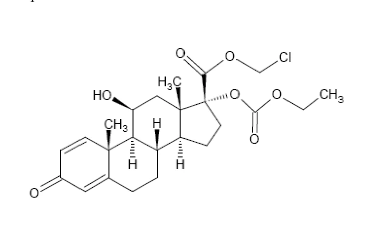
PART II: SCIENTIFIC INFORMATION
PHARMACEUTICAL INFORMATION
Drug Substance
Proper name: loteprednol etabonate
Chemical name: chloromethyl 17α-[(ethoxycarbonyl)oxy]-11β-hydroxy-3-oxoandrosta- 1,4-diene-17β -carboxylate
Molecular formula and molecular mass: C24H31ClO7 Mol. Wt. 466.96
Loteprednol etabonate formula:
Physicochemical properties: Loteprednol etabonate is a white to off-white powder. Loteprednol etabonate is structurally similar to other corticosteroids. However, the number 20 position ketone group is absent. It is highly lipid soluble which enhances its penetration into cells. The suspension is essentially isotonic with a tonicity of 250 to 310 mOsmol/kg. The pH is adjusted to 5.5-5.6.
CLINICAL TRIALS
Study demographics and trial design
Two double-masked, placebo-controlled trials were conducted to evaluate the safety and efficacy of Lotemax™ in controlling anterior chamber cell and flare reaction in patients having cataract surgery with intraocular lens (IOL) implantation.1,2
Both trials were randomized, double-masked, placebo controlled, parallel group multi-center. Both enrolled patients requiring elective cataract removal and posterior chamber intraocular (IOL) lens implantation who, on the day following surgery, exhibited a minimum ACI score (sum of cell and flare reaction) of 3 (moderate, 0 to 9 scale). Patients with history of intraocular or laser surgery on the eye within the past six months or the presence of any ocular pathology other than the cataract (for example vernal conjunctivitis, glaucoma of any kind, giant papillary conjunctivitis, viral or bacterial conjunctivitis, uveitis, retinopathies) were excluded from the trials.
Patients received Lotemax™ or the placebo 4 times a day in the operated eye one day after surgery for up to 14 days.
The evaluation of efficacy for the primary endpoint was the anterior chamber inflammation (ACI) score (the sum of cell and flare ratings) at the final visit (14 days of treatment). Secondary variables assessed for efficacy included: resolution of cell and flare individually, the magnitude of change in the cell and flare ratings, treatment failure and the Investigator’s Global Assessment at final visit.
The patient demographics and completion rates for these 2 trials is outlined in the table below.
| Study | Patients entering/ Completing treatment | Age range (mean) | Percent male/female black/white/other Light/dark Irides | Phacoemulsification Surgery % | Foldable IOL % |
A <125> | 110/99 117/75 | 38-92 (70) | 39/61 4/89/7 58/42 | Lotemax: 91 Placebo: 93 | Lotemax: 44 Placebo: 45 |
B <127> | 102/95 101/69 | 25-99 (71) | 42/58 25/72/3 49/51 | Lotemax: 79 Placebo: 79 | Lotemax: 66 Placebo: 68 |
In both trials by the final visit (last on-treatment observation carried forward (LOCF)), the Anterior Chamber Inflammation (ACI) had resolved in a larger proportion of patients treated with Lotemax™ compared to placebo (P <0.001).
For trial A, by the final visit (LOCF), ACI had resolved in 64% (70/109) of patients in the Lotemax™ group and 29% (33/113) of those in the placebo group (P < .001). The resolution rate and mean change from baseline of the individual components of ACI (cell and flare), as well as other signs and symptoms, was better in the loteprednol etabonate group. The treatment failure rate and the time course of failures were lower in the Lotemax™ group; the differences were clinically meaningful and statistically significant (P < .001). Three patients in the loteprednol etabonate group had an intraocular pressure elevation of 10 mm Hg or more over the preoperative screening value at one or more visits (See Clinical Trial Adverse Drug Reactions).
Resolution of Anterior Chamber Inflammation in Post-Operative Inflammation Trial
| Resolved | |||||||
| Visit | Treatment Group | N at risk | N | % | Treatment effect | 95% C.I. | p value |
| 2(Day 2-6) | Lotemax | 109 | 16 | 14.7% | |||
| Placebo | 111 | 4 | 3.6% | 11.1% | (3.6%, 18.6%) | 0.0043 | |
| 3(Day 7-12) | Lotemax | 102 | 44 | 43.1% | |||
| Placebo | 92 | 17 | 18.5% | 24.7% | (12.2%, 37.1%) | 0.0002 | |
| 4(Days 13 - 20) | Lotemax | 98 | 69 | 70.4% | |||
| Placebo | 76 | 30 | 39.5% | 30.9% | (16.7%, 45.2%) | < 0.0001 | |
Final visit(LOCF) | Lotemax | 109 | 70 | 64.2% | |||
| Placebo | 113 | 33 | 29.2% | 35.0% | (22.7%, 47.3%) | < 0.0001 | |
N at risk is the number of patients with a score > 0 at baseline and a valid on-treatment evaluation for the visit. LOCF is the last valid on-treatment observation, carried forward. Resolved is the proportion of patients at risk for whom the score was 0 at the endpoint. Treatment effect is the difference between treatments in resolution rates (LE - placebo) with investigators pooled. Positive treatment effects indicate that LE is favored over placebo for resolution rate. P-value is from the CMH test for independence of treatment assignment and resolution, controlling for investigator.
The reduction in severity of the ACI scores is shown in the table below for Study A.
| Anterior Chamber Inflammation Score: Change at Each Visit | |||||||
| Visit | Treatment Group | N at risk | Mean Obs. | Chg. | Treatment effect (Median) | 95% C.I. | p value |
1 (Baseline) | LE | 109 | 3.8 | N/A | |||
| Placebo | 113 | 3.8 | N/A | N/A | N/A | N/A | |
2 (Day 2-6) | LE | 109 | 2.2 | -1.6 | |||
| Placebo | 111 | 3.2 | -0.5 | -1.0 | (-1.0, -1.0) | <0.001 | |
3 (Day 7-12) | LE | 102 | 1.2 | -2.6 | |||
| Placebo | 92 | 2.5 | -1.4 | -1.0 | (-2.0, -1.0) | <0.001 | |
4 (Days13-20) | LE | 98 | 0.5 | -3.2 | |||
| Placebo | 76 | 1.2 | -2.7 | -1.0 | (-1.0, -1.0) | 0.027 | |
| Final Visit (LOCF) | LE | 109 | 0.9 | -2.9 | |||
| Placebo | 113 | 2.1 | -1.7 | -1.0 | (-1.0, -1.0) | <0.001 | |
N at risk is the number of patients with a score >0 at baseline with a valid on-treatment evaluation for the visit. LOCF is the last valid on-treatment observation, carried forward. Change is the mean change in severity compared to baseline for that measure. A negative number indicates improvement. Treatment effect is the median difference between treatments, and its 95% confidence interval is estimated by distribution-free methods with investigators pooled. A negative treatment effect indicates that LE is favored over placebo for change. P-value is from the CMH test for equality of treatment group mean ranks for change in severity controlling for investigator.
In study B, the proportion of patients with ACI resolved by the final visit (LOCF) was 55% (56/102) in the Lotemax™ group and 28% (28/100) in the placebo group (p < 0.001). For all the individual components of ACI (cell and flare), as well as other signs and symptoms, the resolution rate and mean change from baseline favored Lotemax™. Expanding the efficacy criterion to include patients with mild inflammation at final visit, the efficacy of Lotemax™ was 93% (95/102), in contrast to 65% for placebo (65/100). The difference in the treatment failure rates, as well the difference in the time-course of
failures was both clinically meaningful and statistically significant, in favor of Lotemax™ (p < 0.001).
Resolution of Anterior Chamber Inflammation in Post-Operative Inflammation Trial
| Resolved | |||||||
| Visit | Treatmen t Group | N at risk | N | % | Treatmen t effect | 95% C.I. | p value |
| 2 (Day 2-6) | Lotemax | 102 | 10 | 9.8% | |||
| Placebo | 100 | 9 | 9.0% | 0.8% | (-7.2%, 8.9%) | 0.8448 | |
| 3 (Day 7-12) | Lotemax | 96 | 33 | 34.4% | |||
| Placebo | 83 | 14 | 16.9% | 17.5% | (5.1%, 30.0%) | 0.0079 | |
4 (Days 13 - 20) | Lotemax | 93 | 54 | 58.1% | |||
| Placebo | 70 | 27 | 38.6% | 19.5% | (4.3%, 34.7%) | 0.0137 | |
| Final visit (LOCF) | Lotemax | 102 | 56 | 54.9% | |||
| Placebo | 100 | 28 | 28.0% | 26.9% | (13.8%, 40.0%) | 0.0001 | |
N at risk is the number of patients with a score > 0 at baseline and a valid on-treatment evaluation for the visit. LOCF is the last valid on-treatment observation, carried forward. Resolved is the proportion of patients at risk for whom the score was 0 at the endpoint. Treatment effect is the difference between treatments in resolution rates (LE - placebo) with investigators pooled. Positive treatment effects indicate that LE is favored over placebo for resolution rate. P-value is from the CMH test for independence of treatment assignment and resolution, controlling for investigator.
The reduction in severity of the ACI scores is shown in the table below for Study B.
| Anterior Chamber Inflammation Score: Change at Each Visit | |||||||
| Visit | Treatment Group | N at risk | Mean Obs. | Chg. | Treatment effect (Median) | 95% C.I. | p value |
1 (Baseline) | LE | 102 | 3.5 | N/A | |||
| Placebo | 100 | 3.7 | N/A | N/A | N/A | N/A | |
2 (Day 2-6) | LE | 102 | 2.2 | -1.3 | |||
| Placebo | 100 | 2.8 | -0.9 | 0.0 | (-1.0, 0.0) | 0.060 | |
3 (Day 7-12) | LE | 96 | 1.4 | -2.2 | |||
| Placebo | 83 | 2.2 | -1.4 | -1.0 | (-1.0, 0.0) | 0.044 | |
4 (Days13-20) | LE | 93 | 0.7 | -2.8 | |||
| Placebo | 70 | 1.4 | -2.3 | 0.0 | (-1.0, 0.0) | 0.145 | |
| Final Visit (LOCF) | LE | 102 | 0.9 | -2.6 | |||
| Placebo | 100 | 2.2 | -1.5 | -1.0 | (-1.0, -1.0) | 0.001 | |
N at risk is the number of patients with a score >0 at baseline with a valid on-treatment evaluation for the visit. LOCF is the last valid on-treatment observation, carried forward. Change is the mean change in severity compared to baseline for that measure. A negative number indicates improvement. Treatment effect is the median difference between treatments, and its 95% confidence interval is estimated by distribution-free methods with investigators pooled. A negative treatment effect indicates that LE is favored over placebo for change. P-value is from the CMH test for equality of treatment group mean ranks for change in severity controlling for investigator.
Both treatments were well tolerated. No clinically significant elevations in intraocular pressure (≥ 10 mm Hg) were seen in the Lotemax™ treatment group in this trial. One
patient in the placebo treatment group met this criterion (See Clinical Trial Adverse Drug Reactions).
Of the four patients whose study participation was terminated due to adverse events, all were in the placebo group.
Thus, Lotemax™ led to a clinically meaningful reduction in the signs and symptoms of post cataract surgery anterior chamber inflammation when compared to placebo in this patient population.
DETAILED PHARMACOLOGY
Results from a competitive binding study indicate that LE has a binding affinity for glucocorticoid (Type II) receptors that is 4.3-times greater than that of dexamethasone and that LE binds competitively to transcortin. In contrast, the LE metabolites, PJ-90 and PJ-91, did not bind to the glucocorticoid receptor.
Primary Pharmacodynamics – Ocular
LE demonstrated anti-inflammatory activity in multiple ocular models of inflammation in rabbits; however, the magnitude of the effect varied depending on the model used and endpoints measured. At the specific doses tested, the anti-inflammatory effects of LE were similar to or less than the effects of the comparator compounds evaluated. In general, these studies were intended to provide proof-of-concept information, and did not include complete dose-vs-response profiles for LE or the comparator compounds that were tested and do not provide definitive information regarding the relative potency of LE vs other anti-inflammatory drugs. A summary of the ocular inflammation models used is shown below.
Ocular Inflammation Models in Rabbits
- i) Paracentesis, nitrogen mustard, Shigella endotoxin, and immune uveitis models
Summary: LE (0.5%) inhibited an increase in protein levels in the aqueous humor in all models, with similar efficacy to dexamethasone (0.1%) and flurbiprofen (0.03%).
ii) Intravitreal endotoxin-induced (E. coli) ocular inflammation model
Summary: LE (1%) demonstrated anti-inflammatory effects that were similar to, or less than those observed with prednisolone (1%). Both agents reduced leukocyte infiltration into aqueous humor. Prednisolone, but not LE, also reduced myeloperoxidase (MPO) activity in iris/ciliary body as compared to placebo.
iii) Acute (endotoxin-induced) uveitis model
Summary: LE (0.5%) demonstrated anti-inflammatory activity, as assessed by conjunctival injection, anterior chamber flare, fibrin, and iris hyperemia, with no significant effect on anterior chamber cells and aqueous protein levels in this model. By several of these measures, anti-inflammatory activity of LE was less than that observed for dexamethasone (0.1%) and/or fluorometholone (0.1%).
iv) Chronic adjuvant-induced immune uveitis model
Summary: Anti-inflammatory effects of LE (1%) was similar to dexamethasone (0.1%), but less than fluorometholone (0.1%) based on conjunctival injection, cornea edema, cornea neovascularization, anterior chamber cells and flare, iris hyperemia, and aqueous protein levels.
- v) Clove oil-induced corneal inflammation model
Summary: Following inoculation of clove oil into the cornea stroma, 0.5% LE was the minimum effective dose in this model, with no anti-inflammatory effects observed at lower doses (0.05% and 0.1%). Higher LE doses (1% and 2%) resulted in maximal anti-inflammatory effects in this model, with LE, 0.5% and 1%, producing equivalent efficacy to prednisolone, 0.125% and 1%, respectively.
Primary Pharmacodynamics – Non-Ocular
The anti-inflammatory effects of LE were also studied in multiple non-ocular models of inflammation in rats and mice.
Non-Ocular Inflammation Models
Model Species Compounds and
Doses Tested
Croton oil-induced ear
edema Rat, Mouse
LE (0.1%)
betamethasone (0.12%) hydrocortisone (0.1%)
DNFB-induced dermatitis Rat LE (0.1%)
hydrocortisone (0.1%)
Cotton pellet granuloma
assay Rat
LE
hydrocortisone
betamethasone
(dose of ≤ 10 mg/pellet for each compound)
Histamine-induced vascular
permeability Rat
LE (0.1%)
dexamethasone (0.1%) hydrocortisone (0.1%)
Carrageenan-induced skin
and paw edema Rat
LE (0.1%)
dexamethasone (0.1%) hydrocortisone (0.1%)
Adjuvant-induced arthritis Rat LE (0.1%)
dexamethasone (0.1%)
Results from these studies support the classification of LE as a topical anti-inflammatory steroid when administered directly to the site of inflammation. Depending on the model, the effects of LE similar to or less than the effects of the other corticosteroids tested following direct application to the inflamed site. LE did not elicit a significant response in the DNFB-induced dermatitis model, and anti-inflammatory effects were not observed in the 2 models where systemic absorption and subsequent distribution to the inflamed site was required (adjuvant-induced arthritis model and carrageenan–induced skin and paw edema model), likely due to the high systemic clearance of LE resulting in low systemic availability in rats.
Secondary Pharmacodynamics
Wound Healing and Scar Formation: The effect of LE on the wound healing and scarring process was assessed in several in vivo and in vitro studies. In rabbits, treatment (2 drops, TID for 15 days, single eye) of full-thickness corneal wounds with LE (0.1%) or dexamethasone (0.1%) resulted in decreased scar formation, inhibition of inflammatory cell infiltration, and inhibition of fibroblast proliferation compared with untreated eyes. The effect was most prominent after day 7, during the time when collagen deposition was evident in untreated eyes. A separate study examined the effect of LE on the corneal wound healing process in rabbits following corneal incision. BID treatment with LE (1%) or prednisolone (1%) for 9 days (17 doses) resulted in a significant decrease in the tensile strength of the resulting scar, which was less than that observed with dexamethasone (0.1%).
Intra-ocular Pressure: Topical ocular treatment of normotensive rabbits with LE (0.1%, 1 dose per hour for 7 hours on two consecutive days) did not result in a sustained rise in IOP during the 55-hr interval following the first administration. In contrast, treatment with dexamethasone (0.1%) with the same dosing regimen produced a statistically significant rise (3-5 mmHg) in IOP, which persisted for 48 hours after the initial dose.
Skin Atrophy and Thymus Changes: The potential effect of LE on skin atrophy and thymus changes were evaluated in rats following topical (dermal) administration at LE doses of 200 mg/rat/day for 14 days or 400 mg/rat/day for 7 days. Both hydrocortisone-17-butyrate and betamethasone-17-valerate caused decreased skin weight and/or skin thickness, as well as a significant decrease in thymus weight. In contrast, LE treatment resulted in a significant decrease in skin weight, but did not affect thymus weight in this study.
Pharmacokinetics
The chemical structure of LE and the two metabolites evaluated in pharmacokinetic studies are shown in the figure below.
Chemical Structure of Loteprednol Etabonate, PJ-90, and PJ-91
O Cl
O
O
O
O
OH
O
OH
OH
O
OH
O
OH
O
O
O
O
OH
O
Loteprednol Etabonate PJ-90 PJ-91
Ocular Pharmacokinetics in Rabbits
The ocular pharmacokinetic properties of 14C-labeled LE (0.5%) were evaluated following topical ocular administration (3 drops per eye at 5-minute intervals) to New Zealand white rabbits. LE was absorbed into ocular tissues, and the presence of metabolites in these tissues suggests that LE is metabolized in ocular tissues following ocular dosing (see table below). Blood levels of LE and its metabolites were not detectable after topical ocular administration in this study.
Mean concentrations of LE and metabolites (nmoles/g) in ocular tissues following multiple topical ocular administration of [14C]LE to albino rabbits
Collection Time (hr) Tissue Analyte 0.5 1 2 4 6 8
LE 30.5 21.5 16.3 11.5 2.5 5.3 Conjunctiva Ma 3.5 1.8 1.2 0.8 0.4 1.0
LE 3.8 2.1 1.3 0.9 0.6 1.1 Cornea M 4.9 4.1 3.2 2.2 1.8 2.2
LE 1.9 1.1 0.7 0.7 0.2 0.9 Iris/Ciliary
Body M 0.4 0.3 0.2 0.3 0.1 0.2
LE 0.027 0.016 0.007 0.003 0.002 0.002 Aqueous
Humor M 0.017 0.023 0.025 0.011 0.007 0.006 a Metabolites. Includes all metabolites contained in aqueous phase following extraction of LE into organic solvent.
The administration of other concomitant ophthalmic drugs during the 30 min prior to or after LE administration did not result in meaningful changes in ocular tissue levels of LE.
Systemic Pharmacokinetics in Rats and Dogs
The systemic pharmacokinetics of LE were assessed following intravenous and oral administration (5 mg/kg) to four mongrel dogs. Blood levels of LE declined rapidly following intravenous administration, showing a biexponential plasma concentration profile. LE was eliminated from the plasma with a total body clearance of 22 L/hr and a half-life of 2.8 hr. LE had a large volume of distribution (37 L), characteristic of lipophilic drugs of this class. No LE was detected in the plasma after oral administration or in the urine after either intravenous or oral administration.
In Sprague-Dawley (SD) rats (5 males/group), following oral administration of [14C]LE (5 mg/kg), levels of [14C]LE in blood were relatively low and constant (20-33 ng/mL) throughout the 5-hr sampling period. Of the tissues analyzed in this study, LE and PJ-91 levels were highest in liver, with maximal concentrations of approximately 1.9 μg/g for LE and 1.3 μg/g for PJ-91. LE levels in liver tended to be higher than PJ-91 levels; however, for other tissues, LE levels tended to be lower than (blood and kidney) or roughly similar to (heart and lung) PJ-91 levels.
Following intravenous administration to SD rats (3/group), LE was rapidly cleared from plasma in a biphasic manner, with half-life estimates of approximately 16-49 min, depending on the dose (see table below). Total clearance of LE from plasma was dose-dependent and decreased with increasing dosage. During the 4-hr collection interval following dosing, approximately 9% of the administered dose was recovered in bile in the form of PJ-91 and PJ-90. Measurable levels of LE and PJ-91, but not PJ-90, were observed in urine, with <4% of the administered dose recovered as intact LE during the 2.5-hr interval after dosing. These results suggest that the liver is an important site for the metabolism of LE and that biliary excretion of the metabolites of LE is a significant route of elimination.
Pharmacokinetic parameter values for LE in plasma following intravenous administration to rats
Dose
(mg/kg)
AUC
(μg•min/mL)
CL
(mL/min/kg)
T½ (min)
MRT (min)
1 9.2 ± 0.4 108.53 ± 4.47 15.92 ± 1.23 17.59 ± 0.95 2 16.0 ± 1.1 125.76 ± 9.01 17.22 ± 1.71 18.34 ± 0.80 5 56.1 ± 6.2 90.28 ± 9.98 29.49 ± 0.00 31.98 ± 0.78 10 159.2 ± 31.3 67.35 ± 11.62 43.41 ± 7.58 48.72 ± 8.95 20 333.2 ± 17.9 60.35 ± 3.09 48.82 ± 1.52 51.79 ± 1.70 Abbreviations: AUC: Area under the concentration-time curve, CL: systemic clearance; T½: apparent terminal phase half-life; MRT: mean residence time.
In Vitro Studies
The in vitro metabolic stability of LE was investigated following incubation with rat, rabbit, and dog plasma, as well as human liver homogenate. LE was rapidly metabolized in rat plasma, with nearly 100% disappearance of intact LE within 30 min; however, no metabolism of LE was evident in rabbit, dog, or human plasma. In human liver homogenate, LE metabolism, while not complete, was more extensive than the other steroids tested.
Metabolic stability of LE and other steroids in human liver homogenate
Compound % Remaining at 30 min
LE 73
Prednisolone 105
Dexamethasone 102
Betamethasone 89
Plasma protein binding and distribution into red blood cells of LE (6.2-18.5 μg/mL) and the metabolite, PJ-91 (5-15 μg/mL), was investigated in vitro in dog blood. LE was highly bound to plasma proteins (mean±SD of 95.3 ± 3.0% bound) over this concentration range. In contrast, PJ-91 was approximately 73% bound to plasma proteins. LE and PJ-91 distributed into red blood cells with a partition coefficient of 7.8 and 0.25, respectively.
Pharmacokinetic Summary
The available pharmacokinetic data from in vivo and in vitro studies indicate that LE is readily absorbed into ocular tissues, with low systemic exposure following topical ocular administration. To the extent that LE reaches the systemic circulation, data from rats suggest that it is extensively metabolized and subsequently excreted via bile and urine. LE is highly protein bound in plasma, and distributes preferentially into the cellular components of blood. Although LE is rapidly hydrolyzed in rat blood, systemic metabolism in humans likely occurs in the liver.
MICROBIOLOGY
This section is not applicable.
TOXICOLOGY
Single-dose toxicity
Acute oral toxicity studies in rats and mice indicate that the MTD for loteprednol etabonate (=LE) is greater than or in the region of 4000 mg/kg bodyweight, which is ~27,000 and ~14,000 times the Lotemax™ clinical dose, respectively. The MTD for both species by the subcutaneous route (rats and mice) was found to be >1333 mg/kg bodyweight (the maximum practical dose by this route). Apparent reductions in spleen size were noted in both species at necropsy following subcutaneous administration and may be treatment related. Combinations of 0.5% LE ophthalmic suspension with sulfacetamide (10% w/v) or Tobramycin (0.3% w/v) were not toxic by the oral route in rats or mice at a dose volume of 20 mL/kg bodyweight and the MTD of the possible secondary metabolite of loteprednol etabonate, PJ-90, was shown to be >100 mg/kg bodyweight when administered subcutaneously in the rat. These data indicate that LE is of a low order of acute toxicity.
Repeat-dose toxicity – sub-chronic studies
Twenty-eight day toxicity studies conducted in the rat by the oral route (0.5, 5, or 50 mg/kg/day) and in the rabbit by ocular administration (0.1 ml/day of LE 0.1%, 0.7%, or 5%) identified the liver as a potential target organ for LE. Increased ALT and glucose levels were identified at the high dosage level in both studies, together with other, less consistent biochemical changes suggestive of hepatotoxic effects.
No histological changes were seen in the liver in either species. This suggests that the observed biochemical changes represent a hypertrophic effect consistent with the liver being a significant site of metabolism of LE, as indicated by the presence of high concentrations of both metabolites of LE in bile fluid. Other changes observed in these studies were generally consistent with the effects which would be expected following administration of high doses of corticosteroids.
The hepatic effects observed in the rat study mainly occurred at the high dose level which represented a multiple in excess of ~400 times the anticipated human dose of Lotemax™, although some evidence of hepatotoxicity was also apparent at the intermediate dose level (>~40 times the anticipated human dose of Lotemax™. In the rabbit study, hepatic effects were restricted to the high dosage group (i.e., ~40 times the equivalent human dose of Lotemax™. The low dose (0.5 mg/kg/day) in rats, which exceeded 4 times the equivalent human dose of Lotemax™, was a no effect level. In rabbits, no significant toxicity was noted at the 0.7% dose, which is equivalent to ~ 6 times the human dose of Lotemax™. Complete systemic absorption of LE to the blood following Lotemax™ administered by ocular route is not expected in humans. See ACTION and CLINICAL PHARMACOLOGY.
No adverse ocular effects were observed following administration of LE at concentrations of up to 5% in 2-hydroxypropyl-β-cyclodextrin, of LE, 0.5% in combination with Tobramycin, 0.3% or of LE, 0.5% in combination with sulfacetamide sodium, 10% for 30 days. Similarly, no adverse effects were apparent following ocular administration of PJ-90, a possible secondary metabolite of LE, for 28 days, however, based on this single study and its limitations, no definitive conclusions can be drawn regarding the potential toxic effect of PJ-90.
Repeat-dose toxicity – chronic studies
In a six-month study, rabbits were exposed to loteprednol etabonate LE 0.5% ocular drops (30µ mL) eight times daily for the first week, and then four times daily thereafter. No significant ocular signs were reported. The average adrenals’ weight in the exposed group was significantly lower, but no corresponding microscopic modifications of the adrenals were observed. Thymus involution was observed more frequently in the treated females. The dose used in rabbits was equivalent to ~5 times the Lotemax™ human dose.
In a one-year study, dogs received 6 drops daily of dexamethasone 0.1%, LE 0.1%, or 0.5% - the latter high dose represents twice the human dose of Lotemax™. An increasing incidence of stromal anomalies ranging from fine haze to crystalline deposits in the cornea of the treated eye in animals receiving LE 0.5% was noted between Week 26 and Week 52. A few of those treated with LE 0.1%, had stromal anomalies, but only at week-52. Some IOP increase (≥5 mmHg) was reported in few animals starting on week-13, however, no clear dose-response or time trend were present. There were no apparent toxic effects on the adrenal glands as confirmed by the histological reports. On the other hand, the number of animals with IOP increase (≥5 mmHg) among those treated with dexamethasone 0.1% was larger and increased with time, and by week-52, almost all dogs treated with dexamethasone 0.1% experienced IOP increase. Also, in contrast to the LE groups, dogs treated with dexamethasone 0.1% had a significant reduction of males’ bodyweight, and adrenals organ weights. The latter was confirmed by the presence of cortical atrophy of the adrenals in all animals treated with dexamethasone. Thymus involution was also more marked in the 5 dexamethasone 0.1% group. Please note that corneal opacities were seen exclusively in dogs and not in any other studied animals (rats, rabbits).
In a six- month study, rabbits exposed to LE 0.5% ocular drops 6 times daily experienced no significant IOP increase, or corneal deposits. However, small adrenal glands were noted in 3/10 animals and were correlated by a lower average adrenals weight and corresponding histological changes (e.g., atrophy). These effects were seen mainly in animal treated for 6 months with the equivalent of ~ 7 times the intended Lotemax™ human dose.
Genotoxicity
Within the limitations imposed by the relative insolubility of LE, no evidence of mutagenic potential was apparent in the four in vitro tests conducted. No evidence of mutagenicity was apparent in the micronucleus test at dose levels in the region of 4,000 mg/kg bodyweight which, although probably slightly less than the maximum tolerated
dose by the oral route in the mouse, equates to an exposure equivalent to ~14,000 times the Lotemax™ clinical dose.
Developmental and reproductive toxicology
In the fertility and general reproductive study in rats clear evidence of parental (F0 generation) toxicity was demonstrated at the high dose levels of loteprednol etabonate (males, 50 mg/kg/day; females 25 mg/kg/day), and to a lesser extent at the intermediate 5 mg/kg/day level which is equivalent to ~40 times the Lotemax™ clinical dose. The fertility and mating performance of the F0 generation was unaffected by treatment. However, pregnancies and pregnancy outcomes were significantly affected (e.g., longer gestation, marked decrease in live foetuses, and poor foetus and pups survival).
Clear evidence of toxicity was observed for Fl generation foetuses and pups produced from F0 animals of the intermediate and high dosage groups. With the exception of slight growth retardation, pups of F0 parents receiving the low dose level (0.5 mg/kg/day,) were unaffected by parental treatment (i.e., ~4 times the Lotemax™ clinical dose). The mating performance of the F1 generation and the F2 generation was unaffected by F0 treatment.
Maternal toxicity was demonstrated in the rabbit embryotoxicity study at 3 mg/kg/day LE (i.e., equivalent to ~50 times the Lotemax™ clinical dose), together with clear evidence of embryotoxicity characterized by slight developmental retardation.. There was also some evidence of teratogenicity as meningocele (major abnormality) in some foetuses and an increased incidence of abnormal left common carotid artery (minor abnormality) were noted. In the 0.5 mg/kg/day group, an increase in the occurrence of abnormal left common carotid artery was suggested, but there were no major adverse effects on embryonic or foetal development at 0.1 or 0.5 mg/kg/day LE. The 0.5 mg/kg/day dose is equivalent to ~8 times the Lotemax™ clinical dose.
In the rat embryotoxicity study, evidence of maternal toxicity was apparent at dose levels of 5, 50 and 100 mg/kg/day and clear evidence of embryotoxicity and teratogenicity was observed for groups receiving 50 and 100 mg/kg/day of loteprednol etabonate. These toxic effects included major abnormalities, such as cleft palate, umbilical hernia, and aortic arches abnormalities. No evidence of major embryotoxicity or teratogenicity was seen at dose levels of 0.5 or 5 mg/kg/day. The latter dose is equivalent to ~40 times the Lotemax™ clinical dose.
In the peri- and post-natal study in rats, maternal toxicity was demonstrated following treatment with loteprednol etabonate during late pregnancy and lactation at dose levels of 0.5, 5 and 50 mg/kg/day. However, no effects on the onset or progress of parturition were observed in any of the treated groups. Maternal treatment elicited clear toxic effects in the offspring at 50 mg/kg/day which included reduced bodyweight, developmental retardation, poor survival and clinical condition, and an increased incidence of umbilical hernia. At 5 mg/kg/day, effects on the offspring were limited to lower birth weight and possibly to the occurrence of umbilical hernia in one pup. There was no apparent toxicity in the offspring at 0.5 mg/kg, which is equivalent to 4 times the Lotemax™ clinical dose.
Carcinogenicity
Long-term animal studies have not been conducted to evaluate the carcinogenic potential of loteprednol etabonate. Loteprednol etabonate was not genotoxic in vitro in the Ames test, the mouse lymphoma tk assay, or in a chromosome aberration test in human lymphocytes, or in vivo in the single dose mouse micronucleus assay.
Delayed contact hypersensitivity study
The sensitizing potential of LE was evaluated in the guinea-pig using a modification of the Buehler test4 using a cream formulation which, presumably, differs in terms of excipients from the intended ophthalmic formulation. There was no evidence to suggest that LE, 0.5% cream had the potential to induce delayed contact hypersensitivity.
REFERENCES
- 1.
Stewart RH, Horwitz B, Howes J, Novack GD, Hart K for the Loteprednol Etabonate
Post-operative Inflammation Study Group 1. Double-masked, placebo-controlled evaluation of 0.5% loteprednol etabonate in the treatment of post-operative inflammation. J Cataract Refract Surg. 1998; 24:1480-1489.
- 2. The Loteprednol Etabonate Postoperative Inflammation Study Group 2. A double-masked, placebo-controlled evaluation of 0.5% loteprednol etabonate in the treatment of postoperative inflammation. Ophthalmol. 1998;105:1780-1786.
- 3. Howes J, Novack GD. Failure to detect systemic levels and effects of loteprednol etabonate and its metabolite, PJ-91, following chronic ocular administration. J Ocular Pharmacol Ther. 1998;14:153-158.
- 4. Ritz HL, Buehler EV. Planning, conduct and interpretation of guinea pig sensitization patch tests. In: Drill VA, Lazar P, editors. Current concepts in cutaneous toxicity. New York: Academic Press; 1980. p. 25–40.